How to prepare for an ultrasound of the stomach?
How reliable the results of the study are depends on whether gastric ultrasound is performed by specialists and on proper preparation for the gastric ultrasound procedure.
You are required to follow a diet for about two days before the procedure.
Avoid: foods that cause gas (rye bread, peas, beans, cabbage, kefir, carbonated mineral water, fresh fruits and vegetables).
- Questions often arise before an ultrasound: is it done on an empty stomach or not, is it possible to eat before an ultrasound of the stomach. The last meal on the day preceding the study should be no later than seven to eight o’clock in the evening.
- On the morning of the ultrasound of the stomach and intestines, do not eat, drink or smoke.
- Patients with severe hunger pains are allowed to drink half a glass of tea and eat a cracker.
How is the procedure done?
The examination is painless and does not imply any violation of the integrity of the skin. There are different methods for doing it.
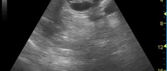
Transabdominal research methods
The transabdominal ultrasound method is an ultrasound examination that is performed using a surface sensor.
The examination is carried out through all layers of the abdominal wall.
- The patient in the ultrasound room must expose the anterior abdominal wall.
- After this, the patient takes a comfortable position on the couch.
- A special gel is applied to the stomach.
- During the procedure, the doctor may ask you to turn on your side or stomach in order to examine the organs from a better projection.
- The doctor can show the patient the progress of the examination on the monitor.
- If preparation for a transabdominal ultrasound was carried out normally, the procedure lasts about half an hour.
This diagnostic method is the most common, since it is completely painless, and during the examination the sensor is not inserted into the body.
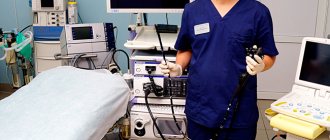
Endoscopic ultrasound
Endosonography is a combination of ultrasound examination of internal organs and endoscopy, which allows you to bring an ultrasound sensor directly to the organ being examined.
This method is used when the necessary data cannot be obtained by transabdominal ultrasound.
- The person lies on his left side and bends his knees slightly.
- The endoscope is carefully inserted through the oral cavity or through the nasal cavity into the lumen of the pharynx and esophagus, which allows the attending physician to examine nearby lungs and lymph nodes using an ultrasound probe.
- By moving the endoscope further into the stomach, the doctor can evaluate the condition of its walls, as well as the nearby spleen and pancreas.
- When the sensor is inserted into the duodenum, the patency and condition of the bile ducts and the head of the pancreas are assessed, where the tumor process is very often localized.
The study is performed using medicated sleep or while taking sedative medications. In particular, medicated sleep is necessary when performing a biopsy of suspicious formations or when performing a study through the colon.
The procedure can take from 20 minutes to 2 hours, depending on the purpose of the examination, as well as the need for additional biopsy or surgery.
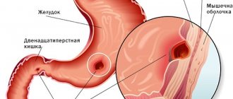
Growing attention to the problem of Barrett's esophagus (BE) is caused by the high incidence of esophageal adenocarcinoma (EA) - a malignant tumor of the esophagus that develops from metaplastic intestinal epithelium of the mucous membrane in BE or (extremely rarely) from the esophageal glands (type I according to the classification of J. Siewert) . Over the past decades, there have been significant changes in the incidence of AP and squamous cell carcinoma of the esophagus. Now, in most cases of esophageal cancer in Western Europe and the USA, it is AP that is detected. In the United States, the incidence of AP has increased by almost 300% over the past 30 years (Fig. 1)
Rice. 1. Increase in the incidence of esophageal adenocarcinoma in the United States and a decrease in the incidence of squamous cell carcinoma (1975–2000) [3]. [12].
In Russia, this trend is not so clearly visible: 7-20% of diagnosed esophageal cancers have histological signs of adenocarcinoma. However, it is difficult to assess the reliability and accuracy of such data: the disease is detected in the late stages of advanced cancer, when it is impossible to determine where the tumor has formed - in the distal esophagus or in the proximal stomach. Among all oncopathologies of the digestive system, esophageal cancer and stomach cancer have the highest mortality rates. The prognosis after the diagnosis of adenocarcinoma is unfavorable - 5-year survival rate does not exceed 10-20%, and improvements in surgical treatment techniques do not make such a significant contribution to improving these indicators [4].
Understanding of the causes of AP and precancerous pathology, the possibility of early detection of precancerous processes and early forms of tumors have improved significantly in recent years. This makes it possible to form groups at increased risk of AP and organize expert centers for the identification and timely treatment of precancerous pathology. According to authoritative experts, adenocarcinoma of the digestive tract most often develops in pathologically altered mucous membranes. The chronic inflammatory process is considered as one of the important links in the chain of processes that ultimately lead to AP [5,6]. The main diseases against which cancer develops significantly more often have also been identified. For A.P. the underlying disease in most cases is BE.
Screening
The risk of developing AP in patients with BE largely depends on the presence of intestinal metaplasia, dysplastic changes in the epithelium and the degree of their severity in the distal esophagus (in the segment of columnar cell metaplasia of the distal esophagus). Based on observational data from 8522 patients included in the Northern Ireland cancer registry (average follow-up of 7 years), the risk of developing adenocarcinoma increased from 0.04 to 0.23% per year in the presence of intestinal metaplasia with goblet cells, and in the presence of epithelial dysplasia low degree in the metaplasia segment - the risk increases more than 9 times. The cumulative risk of developing adenocarcinoma and high-grade epithelial dysplasia is also significantly higher in groups of patients who, at the time of inclusion in the registry, had morphological changes characterized by intestinal metaplasia and low-grade dysplasia (Table 1)
Table 1. Risk of esophageal adenocarcinoma in patients with Barrett's esophagus in the presence of intestinal metaplasia and dysplasia [7].
The risk of developing AP against the background of BE is even more significant in patients with epithelial dysplasia in the area of the segment of columnar cell metaplasia and especially high-grade dysplasia (Fig. 2).
Rice. 2. The risk of developing cancer in patients with a segment of columnar cell metaplasia without intestinal epithelium, with Barrett’s esophagus without dysplasia and with low- and high-grade dysplasia of the mucosal epithelium [8-11].
However, the appearance of AP is preceded by a gradual progression of dysplastic changes with the cells losing signs of differentiation. Progression from mild dysplasia (low-grade dysplasia) to severe (high-grade dysplasia) can occur on average within 29 months, while the subsequent development of adenocarcinoma takes 2 times less time - 14 months [7]. The widespread prevalence of GERD and the identification of the cascade of precancerous events are potentially attractive targets for screening for BE and AP. One of the latest American recommendations (American College of Gastroenterology) suggests that endoscopic examination of the upper digestive tract is necessary for all patients with symptoms of chronic GERD, due to their high risk of BE [12]. The European Society of Gastrointestinal Endoscopy noted that at present screening is advisable only in groups at high risk of developing adenocarcinoma: in the presence of a long history of GERD (more than 5 years), with a combination of such risk factors as the patient’s age over 50 years, male gender, the presence of blood relatives who were diagnosed with AP or BE, high body mass index and other factors [13]. Among all screening methods, endoscopic examination of the upper digestive tract is the “gold standard” in the diagnosis of pathological processes in the mucous membrane of the esophagus, as it allows not only to visually determine the presence of columnar epithelium in the esophagus, but also to perform a biopsy for morphological confirmation of the diagnosis, diagnosis of dysplasia and early cancer . This allows us to consider the endoscopic method the most important screening tool for BE and AP.
Definition
For a long time, the following definition of BE was generally accepted in clinical practice: this is a pathological condition in which part of the squamous epithelium of the mucous membrane of the distal parts of the esophagus is replaced by metaplastic columnar epithelium. A segment of columnar metaplasia should be identified by endoscopic examination, located above the esophagogastric junction or junction (Z-line) and confirmed morphologically by detecting specialized intestinal metaplasia (Fig. 3)
Rice. 3. Barrett's esophagus. a — endoscopic picture when examined in white light; b — endoscopic picture when examined in a narrow-spectrum mode (Narrow Band Imaging, NBI); c — histological picture of columnar cell metaplasia with diffuse intestinal metaplasia with many goblet cells — hematoxylin and eosin staining, ×100; d — Alcian blue staining, ×100. [14]. In 2021, the European Society of Gastrointestinal Endoscopy (ESGE) proposed a more modern definition of BE: it is a segment of columnar metaplastic epithelium of the distal esophagus, at least 1 cm in length, which can be diagnosed by endoscopic examination proximal to the esophagogastric junction and confirmed histologically by the presence in biopsy material of specialized intestinal metaplasia [15].
In accordance with the recommendations of the Russian Gastroenterological Association (RGA), at the moment, to establish a diagnosis of Barrett's esophagus, mandatory histological confirmation of metaplasia of the epithelium of the mucous membrane of the esophagus of the intestinal type is required, since this is the only type of columnar epithelium characterized by an increased potential for malignancy [16].
From this definition it follows that endoscopic and morphological studies are the basis for the correct diagnosis of this pathological condition. Timely diagnosis of BE, dysplastic changes in the mucous membrane and early forms of AP depends on the endoscopist, his knowledge, methodological skills, correct interpretation of identified changes in the mucous membrane, experience and, finally, technical equipment. However, intestinal metaplasia is not always an area of AP growth, and therefore in Japan, to establish the diagnosis of BE, it is only necessary to detect a segment of columnar cell metaplasia in the distal esophagus during endoscopic examination.
Prevalence of gastroesophageal reflux disease and columnar cell metaplasia in the distal esophagus
BE is one of the most serious complications of GERD - a chronic relapsing disease caused by spontaneous, regularly recurring retrograde entry of gastric and/or duodenal contents into the esophagus, leading to damage to the distal esophagus and/or the appearance of characteristic symptoms (heartburn, retrosternal pain, dysphagia). As an independent nosological entity, GERD was officially recognized in the materials on the diagnosis and treatment of this disease, adopted in October 1997 at the Interdisciplinary Congress of Gastroenterologists and Endoscopists in Genval (Belgium) [17]. The results of studies conducted around the world and covering large populations show that symptoms of GERD are experienced by more than 1/3 of the population, and the presence of daily heartburn is 7-10%. Complications of reflux disease and heartburn (the main symptom of GERD) are much more common in whites (12.3% and 34.6%, respectively) and blacks (2.8% and 46.1%, respectively) compared with East Asians (0 and 2.6%, respectively [18]. It is quite difficult to estimate the prevalence of columnar cell metaplasia in the distal esophagus, since more than 80% of cases of the disease remain undiagnosed, as shown by data from an American study of autopsy material [19]. The results of a study conducted in the United States of this The same group of specialists (A. Cameron et al.) show a 28-fold increase in the number of clinically diagnosed cases of metaplastic changes in the distal esophagus for the period from 1965 to 1995. Data from endoscopic examination of the upper digestive tract in patients admitted to clinics with symptoms dyspepsia or for colonoscopy, indicate differences in the prevalence of columnar cell metaplasia of the esophagus depending on ethnic and geographical factors (Table 2).
Table 2. Frequency of detection of Barrett's esophagus during endoscopic examination of the upper digestive tract in patients with dyspepsia
The prevalence of GERD in Russia among the adult population is about 40%, and 45-80% of them have esophagitis: in 10-35% of cases it is severe esophagitis with multiple erosive lesions of the mucous membrane of the distal esophagus. BE of varying degrees of extent is diagnosed on average in 8-15% of patients with esophagitis. AP develops in 0.5% of patients with BE per year with low-grade dysplasia, and in 6% per year with high-grade epithelial dysplasia [26]. Risk factors for BE include middle and old age, male gender. In most patients, this pathology is diagnosed at the age of 50-60 years, in men 3-4 times more often than in women.
Pathogenesis
One of the main causes of the development of GERD and BE is gastroesophageal reflux - the reflux (entry) of gastric contents (primarily hydrochloric acid) into the esophagus. With the development of such reflux, the pH in the distal esophagus shifts significantly towards low values due to the ingress of acidic gastric contents. Prolonged contact of the esophageal mucosa with acid refluxate, which also contains pepsin, contributes to the development of its inflammation. Bile acids and enzymes, which can also be part of the refluxate, can have a strong damaging effect on the esophageal mucosa if the motility of the upper digestive tract is impaired. Esophagitis in some cases is accompanied by a structural restructuring of the epithelium of the esophageal mucosa with the formation of gastric or intestinal metaplasia, which is the background for the development of adenocarcinoma. An analysis of numerous studies shows that the risk of developing cancer in the segment of columnar cell metaplasia is primarily associated with the presence of intestinal metaplasia (incomplete intestinal metaplasia, type II and III) [27, 28]. In the esophagus, metaplastic changes begin with the appearance first of columnar epithelium of the gastric type, and then of the colonic type epithelium. Exposure to acid in the esophagus increases, on the one hand, the activity of protein kinases that initiate the mitogenic activity of cells and, accordingly, their proliferation, and, on the other hand, inhibits apoptosis in the affected areas of the mucous membrane. Approximately 50–80% of cases of dysplasia associated with BE and AP are characterized by mutations in genes involved in cell cycle regulation, DNA repair, and apoptosis [20]. Recent research in this area indicates the important role of the genes (proteins) P53 and P63 involved in the development of squamous epithelial cells. In the esophagus, P63 protein expression is detected only in squamous epithelial cells and is absent in columnar cell metaplasia. In the absence of P63, mucosal stem cells cannot begin differentiation along the path of squamous epithelial cells; as a result of this disorder, columnar epithelial cells are formed [29, 30]. The basis for the origin of intestinal-type epithelial cells can be both the stratified squamous and cuboidal epithelium of the ducts of the glands of the submucosal layer of the esophagus, and the cardiac-type epithelium in the distal esophagus, exposed to refluxate [31, 32]. Among the factors influencing the processes of carcinogenesis in the area of the esophagogastric junction, it is also necessary to take into account smoking, alcohol, increased body weight and bile reflux. Dynamic observations of patients with BE showed that the development of adenocarcinoma occurs through a multi-stage pathological process. This process is characterized by an increase in the degree of dysplasia, a pathology that precedes adenocarcinoma. An important promoter of this process is nitric oxide, which can accumulate in pathologically altered tissues of the distal esophagus and cause genetic changes. Genetic changes in cells occur parallel to the transition from metaplasia to dysplasia and then to adenocarcinoma [33].
Diagnosis and screening of Barrett's esophagus and esophageal adenocarcinoma
Endoscopic examination is key in diagnosing BE: while other methods (x-ray, scintigraphy) can only suggest this diagnosis, the endoscopic method can establish it with a high degree of probability. An endoscopic examination determines the extent of changes in the mucous membrane, the ratio of the zone of changes in the mucous membrane along the length to the esophagogastric junction, as well as the proximal border in relation to the incisors. In this case, the spread of the metaplasia zone is clearly visualized in the form of foci of hyperemia (“tongues of flame”) against the background of the “pearly white” epithelium of the esophagus. One of the important tasks of endoscopic examination is obtaining biopsy material. The objectives of the morphological study are to confirm metaplasia of the esophageal mucosa, identify areas of dysplasia and foci of adenocarcinoma.
From an endoscopic perspective, accurate diagnosis of BE presents several challenges. One of them is to determine the key landmarks - the zone of the esophagogastric junction or junction (EGJ), the Z-line and the boundaries of the segment of columnar cell metaplasia. Another is the accuracy of performing a biopsy in the focal distribution of areas of intestinal metaplasia and dysplasia in the segment of columnar cell metaplasia of the esophagus and the difficulties of endoscopic diagnosis of these foci [34].
Key guidelines for endoscopic diagnosis
1. The zone of the esophagogastric junction (junction) is the area of connection between the muscular layer of the esophagus and the muscular layer of the cardia of the stomach. It corresponds to the boundary between the tubular structure of the esophagus and the proximal part of the stomach with longitudinal folds. This anatomical boundary is defined in the region of the proximal edge of the longitudinal folds of the gastric mucosa (Fig. 4).
Rice.
4. Endoscopic picture of Barrett's esophagus. The arrows indicate the proximal edge of the folds of the gastric mucosa, corresponding to the area of the esophagogastric junction. Another additional landmark of the esophagogastric junction is the distal border of the longitudinal vessels of the mucous membrane visible during endoscopy (palisade or longitudinal intramucosal vessels)
. These vessels were first described by J. Carvalho in 1963 and are veins located in the mucosa above the muscularis propria. In the stomach they are located in the submucosal layer and extend into the superficial layers of the mucous membrane only in the area of the border of the stomach and esophagus. In the mucous layer of the esophagus, they run proximally for about 2 cm parallel to each other in the form of a “picket fence” and then plunge back into the submucosal layer, forming larger veins. The Japanese Society for the Study of Diseases of the Esophagus recommends defining the esophagogastric junction precisely along the distal edge of these longitudinal vessels (Fig. 5)
Rice. 5. Longitudinal vessels of the mucous membrane of the distal esophagus (examination in the narrow-spectrum NBI mode). The arrows indicate the distal edge of the palisade vessels, corresponding to the area of the esophagogastric junction. [35].
2. Jagged line or Z-line
- the border between the pale pink stratified squamous epithelium of the esophagus and the brighter and darker columnar epithelium of the stomach (see Fig. 3). The letter Z stands for zero - this is the zero mark of the zone where the squamous epithelium of the esophagus ends [36]. Normally, this uneven line coincides with the anatomical border of the esophagus and stomach. In the presence of columnar cell metaplasia in the distal esophagus, the Z-line does not coincide with the area of the esophagogastric junction.
3. In the case when the Z-line is located above the anatomical border of the esophagus and stomach (AGB), a segment of columnar cell metaplasia located between these two landmarks is determined. Modern diagnosis of the boundaries and extent of the segment of columnar cell metaplasia of the distal esophagus is based on The Barrett's Prague C&M Criteria, developed by The international working group for the classification of oesophagitis at the 12th European Gastroenterological Week in Prague in 2004. These criteria involve determining the proximal border of the circular segment of metaplasia, as well as determining the maximum extent and the uppermost limit of the longest “tongue” of metaplasia running from the circular segment to its upper edge (M value). The length of the circular segment of columnar cell metaplasia is measured from the edge of the gastric folds (GPL) to its proximal level (C value) (Fig. 6).
Rice. 6. Prague criteria for the diagnosis of Barrett’s esophagus [37]. a — the length of the circular segment of metaplasia is 2 cm, the length of the longest segment of metaplasia is 5 cm; endoscopic conclusion - “Barrett's esophagus C-2, M-5”; b, c - an endoscopic example of a correct endoscopic conclusion after the correct definition of the Prague criteria for Barrett's esophagus - “Barrett's esophagus C-1, M-6”. Small islands of metaplasia located proximal to the common segment, separate from it and unrelated to it, are not taken into account.
The presence in a patient of an axial esophagogastric hernia of the esophageal opening of the diaphragm (HH) significantly changes the position of the key diagnostic guidelines for P.B. Therefore, the diagnosis of this pathological condition is also an important element of endoscopic examination and involves determining the narrowing corresponding to the POD, located distal to the PVC zone [38].
Diagnosis of intestinal epithelium in the segment of columnar cell metaplasia
Histological examination in the BE zone reveals three types of glandular epithelium: epithelium of the fundus of the stomach (integumentary pit), epithelium of the cardia (or transitional type) and specialized (special) intestinal epithelium with goblet cells [39]. Verification of P.B. involves morphological confirmation of the presence of a specialized, intestinal-type epithelium in the distal segment of the esophagus. If histological examination reveals only cells of the fundic or cardiac types, one should not talk about BE, but only about columnar cell metaplasia of the esophagus, which is not associated with a high risk of AP. Until recently, endoscopic examination of a metaplastic segment of the distal esophagus with so-called “blind” biopsies at four points around the circumference of the esophagus and along each centimeter along the length of the segment was the “gold standard” for diagnosing P.B. However, the results of recent studies have shown that the accuracy of such diagnosis of intestinal metaplasia foci is 48.2% [40].
Is it possible to increase the efficiency of diagnosis and dynamic monitoring of patients with BE in order to timely detect not only specialized intestinal epithelium, but also dysplastic changes in the epithelium, as well as early cancer?
Currently, it is relevant to develop new and determine the effectiveness of existing methods for staining the mucous membrane of the distal esophagus, which can effectively diagnose metaplastic, dysplastic changes, as well as early forms of cancer. In addition, there are a number of new optical endoscopic techniques that make it possible to avoid performing “blind” biopsies. Among these techniques, the most effective are chromoendoscopy, magnifying and narrow-spectrum endoscopy.
Chromoendoscopy
The methylene blue staining technique, based on the absorption of the dye by metaplastic intestinal epithelium, is effective in diagnosing foci of intestinal metaplasia in the segment of columnar cell metaplasia of the distal esophagus. It is this technique that allows you to objectively assess the localization, size and distribution of foci of intestinal metaplasia (Fig. 7, a).
Rice. 7. Foci of intestinal metaplasia. a - after staining with a 0.5% solution of methylene blue, they look like blue spots against the background of pink gastric epithelium, which does not absorb the dye; b — false positive results of chromoscopy, methylene blue dye is adsorbed in the area of linear erosions and ulcers. And the staining technique with indigo carmine, which allows using a dye to highlight and emphasize structural surface changes in the mucosa, helps in the diagnosis of focal lesions of the mucous membrane, including early cancer. Performing a targeted biopsy of areas stained with methylene blue followed by a morphological examination of gastrobiopsy specimens allows one to assess the type of intestinal metaplasia and timely diagnose foci of epithelial dysplasia occurring against the background of metaplasia and early forms of cancer. However, chromoscopy techniques are not highly specific for metaplastic and dysplastic changes in the epithelium of the distal esophagus. Methylene blue dye can stain not only foci of intestinal metaplasia, but also be adsorbed in the area of erosions and ulcers, which leads to false positive diagnostic results (see Fig. 7, b).
Magnifying endoscopy
Magnifying endoscopy allows you to study in detail the microarchitecture of the mucous membrane of a segment of columnar cell metaplasia of the esophagus using an optical 115-fold magnification of its surface, to determine the types of patterns corresponding to metaplastic and dysplastic changes in the epithelium. This technique, in combination with 0.5% methylene blue chromoscopy, showed high specificity and sensitivity not only in diagnosing foci of intestinal metaplasia, but also dysplastic changes in the epithelium in patients with BE and made it possible to determine the types of microstructure of the surface of the mucous membrane (Fig. 8),
Rice. 8. Magnifying endoscopy (115x optical magnification) of a segment of columnar cell metaplasia of the distal esophagus. The type of pattern of “longitudinal ridges” corresponds to the intestinal epithelium (Barrett's esophagus). corresponding to these pathological changes [41].
Narrow band endoscopy
Narrow-spectrum endoscopy is one of the most modern endoscopic techniques registered by the Russian Ministry of Health and approved for use in the Russian Federation. Endoscopic examination with narrow band
) is based on the use of special narrow-spectrum optical filters that change the spectrum of the light flux. The depth of penetration of the light flux depends on the wavelength; light waves of a longer spectrum (for example, red) penetrate deeper into tissue, while the visible blue spectrum (shorter waves) is able to penetrate only the superficial layers of tissue, which allows for more detailed examination their microstructure. Thus, the use of special narrow-spectrum filters in this system, transmitting light fluxes with a wavelength of 415 and 445 nm and delaying light waves that penetrate deeper into the tissue, allows for improved visualization of the surface of the mucous membrane. In addition, narrow-spectrum light waves are well absorbed by tissue hemoglobin, which makes it possible to study the microvascular pattern of the capillaries of the mucous membrane and submucosal layer of the esophagus. Using this technique, it is possible not only to increase the efficiency of determining key landmarks for the diagnosis of BE (longitudinal vessels), but also to identify the smallest disturbances in the architectonics of the epithelium, characteristic of metaplastic, dysplastic changes and initial forms of cancer (Fig. 9).
Rice.
9. Barrett's esophagus. a — diagnostics in the normal light spectrum; b - narrow-spectrum endoscopy with optical image magnification - clearer visualization of a segment of metaplasia of the distal esophagus with a violation of microarchitecture and vascular pattern, areas of intestinal epithelium and dysplasia (indicated by an arrow). Using magnifying and narrow-spectrum endoscopy, we studied the pattern of various sections of the mucous membrane in the segment of columnar cell metaplasia of the distal esophagus, which made it possible to determine the types of epithelial pattern that do not have signs of dysplasia, as well as structural changes corresponding to dysplasia and early esophageal cancer (Fig. 10).
Rice. 10. Types of pattern of the epithelium of the distal esophagus without neoplastic changes. a - oval pattern of pits - epithelium of the cardia of the stomach in the distal segment of the esophagus; b - large and elongated oval pattern - “oval ridges” - characterizes intestinal metaplasia; c - pattern of longitudinal ridges - intestinal metaplasia; d - pathological vessels and destroyed type of epithelial surface pattern - high-grade dysplasia. The results obtained and the correlation of endoscopic findings with histological data showed high specificity and sensitivity of the methods in diagnosing the types of epithelium of the distal esophagus [42]. This allows us to talk about a new direction in endoscopic screening of BE - “optical biopsy” - and timely, highly effective detection of precancerous changes and early forms of cancer [43].
Endoscopic examination using optical image magnification (more than 100x) in combination with a narrow-spectrum contrast examination mode allows for accurate diagnosis of pathological changes in the epithelium, identifying areas of gastric and intestinal metaplasia and detecting pathological areas of dysplasia and adenocarcinoma. The specificity and sensitivity of the technique for metaplasia are 92 and 95%, respectively.
Treatment of patients with Barrett's esophagus
Most patients with BE have symptoms caused by reflux of gastric contents into the esophagus and characteristic of GERD: heartburn, regurgitation, chest pain, and sometimes extraesophageal manifestations (laryngitis, chronic cough and bronchial asthma). Treatment of patients with BE has two main directions: treatment of symptoms and manifestations of GERD, as well as reducing the risk of developing AP. Modern principles of effective therapy for any form of GERD and, in particular, erosive esophagitis, are reflected in the recommendations of the Russian Gastroenterological Association and consist of two main stages: induction of remission and maintenance of remission of erosive esophagitis. The first stage of treatment involves the administration of proton pump inhibitors (PPIs) in standard dosages for 4 weeks for single erosions and for 8 weeks for multiple erosive lesions of the mucous membrane. Maintenance therapy involves taking PPIs at half the dosage for 26-52 weeks. PPIs create optimal pH conditions for healing erosions and reducing hyperproliferation in metaplastic epithelium. This tactic makes it possible to achieve rapid improvement in clinical symptoms, positive dynamics of inflammatory changes determined by endoscopy, and reduce the time and costs of the treatment course. PPIs are the basis of both course and maintenance therapy for all forms of GERD and are 2 times more effective than histamine H2 receptor blockers in total clinical effectiveness, and 3 times more prokinetic than blockers [44]. After long-term use of drugs of this group, patients with BE experience a decrease in proliferation markers. And although it is believed that BE, as a rule, does not undergo reverse development, in some cases it is possible to achieve partial regression of a limited area of intestinal metaplasia. In one of the few randomized studies of 68 patients with BE treated for 24 months with omeprazole 40 mg (group 1) and ranitidine 150 mg (group 2) twice a day, a small but statistically significant regression of the metaplasia segment was established . A decrease in segment length and area by 8% was recorded only in group 1 of patients receiving omeprazole. No changes were observed with ranitidine [45].
Preventing the progression of metaplastic changes and neoplasia in patients with BE remains a complex and poorly understood problem. Two studies showed that in 230 and 80 patients with GERD who received long-term therapy with omeprazole in various daily doses (from 20 to 80 mg), during follow-up (average 6.9 years) in 12 and 14.5% cases, accordingly, metaplastic changes were found in the distal esophagus, characteristic of BE [46, 47]. On the other hand, results of follow-up of 350 patients with BE for an average of 4.7 years showed that the risk of dysplastic changes was 5.6 times higher in those who did not receive regular PPI therapy (for at least 2 years) in throughout the entire observation period [48]. The results of work in this area generally indicate that PPI therapy cannot in all cases prevent metaplastic and dysplastic changes in the distal esophagus in patients with GERD. Further research is needed in this area, improving the mechanisms for recording the smallest structural changes in the mucous membrane, which is possible using new optical endoscopic techniques.
In case of ineffectiveness of drug therapy, as well as when dysplastic changes in the mucous membrane are detected, surgical and endoscopic treatment methods can be used. A retrospective analysis of the results of surgical operations—fundoplications—did not show significant advantages of this technique compared to drug therapy with PPIs. Fundoplication does not prevent the onset of BE in patients with GERD, nor its progression to AP [49]. Endoscopic techniques for ablation and resection of metaplastic and dysplastic changes in patients with BE are an alternative to surgical resection, which has a high risk of complications (Fig. 11)
Rice. 11. Stages of endoscopic argon plasma ablation of a segment of Barrett’s esophagus with low-grade dysplasia detected and confirmed by an expert morphologist without a visible pathological area. a, b - diagnosis of Barrett's esophagus when examined in a standard white light mode and using a narrow-spectrum mode (NBI)
Rice. 11. Stages of endoscopic argon plasma ablation of a segment of Barrett’s esophagus with low-grade dysplasia detected and confirmed by an expert morphologist without a visible pathological area. c—f — stages of hybrid argon plasma ablation using the new ERBE instrument (Germany), the first operation in Russia is performed by Dr. Thorsten Beina (Dusseldorf, Germany). [50, 51].
Endoscopic treatment should be accompanied by adequate antisecretory PPI therapy to ensure effective healing of mucosal defects in the area of distant foci of metaplasia or dysplasia and to create conditions for the appearance of stratified squamous epithelium of the esophagus in these areas. Despite the high efficiency and relative safety of argon plasma and electrocoagulation, as well as laser destruction, these ablation techniques do not always lead to histological eradication of intestinal metaplasia and cannot prevent the subsequent development of dysplastic changes in 100% of cases [52].
The algorithm for managing patients with BE in the presence of epithelial dysplasia is defined in the recommendations of the European Society of Gastrointestinal Endoscopy. Basic provisions of the algorithm:
- prophylactic endoscopic therapy, such as ablation, should not be performed if the patient has BE without dysplasia (due to the extremely low risk of developing adenocarcinoma
What does a stomach ultrasound show?
What does an ultrasound of the stomach show in adults:
- the presence (or absence) of gastritis or ulcers;
- malignant tumor;
- narrowing of the pylorus, if it is sufficiently pronounced;
- intestinal obstruction (this requires ultrasound examination of the entire gastrointestinal tract);
- any abnormal phenomena in the structure of the organ under study.
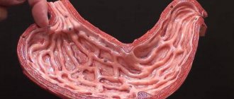
Decoding the result
When performing an ultrasound of the stomach, you can normally see the following:
- the organ in section usually looks like an oval ring-shaped formation with a rim, the nature of which is echo-negative, and the center is echo-positive;
- the walls of the stomach in the proximal sections have a thickness of about 5 mm, and in the pyloric section - about 7 mm;
- the presence of 5 layers in the wall of the stomach, differing in the degree of echogenicity;
- the serous membrane, which is located outside the stomach, is hyperechoic;
- the muscle sheath is hypoechoic, its thickness is about 2.5 mm;
- the submucosal membrane (with average echogenicity) measures about 3 mm;
- the mucosal plate, consisting of muscles, is usually low hypoechoic, its thickness is no more than 1 mm;
- the mucosa has a thickness of about 1.5 mm, its character is hyperechoic;
- A glass of liquid drunk by the patient is removed from the stomach in about 1/3 of an hour, and the primary withdrawal of liquid is normally about 180 seconds.
It is important to assess not only the thickness of the stomach walls, but also how uniform they are.
Can stomach cancer be seen on an ultrasound?
In the early stages, stomach cancer cannot be seen on ultrasound. This is due to the fact that at this stage of the disease, changes in the stomach are subtle and very difficult to identify.
In the later stages, changes in the body are already quite extensive. An ultrasound of the abdominal cavity will allow us to judge the presence of metastases and their size.
However, the sensitivity of the scanner is limited, so obvious tumors are detected only when they reach a large size.
Can a stomach ulcer be seen on an ultrasound?
Is a stomach ulcer visible on an ultrasound? The area where the ulcer is will not reflect ultrasound waves. Having noticed such a hole, the specialist will understand that the patient has an ulcer. However, such an indication on the disease screen is considered conditional. Additional examination will be required.
Pathology of the esophagus on ultrasound
If the esophagus is affected, the patient complains of difficulty swallowing: “Food stops somewhere in the chest and does not pass.” Pain when swallowing food is less common. Esophageal vomiting appears some time after eating and is not accompanied by nausea; vomit is poured out without gagging, consists of ingested food mixed with saliva and mucus, and sometimes has a putrid odor.
GER and GERD on ultrasound
Gastroesophageal reflux is the reflux of stomach contents into the esophagus when the esophagus relaxes outside the act of swallowing. An ultrasound is performed with the patient lying on his back; the head end of the couch can be lowered, then the fluid presses on the esophagogastric junction. On ultrasound, we are interested in the outer diameter of the esophagus, wall thickness, abdominal length, and His angle. See more details here.
GER is considered physiological when reflux occurs no more than 50 times a day, with a total duration of less than 60 minutes, and the wall of the esophagus is not damaged. In infants, due to the immaturity of the muscular layer and fundus of the stomach, frequent reflux from the stomach into the pharynx and oral cavity when air is expelled. Ripening occurs at 6-12 months.
Drawing. On ultrasound, the esophageal-gastric junction of a baby: normally, outside the act of swallowing, the LES is closed (2), with GER, the reflux of milk from the stomach into the esophagus is determined - a hyperechoic column (3). Up to 4 months of life, GER after feeding is considered normal.
With gastroesophageal reflux disease, reflux from the stomach into the esophagus occurs due to weakness of the sphincters, with increased intra-abdominal pressure and hiatal hernia. With GERD, frequent acid reflux is always accompanied by reflux esophagitis. In case of severe reflux, ultrasound reveals more than 2 refluxes in 5 minutes, the duration of the episode exceeds 9.5 seconds. Ultrasound signs of esophagitis include wall thickening >5 mm and an enlarged lumen of the esophagus.
Chalazia of the esophagus and chalazia of the cardia on ultrasound
Chalazia of the esophagus - weakened peristalsis of the esophagus makes it impossible to swallow solid food, in the complete absence of any obstructions in the esophagus. A weakening of peristalsis most often results from esophagitis, and less commonly from infections, botulism, lead poisoning, and organic lesions of the central nervous system. Food moves under the influence of gravity, and there is stagnant content in the esophagus.
Chalazia cardia - weakening of the esophagus, the lumen of the distal esophagus gapes - 6-14 mm, the contents of the stomach flow into the esophagus at the time of swallowing, accompanied by reflux esophagitis. Gaping of the LES often occurs in infants and is manifested by persistent regurgitation after eating. Up to 4 weeks, cardia chalazia in an infant is physiological.
Cardiospasm and achalasia of the cardia on ultrasound
Cardiospasm is an increased tone of the lower third of the esophagus and impaired relaxation of the esophagus during swallowing. The causes of cardiospasm are functional disorders of the nervous system, mental trauma; occasionally, reflex spasm of the esophagus due to stomach ulcers or helminthiasis.
More pronounced changes are caused by damage to the neuromuscular plexuses of the lower 2/3 of the esophagus - achalasia cardia. With achalasia cardia, there are no peristaltic waves in the distal esophagus, and the NSP cannot completely relax when swallowing.
Achalasia cardia can be a symptom of a tumor of the upper stomach, high vagotomy with gastrectomy, epiphrenic leiomyoma of the esophagus, ulcer, subcardial diverticulum. Idiopathic achalasia is a rare, slowly progressive disease.
Petrovsky B.V. (1957) distinguishes four stages of the disease:
- Stage I – functional intermittent spasm of the cardia, dilatation of the esophagus is not observed;
- Stage II – stable spasm of the cardia with mild dilatation of the esophagus;
- Stage III – cicatricial changes in the muscular layers of the cardia with pronounced dilatation of the esophagus;
- Stage IV – pronounced stenosis of the cardia with dilatation of the esophagus and esophagitis.
On ultrasound, the thoracic esophagus is dilated, the esophageal-gastric junction is sharply narrowed - the opening is no more than 3-5 mm, the passage of fluid into the stomach is slow, and there is stagnant content in the lumen. The muscular layer of the esophagus in the early stages is slightly thickened, in the later stages it grows significantly. Thickening of the mucous membrane indicates congestive esophagitis.
Drawing. A child complains of a night cough for six months. On ultrasound, the ESP is closed, the overlying sections are expanded to 27 mm, passage into the stomach began only in a standing position after taking 300 ml of liquid: longitudinal section at the level of the ESP (1), transverse section in the middle part of the esophagus (2), transverse section of the esophagus behind the left lobes of the thyroid gland (3), radiography with barium (4). With endoscopy, no pathology is determined. Conclusion: Cardiac achalasia.
Drawing. Child with prolonged cough, dysphagia, weight loss. Ultrasound of the esophagus: on a longitudinal section (1) the lower third is significantly expanded, at the esophagogastric junction it narrows in the form of a funnel; on the cross section from the suprasternal approach (2), the thoracic region is expanded. On a barium x-ray (3), the esophagus is significantly dilated, in the area of the esophagus it is narrowed like a bird’s beak. Conclusion: Achalasia of the esophagus.
Drawing. On ultrasound, the esophagus is closed, the overlying sections are significantly expanded, the passage of fluid is delayed, the wall of the esophagus is thickened due to the muscular and muco-submucosal layers. Conclusion: The echo picture may correspond to achalasia of the cardia, indirect signs of esophagitis.
Hiatal hernia on ultrasound
A hiatal hernia is a prolapse of part of the stomach through a weakened diaphragmatic ring into the chest cavity. Occurs in 15% of the population over 50 years of age. Displacement of the cardia leads to its insufficiency, GERD. There are sliding hernias and paraesophageal hernias. With a sliding hernia, the pancreas and the fundus of the stomach freely enter the chest cavity and return, and with a paraesophageal hernia, the pancreas remains under the diaphragm, and part of the stomach prolapses next to the thoracic esophagus. Children with a congenital short esophagus (chest stomach) have a hiatal hernia and most of the stomach is in the chest cavity. Along with the stomach, intestinal loops may be located in the chest cavity.
Drawing. Classification of esophageal hernia: type 1 - sliding hernia, type 2 - fixed paraesophageal hernia, type 3 - mixed hernia, type 4 - intestines and other abdominal organs move into the chest cavity along with the stomach.
An ultrasound shows a direct sign of a sliding hiatal hernia - the esophagus flows into the stomach above the diaphragm, on inhalation, part of the stomach enters the chest cavity, and on exhalation it returns back. Normally, the esophageal-gastric junction is at the level of the diaphragm opening of 7-10 mm. With a hernia, the diameter of the intestinal tube at the level of the diaphragm opening is 17-21 mm. The diameter of the esophagus is 11.5-13.5 mm - there is a possibility of a hernia; more than 14.5 mm - the probability of a hernia is very high; in children of the first year, 10-12 mm requires close attention.
Drawing. Esophageal hernia on ultrasound (1, 2): funnel-shaped expansion of the distal esophagus, at the level of the diaphragm, the outer diameter is more than 17 mm (normal is 7-10 mm), lumen is 11-15 mm, the abdominal section is shortened (normal in adults >2 cm) , His angle >90° (normal <90°). Normally (3), the abdominal esophagus is surrounded by a hyperechoic fat pad (arrows); its absence may indicate a sliding hiatal hernia.