Intestinal endoscopy, as a diagnostic method, is used to examine the mucous membrane of the organ for the presence of pathological processes and formations. The procedure is carried out using special equipment - an endoscope, which is a flexible conductor equipped with a “flashlight” and a miniature video camera that transmits an image to a monitor at multiple magnification. Endoscopic examination is a minimally invasive procedure that rarely causes side effects or complications.
What is intestinal endoscopy and its types
Endoscopy of the gastrointestinal tract (GIT) is carried out using endoscopes - flexible tubes with an optical system and displaying images on a monitor. Different parts of the gastrointestinal tract are examined using different endoscopes and on this basis are divided into types:
- Esophagogastroduodenoscopy
(EGDS) - the duodenum is examined by introducing a fiber gastroscope through the mouth after the esophagus and stomach. - Enteroscopy
– examination of the small intestine with an enteroscope:
- Jeunoscopy - the upper parts of the small intestine (jejunum) is performed with the introduction of an enteroscope through the mouth;
- Ileoscopy - lower parts of the small intestine (ileum) is performed with the introduction of an enteroscope through the anus.
- Colonoscopy – colonoscopy of the large intestine through the anus.
- Anoscopy
– lower ampullary rectum and anal canal using an anoscope. - Sigmoidoscopy
– rectum and lower parts of the sigmoid using a sigmoidoscope. - Capsule endoscopy
– small and large intestine using a capsule video camera taken inside.
Capsule endoscopy research technique
The doctor pre-consults the patient, talks about the technique and explains how to prepare for this study. 3 days later, after preparing the gastrointestinal tract, a study is carried out: at 9-10 o’clock the capsule is placed, at 11 o’clock the study begins, then the patient goes home or to work, at 23 o’clock the study is completed and the device is turned off, which the patient carries out independently at home. The next day, a second visit to the clinic, handing over the device and deciphering the results. The capsule is disposable and is not given to the clinic. It comes out on its own through the anal canal and is disposed of in the toilet.
The capsule is activated when the doctor removes it from the package. But during research it is turned on only where necessary. Next, the patient swallows a PillCam™ COLON system capsule (Israel), which is equipped with two cameras recording images with a wide viewing angle of 172° for each camera, a radio transmitter and a battery. A recording device is attached to the patient's belt, which is highly sensitive to receiving the signal coming from the capsule. The capsule moves along the digestive tract, thanks to the peristaltic waves of the intestine, and produces up to 35 pictures per second. During the study it is not necessary to be in the clinic; you can freely manage your personal time. At the end of the study, you must return the recording device to the doctor. The doctor's report can be obtained after a few hours.
Indications and contraindications for
Colon endoscopy is prescribed for:
- suspected tumor;
- chronic ulcerative colitis, Crohn's disease;
- intestinal obstruction;
- the appearance of black or bloody stool;
- prolonged diarrhea alternating with constipation;
- increased gas formation;
- anemia of unknown origin;
- causeless loss of body weight;
- abdominal pain.
Endoscopy is mandatory if internal bleeding is suspected.
Contraindications:
There are no absolute contraindications
, it all depends on how vital the examination is. Relative contraindications are:
- severe general diseases;
- psychoses;
- acute intestinal diseases;
- third trimester of pregnancy.
Advantages of video capsule endoscopy:
- disposable capsules eliminate the risk of infection;
- the study does not require sedation (medicated sleep) and is absolutely painless;
- no air supply to the intestines is required (the study is carried out absolutely comfortably);
- this study is even more minimally invasive than standard endoscopic studies;
- high image quality ensures diagnostic efficiency.
Outpatient procedure - does not require long-term hospitalization.
How is the test done and is it painful?
From this section you can learn how intestinal endoscopy is done and what kind of procedure it is.
Preparation:
- Before the procedure, exclude from the diet foods that cause increased gas formation: cabbage, peas, beans, bananas and other foods. On the day of the study, you should not eat or drink;
- cleansing enemas are done the day before and in the morning before the procedure;
- Instead of enemas, you can take the laxative Fortrans, Moviprep and others.
Carrying out a diagnostic procedure with the introduction of an endoscope through the oral cavity:
- An anesthetic spray is applied to the throat area of an adult;
- A mouthguard is placed in the oral cavity to prevent compression of the endoscopic tube;
- the patient lies on his side, swallows the probe, and as it moves, the doctor assesses the condition of the mucous membranes of the gastrointestinal tract;
- the probe is removed through the mouth.
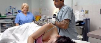
Carrying out a diagnostic procedure with insertion of an endoscope through the anus. Patients often ask whether it hurts. The procedure is painful, so it is often performed under general anesthesia, but it can also be performed under local anesthesia. Algorithm:
- the patient lies on the left side or is in the knee-elbow position;
- First, a solid tip is inserted into the rectum, and then a flexible endoscope is inserted through it;
- After the examination is completed, the endoscope is removed.
Esophagogastroduodenoscopy
As a rule, esophagogastroduodenoscopy takes no more than 10 minutes. Before the procedure, the oral cavity is irrigated with lidocaine - a local anesthetic that helps reduce the sensitivity of the mucous membrane. During the manipulation, the patient should breathe through the mouth, remain calm and unquestioningly comply with all requests of the medical personnel.
At the end of the diagnostic procedure, the patient is given a conclusion containing information about the state of the anatomical structures, the appearance of the mucous membrane, the presence of pathological foci - their location, number, shape, size. Having received a doctor’s conclusion, the patient should not search for information on the Internet and draw hasty conclusions - it is necessary to visit a gastroenterologist who will make a competent diagnosis and choose rational treatment tactics.
Being minimally invasive and highly informative medical services, endoscopic operations and manipulations are widely used in various areas of medicine. During the examination, endoscopes come into contact with mucous membranes, tissues and cavities of the human body and carry certain risks of contact infection of the patient.
Sanitary rules SP 3.1.3263-15 “Prevention of infectious diseases during endoscopic interventions” have defined new requirements for the organization and processing of endoscopic devices and instruments for them. This document summarizes at the current level the approaches of domestic and foreign scientists to the problem of disinfection of endoscopic equipment. To ensure the infectious safety of endoscopic procedures, in accordance with the requirements of MU 3.1.3420-17, sterilization and high-level disinfection (HLD) of endoscopes is provided. The concept of TLD in the Russian Federation was first introduced in 2003 and is applicable only to endoscopic equipment - flexible endoscopes. This level of disinfection is sufficient and necessary to prevent the transmission from patient to patient of pathogenic and opportunistic microflora, all vegetative bacteria, viruses, fungi, mycobacteria and some bacterial spores.
Thanks to strict adherence to standards and algorithms for processing endoscopic equipment and the presence of an effective infection control system in the EXPERT clinic, the possibility of transmission of infectious agents is considered insignificant. Information about each endoscope processing cycle is necessarily recorded in journals that comply with the regulations.
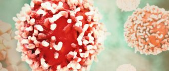
What can be discovered during endoscopy
During the examination, the endoscopist sees the wall of the gastrointestinal tract from the inside. This allows him to clearly examine the condition of the mucosa, any changes in its color, structure, and the presence of space-occupying formations. During the examination, the following is revealed:
- polyps, benign and malignant tumors; neoplasm tissue is taken by biopsy for histological examination;
- inflammatory diseases of the gastrointestinal tract;
- diverticula, strictures, congenital anomalies of various parts of the gastrointestinal tract.
FAQ
What is a video capsule and can it be reused?
The endoscopic capsule PillCam™ COLON system (Israel) is made of biologically inactive materials and is absolutely safe. The capsule size is 11 x 26-31 mm, weight - 4 g, there are built-in cameras inside it that take from 4 to 35 frames per second depending on the speed of movement through the intestine, 4 light sources, a radio transmitter and batteries designed to operate in for 11 hours. The capsule is disposable.
Can the capsule be retained in the intestines and what to do if this happens?
Patients are always worried when they hear about the risk of capsule retention. The fact is that such a risk is small (this occurs in approximately 0.1% of studies), and in each case it depends on the clinical situation. In a “healthy” intestine, the capsule’s progress may slow down, but it never stops there for long. The capsule lingering in the intestine helps to identify the “problem” and clearly indicate the location of the pathologically changed area. In addition, it is possible to remove the capsule without resorting to surgery using balloon-assisted enteroscopy. But it is also possible that a pathological formation of the intestine that causes capsule retention may require surgery.
Typically, foreign bodies of the gastrointestinal tract up to 7 cm in size come out on their own, and the capsule size is only 1x3 cm and has a very smooth rounded surface.
Is it necessary to monitor the exit of the capsule from the intestine?
Many people believe that the capsule itself is necessary to view the data. This is wrong. All recording of transmitted data is carried out on a special recording device. In most cases, there is no need to track the exit of the capsule from the body. It is disposed of naturally in the toilet.
Is it possible to examine the stomach with a small intestinal capsule?
When analyzing a video recording made by a video capsule, an endoscopist may notice changes in the gastric mucosa. But as a rule, the gastric mucosa can only be partially examined. This examination is not considered diagnostically accurate. For a full examination of the upper digestive tract, it is better to perform a standard gastroduodenoscopy.
Is it possible to examine the large intestine with a small intestinal video capsule?
A detailed examination of the colon in this case is hampered by several reasons, such as incomplete preparation of the colon, the need for additional stimulation of the gastrointestinal tract for better advancement of the capsule, and the absence of a second chamber on the small intestinal capsule. Thus, for an adequate, complete examination of the colon, it is recommended to perform a standard video colonoscopy or use a colon capsule.
Advantages and disadvantages
Endoscopy under anesthesia has the following advantages:
- there are no painful or unpleasant sensations;
- During the study, extensive surgical procedures can be performed;
- the doctor can fully concentrate on his actions and not be distracted by the patient’s anxiety;
- the study can be carried out longer.
Disadvantages include an increase in the cost of the examination due to the involvement of additional specialists, the need for follow-up, and more complex preparation.
The existing disadvantages of this procedure are insignificant compared to the significance of the method in diagnostic practice. In many developed countries, endoscopy of the large intestine is a mandatory method for medical examinations after 40 years, as it plays an important role in the prevention of cancer. Detection of tumors in the early stages and precancerous processes, such as polyps, allows timely treatment to begin, which significantly increases the life expectancy of such patients.
The Medicenter network of clinics performs a wide range of endoscopic examinations using general anesthesia. Experienced anesthesiologists and modern medications will help you get rid of discomfort and fear before the examination.
Disadvantages of methods of preparation for colonoscopy
The main disadvantage of the traditional method of preparation is the need to use cleansing enemas and take an oil laxative. The disadvantage of the scheme using Fortrans is its relatively high cost. With the traditional method, the frequency of cases of inadequate preparation is higher than when using the Fortrans scheme. However, this is largely explained by the inability of a number of patients to do cleansing enemas or taking less effective laxatives (bisacodyl, senade, etc.) instead of castor oil. Castor oil is not recommended for patients with gallstone disease, because it can provoke an exacerbation.