Indications
The main indications for performing an abdominal ultrasound include:
- pain localized in the abdominal area;
- pain in the hypochondrium;
- abdominal trauma;
- bitterness in the mouth, belching, feeling of heaviness and bloating;
- problems with stool, increased gas formation;
- control in the presence of chronic or acute diseases of acute respiratory disease;
- nausea with fever, vomiting;
- pain on palpation or abnormalities on palpation;
- symptoms of neoplasms (tumors);
- the period before surgery or to mark the biopsy procedure;
- developmental abnormalities of the abdominal organs;
- burning and pain during urination;
- inflammatory and infectious processes.
Scanning Features
First, the victim will be asked to remove all metal jewelry such as piercings from the abdominal area. Only after this the person lies down on a medical couch, exposing his stomach.
The doctor, using a special gel for better contact with the skin, passes the sensor through different parts of the stomach. To achieve effective visualization, the victim will have to listen to the diagnostician, who will ask him to change his position several times during the manipulation.
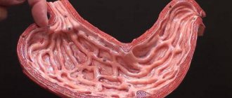
Each of the provisions is designed to identify possible pathologies in different parts of the stomach. We are talking about the bottom, the body of the organ, as well as the antrum, the initial segment of the duodenum. Taken together, this makes it possible to monitor the thickness of all the walls of these internal organs, as well as potential deformations or other pathologies.
If the device belongs to a new generation of medical equipment, then it has a Doppler ultrasound mode. With its help, the expert will receive an information summary of the blood supply to the lower part of the stomach. Additionally, the technique will tell you how the evacuation function works.
To carry out the last measurement, first examine the antrum on an empty stomach, and then, to stimulate peristalsis, suggests drinking 400 ml of mineral water without gas. After preparation, the antrum should be scanned every 15 minutes.
The average statistical indicator is a decrease in its volume by approximately 50% relative to the original level. In this case, the evacuation of incoming water must fit within a radius of 20 to 75 minutes.
How often should an abdominal ultrasound be done?
It is recommended to undergo an ultrasound examination of the abdominal organs for the purpose of preventive monitoring of health status once every 2 years. For those patients who have chronic diseases of internal organs, ultrasound examination should be performed at least once every six months. In some cases, screening is carried out more frequently to monitor the dynamics of changes in organs. There is no need to be afraid of this. Ultrasound is an absolutely safe and harmless diagnostic method and does not harm the patient’s health.
Previous NextAdvantages
- The study is carried out transabdominally, that is, the device’s sensor sends ultrasonic waves and records their reflection from the internal organs through the abdominal wall. Ultrasound is absolutely painless and does not cause any discomfort, so it is often prescribed if endoscopy is not possible.
- The technique allows you to control the progress of other diagnostic and therapeutic procedures, since the image is constantly broadcast on the screen, in real time.
- Ultrasound diagnostics does not create radiation exposure for the patient, so it can be performed regardless of age, even for children. It is allowed for pregnant women and during breastfeeding.
What organs are included in an abdominal ultrasound?
An ultrasound scan of the abdominal cavity can clearly assess the condition of the following organs:
- liver, porta hepatis, lobe sizes;
- gallbladder, duct of Wirsung;
- biliary tract;
- pancreas;
- spleen;
- kidneys and adrenal glands;
- retroperitoneal space;
- lymph nodes;
- portal vein.
Unfortunately, ultrasound of the abdominal cavity does not visualize the intestine well. Diagnosis of this organ using ultrasound is not very informative, and it is better for the patient to undergo an MRI of the gastrointestinal tract, virtual colonoscopy or enterography.
What will an ultrasound of the abdominal organs show?
Ultrasound of OBP will show well:
- inflammatory and necrotic liver lesions;
- hepatitis;
- cysts and tumors of organs, polyps;
- formation of stones, inflammation or scarring in the gallbladder and ducts;
- cholecystitis;
- damage to the kidneys and adrenal glands;
- acute and chronic pancreatitis of the pancreas;
- enlarged lymph nodes in the abdominal cavity;
- enlarged spleen;
- abnormal arrangement of organs.
Unfortunately, ultrasound examination is not informative enough to judge 100% the malignancy and benignity of a space-occupying lesion. Therefore, if tumors are detected, the doctor will refer the patient for further examination using an MRI of the abdominal cavity with contrast.
What do the abdominal organs look like on an ultrasound?
How it goes
The ultrasound examination of the stomach and intestines itself requires 15-20 minutes; preparing a conclusion requires another 10 minutes.
The doctor applies a special gel to the skin of the examined area, which improves the glide of the sensor and facilitates the passage of ultrasound waves. Next, the specialist studies the size and location of the gastrointestinal tract organs, their condition, the presence of pathological formations, and the quality of blood flow in the vessels. To obtain the most objective information, ultrasound is performed in different positions of the patient’s body (lying on his back, on his side, sitting, standing).
Ultrasound of the abdomen and bladder
A conventional ultrasound examination is sufficient to evaluate the structure of the liver, pancreas, gallbladder, bile ducts, spleen, and if there is a need to examine the kidneys, then the structure of the kidneys and adrenal glands. If there is a need to assess the presence of urinary excretion disorders and the condition of the bladder, then you should sign up for a comprehensive ultrasound of the abdominal cavity, kidneys and bladder. Then the sonologist will be able, in addition to the organs of the abdominal cavity and retroperitoneal space, to evaluate the structure of the bladder:
- does it contain salts, stones;
- unevenness of some wall;
- the presence of neoplasms and inflammatory processes.
What will an abdominal ultrasound not show?
When the question arises about the need to examine the intestines, stomach, duodenum, or colon, it is best to use endoscopic methods such as gastroscopy, endosonography, colonoscopy or virtual colonoscopy using MRI or CT instead of abdominal ultrasound. These forms of diagnostics make it possible to more accurately determine the condition of the walls and mucous membranes of these organs. An ultrasound examination can only indicate indirect signs that the patient has disorders of the mucous membrane, and its informativeness and diagnostic value of data is significantly lower than that of colonography or gastroscopy.
| Ultrasound service | Price according to Price, rub | Promotion price, rub |
| Ultrasound of the abdominal organs and retroperitoneal space (liver, gall bladder, pancreas, spleen, stomach) | 1500 rub. | |
| Ultrasound of one organ (liver, gall bladder, spleen, pancreas, bladder, adrenal glands) | 800 rub. | |
| Ultrasound of the abdominal organs and kidneys | 1700 rub. | |
| Ultrasound of the abdominal organs + ultrasound of the kidneys + ultrasound of the bladder | 2000 rub. | |
| Kidney ultrasound | 800 rub. | |
| Comprehensive ultrasound (ultrasound of the abdominal organs + ultrasound of the kidneys + ultrasound of the thyroid gland) | 2400 rub. | 1999 rub. |
| Comprehensive ultrasound (ultrasound of the abdominal organs + ultrasound of the kidney + ultrasound of the thyroid gland + pelvic ultrasound with an abdominal probe + ultrasound of the mammary glands) | 4200 rub. | 2999 rub. |
| Comprehensive ultrasound (ultrasound of the abdominal organs + ultrasound of the kidneys + ultrasound of the thyroid gland + ultrasound of the prostate gland with an abdominal probe) | 3300 rub. | 2499 rub. |
| Comprehensive body diagnostics (MRI of the thoracic spine, MRI of the lumbar spine, ultrasound of the abdominal organs, ultrasound of the kidneys, ultrasound of the bladder, consultation with a neurologist, consultation with a therapist) | 11700 rub. | 7000 rub. |
Ultrasound of the pancreas
The pancreas is very important in the digestive system. Many diseases negatively affect the functioning of this organ, which negatively affects the body as a whole. One of the effective ways to check the pancreas is ultrasound diagnostics (ultrasound). This technique is completely safe and can be performed on both adults and children.
Location of the pancreas in the human body
The pancreas is located behind the stomach on the left side, fits very tightly to the duodenum, and is also under the reliable protection of the ribs. Why is the role of the pancreas so important in digestion? The fact is that it secretes about two liters of pancreatic juice per day, thanks to which the process of digesting food is carried out.
The thickest part of the organ is the head, then it passes into the body and tail, the end of which is located near the spleen. The pancreas is covered by a special membrane. It is worth noting that the health of the organ is directly related to the urinary tract.
When is an ultrasound scan of the pancreas prescribed?
The pancreas must be checked by ultrasound diagnostics if the patient:
- Complains of pain in the area of the left ribs, but the doctor cannot determine the cause by palpation;
- He is losing weight sharply.
If there is pathology in laboratory tests, ultrasound diagnostics of the pancreas is mandatory. In addition, diagnosis of the gland is prescribed after suffering from diseases such as hepatitis A, C, B.
Other situations in which an ultrasound scan of the pancreas is required:
- Feeling of bloating;
- Bitter taste in the mouth;
- Yellow skin tone;
- Problems with stool;
- Abdominal trauma;
- Detection of tumors.
As part of the pancreatic examination, a complete diagnosis of the abdominal cavity is carried out. The study allows us to identify pathologies at the very initial stage. This will help to prescribe treatment as soon as possible and avoid the development of the disease. After all, gland diseases directly affect the health of other organs.
An ultrasound examination of the pancreas is recommended once a year for all people over 25 years of age.
What is included in preparation for an ultrasound.
To obtain a reliable result during diagnosis, you must follow all the rules for preparing for it. If the rules are ignored, the results of the study may be distorted or difficulties may arise during the ultrasound. First of all, you should completely cleanse the intestines through an enema or taking laxatives. Two days before the test, it is allowed to take sorbents that help reduce the formation of gases.
Two days before the ultrasound, you should completely remove from the diet:
- alcoholic drinks and soda;
- fresh fruits/vegetables;
- dairy products;
- meat;
- eggs;
- black bread.
You can eat cereals, fruits that have undergone heat treatment, and honey.
Eating later than 12 hours before the test is unacceptable.
It is better to plan the examination for the first half of the day, because eating and drinking before the examination on this day is not allowed. Chewing gum and smoking are also prohibited. If you take medications, be sure to ask your doctor how to take them before your ultrasound.
What you need to keep in mind if you need to check your pancreas:
In some cases, a special diet is prescribed only by a doctor; it depends on the individual characteristics of the body;
If you are overweight, the enema should be done 2 times: on the eve of the procedure and on the day of the study.
If all recommendations are not followed, the ultrasound result will lose its information content by 50%.
How is an ultrasound of the pancreas performed?
The pancreas is diagnosed using ultrasound within 30-60 minutes. The procedure is performed lying on your back. A special gel is applied to the stomach. Then the ultrasound doctor begins to conduct the examination using a special sensor. If necessary, the doctor may ask the patient to retract or inflate the abdomen.
During the ultrasound, the patient will need to lie first on the right side and then on the left side; in some cases, the doctor asks the patient to stand on his feet. This is required in order to examine the pancreas as thoroughly as possible.
Immediately after performing the ultrasound, the doctor gives the patient a transcript and, if necessary, comments on certain points in order to focus the patient’s attention on them.
Diseases that can be detected through pancreatic ultrasound diagnostics.
The appearance of pancreatitis in the acute stage is indicated by a decrease in the echo signal. In this case, the gland becomes completely white. If the picture becomes less intense, this may indicate the presence of pancreatic edema.
The presence of tumors in the pancreas can be indicated by deviation of its tail. If the echo signal becomes stronger, this may indicate a chronic stage of pancreatitis or the presence of oncology. In places where it appears, the color of the pancreas will differ from the main one.
If the size of the liver or gallbladder increases, this may also indicate the presence of neoplasms. To identify its etiology, a piece of tissue is sent for histological examination.
Abscesses that contribute to the appearance of cavities with cloudy exudate indicate pancreatic necrosis. If the Wirsung duct is dilated, this may indicate an inflammatory process in the gland.
It is worth noting that many dangerous diseases may not manifest themselves at all in the early stages, but can be diagnosed through ultrasound. Therefore, in no case should you ignore the study if it was prescribed by the doctor.
What indicators are considered normal?
Ultrasound of the pancreas allows you to assess the health of the organ. The following are normal pancreatic values in adult patients:
- The echo signal is average, but becomes higher as the patient ages;
- The structure is holistic, homogeneous, reminiscent of the liver. The appearance of small inclusions is allowed;
- All parts of the pancreas should be clearly visible;
- The diameter of the Wirsung duct should be 1.5-2.5 mm, it should not be dilated;
- There is no deformation in the vascular pattern;
- Normal size indicators: body from 8 to 18 mm, head 18-28 mm, tail from 22 to 29 mm.
Normal indicators for an adult differ from those typical for a child.
Once again, we would like to note that the diagnosis of the pancreas is carried out as part of a comprehensive ultrasound diagnostic of the abdominal cavity. This is due to the fact that it is quite difficult to identify diseases of the pancreas using ultrasound, but it is possible to diagnose diseases of the organs bordering it, which makes it possible to assess the general condition of the abdominal cavity. If abnormalities are detected during an ultrasound, the doctor prescribes additional tests.
Ultrasound diagnostics of the pancreas makes it possible to safely and in a short period of time detect the disease at an early stage and prevent it in a timely manner.
Ultrasound of the pancreas in N. Novgorod
Diagnosis of the pancreas in our city is carried out at the VIP Academy clinic. The study is performed by doctors with more than 15 years of experience. All of them are certified specialists. The clinic has two high-tech ultrasound machines on which examinations are performed. The equipment makes it possible to most accurately assess the condition of the pancreas and nearby organs. The cost of diagnostics is the city average.
You can find out more detailed information about the study, as well as sign up for an ultrasound scan, from the administrators of the VIP Academy clinic. Phone number: +7 (831) 270-00-00.
Contraindications
Ultrasound diagnostics of the abdominal organs has a number of contraindications for which such examination is not recommended. Such contraindications include:
- acute infectious diseases;
- open wounds;
- various skin lesions;
- after endoscopy of the gastrointestinal tract;
- after pneumoperitoneum;
- after colonoscopy.
If there are such contraindications, the abdominal ultrasound procedure should be postponed for several days - 2-5 days.
How to properly prepare for an abdominal ultrasound?
The main task when preparing for an abdominal ultrasound is to try to remove excess air from the intestines. Before undergoing the procedure, you must follow some nutritional rules. In order to prevent gas formation, you need to:
- A few days before the examination, stop eating bread and dairy products, and avoid carbonated drinks.
- Legumes or peas must be excluded.
- Vegetables and fruits should also be removed from the diet.
- It is worth giving up juices, confectionery and, of course, eliminating alcohol intake.
The presence of gases in the body will interfere with a clear image and make it difficult for the doctor to make a correct diagnosis.
If you want to properly prepare for an abdominal ultrasound, you should introduce some foods into your diet. The following will be useful: beef, chicken, low-fat fish, porridge cooked in water without adding butter. It is advisable to steam the food or eat it boiled. You can take some enterosorbent if there are no contraindications. It is advisable to empty your bowels naturally 2 hours before the procedure or do a small cleansing enema at home. The procedure itself is best done on an empty stomach or at least 6 hours after eating. Smoking patients must abstain from their bad habit for 4 hours before the examination. Author: Telegina Natalya Dmitrievna
Therapist with 25 years of experience
Preparation for the procedure
You need to prepare for scanning in advance. Firstly, a diet must be followed for 2-3 days: confectionery products, alcohol, raw vegetables, carbonated and dairy drinks are excluded from the diet. Secondly, it is necessary to prevent possible gas formation in the intestines. To do this, you can drink Festal or activated carbon, and people prone to constipation are recommended to drink laxatives. Before the procedure, you should not eat or drink (it is best to perform an ultrasound in the morning), or smoke. If the patient is suffering from “hunger pains,” then you can drink some unsweetened tea and eat a cracker.