Questioning
Dysphagia (difficulty swallowing) with solid foods is usually observed with carcinoma, benign stricture, esophageal membrane; when eating solid and liquid food - indicates a violation of motility of the esophagus (achalasia, diffuse spasm of the esophagus and other motility disorders), damage to the esophagus by caustic alkalis or acids (burn of the esophagus), severe peptic esophagitis, infectious lesions of the esophagus (HIV infection, cytomegalo- and herpes viruses, fungal infections), diffuse spasm of the esophagus
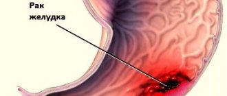
Heartburn and pain behind the sternum when food passes through the esophagus are typical symptoms of cancer and reflux esophagitis.
Indications for laboratory diagnostics of the gastrointestinal tract
In addition to acute manifestations in the form of severe pain, bleeding and critical disorders, primary and secondary symptoms for conducting a diagnostic examination of the gastrointestinal tract can be a whole range of direct and indirect indications. The main clinical syndromes are:
- heartburn;
- flatulence;
- private constipation and diarrhea;
- critical changes in stool consistency;
- chest pain (non-cardiac origin);
- painful discomfort in the iliac region and tenesmus;
- discharge of mucus with feces;
- decreased appetite;
- weight loss.
Moreover, in some patients, the development and course of gastrointestinal diseases may be accompanied by other symptoms: constant or periodic diffuse epigastric pain, aversion to food, belching, dysphagia and vomiting. Changes in microflora, yellowing of the skin, sleep disturbances, and nervous irritability may also occur.
X-ray examination of the esophagus
X-ray examination currently remains the main indicative diagnostic method. With various rotations of the patient around the vertical axis, a contrast X-ray examination is performed with an aqueous suspension of barium sulfate, and if perforation is suspected, with a water-soluble contrast. Pay attention to the nature of the contours, peristalsis, relief of the mucous membrane, and the function of the esophageal sphincters. Examination of the patient in a horizontal position with the foot end of the X-ray table raised allows us to identify dysfunction of the lower esophageal sphincter (reflux of contrast material from the stomach into the esophagus).
Methods of laboratory diagnostics of the gastrointestinal tract
To obtain an objective clinical picture of the nature of the disease and to confirm diagnoses, it is often necessary to carry out a differential diagnosis using invasive and non-invasive methods. The Northwestern Center for Evidence-Based Medicine offers laboratory, instrumental and functional examinations without a queue or registration.
The services of our center will help identify pathologies, determine the dynamics of recovery, confirm the effectiveness of therapy and prevention, as well as diagnose neoplasms and helminth infestations. We offer our clients both traditional diagnostic methods and the most innovative examination methods, and the latest results coding system and automated information support eliminate the possibility of errors and confusion in the analyzes.
Where to get tested
The Northwestern Center for Evidence-Based Medicine, having a powerful laboratory base and the latest generation diagnostic equipment, offers regular and potential clients to undergo diagnostics of acute, chronic and infectious diseases of the gastrointestinal tract.
At SZTsDM JSC you can also order a call from a nurse to collect biological material at home. Contact us, we guarantee high competence of specialists, reliability of diagnosis and complete confidentiality.
Radiation diagnostics (CT)
A modern method that combines digital technologies with x-ray examination. The SZTsDM has a latest generation tomograph installed, which made it possible to extremely reduce the impact of radiation on the patient, obtain diagnostic 2D and 3D images of excellent resolution and analyze them from any angle.
CT allows you to non-invasively identify tumors and metastases that are not diagnosed by ultrasound, as well as determine:
- inflammatory and erosive processes;
- elasticity and thickness of the walls of the esophagus, stomach and intestines;
- congenital pathologies and postoperative recovery dynamics.
The study was well tolerated by patients of all ages. Requires minimal preparation.
Prevention and recommendations
Most gastrointestinal diseases can be quite easily prevented. It is enough to limit the consumption of fatty and fried foods, introduce fermented milk products, vegetables and dishes rich in fiber into the diet.
Drinking clean water is also of no small importance in the prevention of diseases of the gastrointestinal tract. And:
- healthy sleep;
- cardiological and strength physical activity;
- timely treatment of giardiasis, dysbacteriosis, helminthic infestations;
- taking medications strictly according to the instructions and according to the recommendations of doctors;
- giving up bad habits (smoking, abuse of strong brewed tea and coffee, alcohol, including low-alcohol drinks).
Laboratory diagnostics of the gastrointestinal tract
General blood analysis.
An invasive clinical study that allows you to obtain detailed information about the main indicators of the patient’s capillary or venous blood:
- number of leukocytes, platelets and red blood cells;
- hemoglobin and ESR levels;
- color index.
The assay is widely used to provide an overall clinical picture and monitor therapy or disease progression. SZTSDM specialists use high-quality consumables and perform blood sampling as comfortable as possible for the patient. Combined with full compliance with all medical and sanitary standards, it guarantees maximum reliability of the result.
Examination of stool for the presence of protozoa.
This microbiological analysis makes it possible to detect cysts of protozoan microorganisms in the feces, which enter the human body with poorly washed vegetables, poor-quality water, traces of soil substrates or from an infected carrier (animals, people, birds). Microspopia can accurately detect giardiasis, balantidosis, amebiosis and toxoplasmosis, which affect the gastrointestinal tract and suppress the immune system.
Examination of stool for worm eggs.
As a rule, microscopic analysis for helminths reveals the presence of worms and eggs of parasites such as ascorids, necator, pinworms or hookworms in feces. But in the SZDCM laboratory, a full-fledged study is carried out, which makes it possible to accurately identify the facts of infection of children and adults with various genera of parasitic helminths and cross-reactions with the antigens of other parasites, which means that the patient will be prescribed correct and effective treatment.
Analysis of microflora for dysbacteriosis.
Bacterial research reveals quantitative and qualitative changes in intestinal biocinosis. Allows you to determine the increase or decrease in pathogenic and healthy microflora (bacteroides, bifidobacteria, candida, Klebsiella, clostridia, Proteus, Escherechia, Pseudomonas aeruginosa, strepto-, entero- and staphylococci) and initially determine the degree of dysbiosis.
It should be noted that for each age group special methodologies are used for interpreting the results of this analysis. In this case, the study can also be prescribed to newborns, people with allergic manifestations and patients who have undergone intensive anti-inflammatory, hormonal and chemotherapy.
Coprogram.
A comprehensive chemical, macro- and microscopic laboratory study of a wide range of stool, widely in demand among clinicians and easily interpreted. The analysis assumes:
- determination of enzyme activity of the digestive organs, evacuation capacity of the stomach and intestines;
- study of pH reaction, state of microflora, color and consistency of feces;
- detection of bile pigments, fatty acid salts and mucus.
This non-invasive study also involves identifying inflammatory processes and conducting an analysis for the presence of hidden traces of pus and blood in the stool (Gregersen reaction). It allows you to accurately detect altered erythrocyte hemoglobin and cloudy exudate and thereby identify hidden bleeding and pathology of the gastrointestinal tract.
Antigen screening
rotavirus, norovirus, astrovirus. The analysis is carried out using the polymerase reaction method (PCR) and enzyme immunoassay. This ensures high analytical accuracy and makes it possible to identify infectious pathogens of gastrointestinal diseases in the stool with maximum reliability.
As a rule, infectious agents are actively transmitted through the oral-fecal route from an infected person, so it is extremely important to correctly and timely identify pathogens. The study will determine the etiological factor of acute intestinal infections and will allow a differentiated approach to the treatment of inflammatory processes.
go to analyzes
Functional diagnostics of the gastrointestinal tract
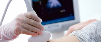
Ultrasound examination (ultrasound)
. A modern and highly informative method of non-invasive diagnostics. Its principle is based on the Doppler effect (a change in the frequency and wavelength of ultrasound as a result of perception or reflection by matter), which makes it safe for adults and children and allows the examination to be performed quickly and without prior preparation of the patient.
Ultrasound of the gastrointestinal tract can be prescribed:
- children;
- pregnant women;
- people after chemotherapy and radiation therapy.
Depending on the doctor’s recommendations and symptoms, the procedure may involve a comprehensive examination of the abdominal cavity or only the stomach and intestines, including the cecum, sigmoid, rectum, ascending and descending colon. In some cases, an examination of the intestines is carried out with the introduction of a rectal sensor. Ultrasound of the stomach is inferior in information content to gastroscopy. But given the non-invasive nature of the procedure, it allows diagnosis even in infants and people with inflamed and damaged food tracts.
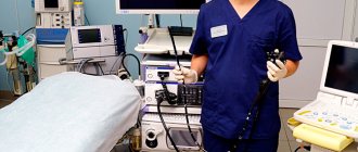
Colonoscopy.
An endoscopic method for diagnosing the condition of the entire large intestine, the length of which reaches more than a meter. It is characterized by the highest information content and allows you to:
- visually determine the nature of the development of ulcers and the relief of the internal walls of the entire large intestine;
- detect even microscopic tumors and polyps;
- perform a biopsy.
Colonoscopy allows early diagnosis of ulcers and neoplasms. Therefore, doctors recommend that even in the absence of obvious symptoms, people over 50 years old should be examined every 5 ÷ 7 years.
Diagnosis is based on the introduction of an endoscope and is associated with flushing and distension of the intestines, which causes significant discomfort and pain. Therefore, it is carried out after deep cleansing and using anesthesia.
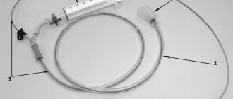
Endoscopy.
A modern method that offers wide possibilities for examining the mucous surfaces of the gastrointestinal tract. Allows:
- take a tissue sample for laboratory testing;
- determine the narrowing and expansion of the esophagus;
- diagnose fibrous layers.
Diagnosis is based on the introduction of a flexible endoscopic sensor into the esophagus naturally. The endoscope is equipped with fiber optic illumination and transmits a high-quality magnified image to the monitor. Thus, gastroendoscopic diagnostics provides a complete clinical picture of the presence of erosions, ulcers, and inflammatory processes on the mucous membrane of the stomach, esophagus and duodenum. Allows you to diagnose gastrointestinal cancer in the early stages and objectively monitor the dynamics of pathology or remission. It should be noted that the procedure is strictly carried out only on an empty stomach.
Laparoscopy.
A minimally invasive method of examining the abdominal cavity, performed using a rigid endoscope. Widely used in the postoperative period to diagnose complications and in cases where X-ray and other clinical and laboratory methods have proven ineffective.
It is highly informative, reliable and technically simple, but requires local or general anesthesia. Applicable for children and older patients.