We have already written several times that gastroscopy is one of the most important examinations for diseases of the gastrointestinal tract. However, its implementation is associated with certain discomfort for the patient.
No, we are not talking about pain; there are no painful sensations during FGDS. But due to the fact that the gastroscope tube is inserted into the stomach through the esophagus, profuse salivation begins and vomiting may occur. Moreover, the procedure requires certain preparation from the patient. If he cannot comply with several conditions, then the gastroscopy will have to be redone and again find himself in an environment that is not the most comfortable for him.
Therefore, if the doctor has prescribed an FGDS for you, then listen carefully (or better yet, write down) what requirements need to be met before the procedure. In this article we will talk about the most general principles of preparation for fibrogastroscopy, but in each individual case there may be some differences, so the advice of the attending physician must be followed first.
How to prepare for gastroscopy
The main recommendations concern nutrition. FGDS should be carried out on an empty stomach, therefore, as a rule, the examination is prescribed in the morning.
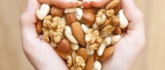
You need to have dinner 3-4 hours before bedtime (so that your last meal is 10-12 hours before the procedure). It is advisable to refrain from heavy foods (fried meat, nuts, chocolate, mushrooms). You should also exclude foods that cause increased gas formation (milk, legumes, cabbage, grapes, apples, carbonated drinks).
In preparation for gastroscopy, you should give up alcohol and spicy foods for several days. It is not recommended to smoke immediately on the day of the procedure - smoking promotes the production of mucus in the stomach, and this can lead to an exacerbation of the gag reflex. But there is no strict prohibition; you decide for yourself whether to give up tobacco or not.
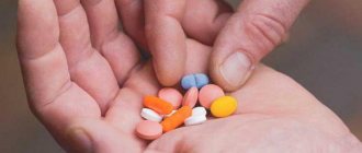
You should also tell your doctor in advance about all the medications you take regularly. Some of them may need to be eliminated for a while.
Preparation for gastroscopy
Gastroscopy requires special preparation, which the patient is warned about at the time the procedure is prescribed.
1. If time permits, you should exclude chocolate, nuts, seeds and spicy foods from your diet two days before the procedure. It is advisable to stop drinking alcoholic beverages at the same time.
2. The last meal before the diagnostic procedure should take place no later than 18:00 the previous day. Dinner should be hearty, but not contain food that is difficult to digest.
3. Before carrying out, you should exclude foods rich in fiber, heavy salads dressed with mayonnaise, large amounts of whole grain bread, fatty meat or fish, and cheeses. It is best to have dinner with a portion of green salad with a small amount of white chicken meat, steamed chicken cutlets, buckwheat porridge, and low-fat cottage cheese. You should avoid legumes and pearl barley, but a portion of mashed potatoes or steamed broccoli may well become the basis of the evening menu on the eve of the diagnosis.
Preparation on the day of gastroscopy
- On the day of the test, you should refrain from eating or drinking anything. You can drink some water no later than 2-4 hours before the start of the test.
- If the patient constantly takes medications in capsules or tablets, you will have to postpone taking them, since any foreign objects in the cavity of the organ being examined can distort the picture.
- Gastroscopy is accompanied by an increased gag reflex, and therefore food from the stomach can not only stain clothes, but also enter the upper respiratory tract when inhaled during regurgitation. In addition, taking medications before manipulation is accompanied by excessive formation of gastric juice. In a situation “on an empty stomach,” this can aggravate pathological processes. For the same reason, it is not recommended to smoke before gastroscopy of the stomach. This concludes the preparation, then we proceed directly to the research.
It is very important to warn your doctor about any allergic reactions you have, especially if they occur to medications. Before starting the procedure, the doctor performs premedication, or anesthesia of the root of the tongue and pharynx using an aerosol. This reduces unpleasant and painful sensations and partially reduces the manifestations of the gag reflex, thereby simplifying the doctor’s task, but can cause an allergic reaction in the patient.
For gastroscopic examination of the gastrointestinal tract, you should take the results of previous manipulations, if any, as well as x-rays, test data and other materials from previously performed diagnostic studies of the stomach and duodenum.
Performing gastroscopy
Immediately before the procedure, the doctor will ask you to sign a consent form for the manipulation. Be sure to discuss with your doctor the likelihood of consequences, as well as the risks of the examination.
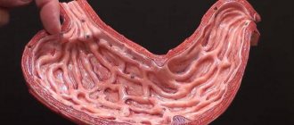
Gastroscopy of the stomach is a procedure that allows you to examine the stomach and esophagus using a special probe, which is a special optical tube with a video camera (endoscope). The terminal part of the device is inserted through the mouth and then gradually moves down into the stomach.
Gastroscopy is performed in a specially equipped room and is performed by an endoscopist specializing in endoscopy and gastroenterology (gastroenterologist).
Before starting, the root of the tongue is anesthetized with a spray - an anesthetic or rinsed with an anesthetic solution. This allows the muscles of the pharynx to relax and reduces the intensity of the gag reflex, which will allow the doctor to later easily insert the endoscope into the esophagus.
You will be placed on your side, usually on your right. Then a special mouthpiece is inserted into your mouth in order to protect your teeth from damage by the endoscope and the endoscope itself from biting, since it is quite expensive equipment. After this, the tip of the endoscope is inserted into the mouth, gently pressing on the tongue as it moves forward. The doctor will then ask you to take a sip to move the endoscope into the esophagus. Since the endoscope is significantly smaller in diameter than a bolus of food, you should not have any problems with swallowing or breathing.
During the procedure, your doctor will advise you not to swallow unless necessary. If saliva accumulates in the mouth, the nurse assisting the doctor during the examination will remove it using suction.
The endoscopist will gradually advance the endoscope through the digestive tract while looking through the eyepiece or video monitor to assess the condition of the walls of the esophagus, stomach and duodenum. If problems arise with examining the walls of organs, air or water is supplied through a special channel in the endoscope into the lumen of the stomach, which, washing the walls, clears the area to be examined or cleans the endoscope lens. After this, the liquid and air are removed using suction.
A camera connected to endoscopic equipment allows you to record the entire period of the study on video for subsequent detailed assessment of the findings. Through a special channel in the endoscope, the doctor can pass tiny endoscopic instruments (forceps, loops, staples), which will allow him to perform a biopsy or remove pathological growths of the mucous membrane. The biopsy procedure is absolutely painless.
How to swallow a tube
This question is no longer directly related to preparation for fibrogastroscopy of the stomach, but will help you better understand how the procedure goes.
To prevent the patient from accidentally squeezing the hose with his teeth, a special mouthpiece is inserted into the mouth. Next comes the most unpleasant moment for many - swallowing the hose. However, with the advent of local anesthesia, this process is no longer so uncomfortable - before the procedure begins, the larynx is treated with a lidocaine solution.
After the end of the FGDS, you will not be able to eat for some time, and there may be discomfort in the abdomen.
In general, gastroscopy is a widely used and proven diagnostic that helps to effectively treat many diseases. In the Medicenter clinic network, all endoscopic examinations are performed by doctors of the highest qualification category. The procedure is as comfortable as possible for the patient. It is also possible to do an FGDS under general anesthesia.
Indications
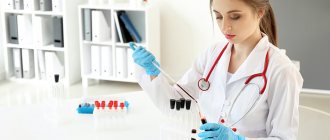
Duodental intubation is necessarily prescribed to patients when it is difficult to prescribe treatment due to the vagueness of symptoms and their similarity to pathologies of the kidneys or gallbladder. They may experience difficulty urinating, bitterness in the mouth, pain on the right side, high blood pressure and nausea. The procedure is recommended for patients with the possible presence of echinococcosis or Giardia. It should be noted that with the help of probing, the condition of the liver and gallbladder ducts is determined, allowing the elimination of stagnant and accumulated bile. During the analysis, the doctor may inject medications into the damaged organ. Bile is collected in 3-5 stages, the most effective is fractional collection (five-phase). Determining the type of pathology is possible based on the presence of bile and its shade at each stage.
Duodenal sounding
Duodenal sounding is carried out using a thin rubber probe with a metal or plastic olive at the end. It is more preferable to use a double probe, one of which serves to pump out gastric contents.
The study is carried out in the morning on an empty stomach. With the patient sitting, a duodenal tube is inserted (in the same way as a thin gastric tube). When the “40 cm” mark is at the teeth, the probe is advanced another 10-15 cm, a syringe is connected to it and the gastric contents are aspirated. After this, the patient swallows the probe up to the “70 cm” mark.
Next, the study continues with the patient positioned on the right side; a soft cushion or pillow is placed under the pelvis (in this position, the passage of the probe to the pylorus and into the duodenum is facilitated), and a warm heating pad is placed under the right hypochondrium. The outer end of the probe is lowered into the test tube, and the rack with test tubes is placed on a low bench at the head. In this position, the patient gradually (within 20-60 minutes) swallows the probe to the 90 cm mark. As soon as the olive passes from the stomach to the duodenum, a yellow liquid begins to flow into the test tube - duodenal contents, colored with bile.
There are five phases of fractional duodenal intubation.
The first phase is the release of duodenal contents (portion A) from the moment the probe enters the duodenum until the introduction of one of the cholecystokinetic agents. This portion of duodenal contents is a mixture of bile. pancreatic, intestinal and, partially, gastric juices and does not have much diagnostic value. Bile from portion A is collected within 10-20 minutes.
The second phase is the phase of complete cessation of bile secretion due to spasm of the sphincter of Oddi, which occurs as a result of the administration of a cholecystokinetic agent (30-50 ml of a warm 33% solution of magnesium sulfate through a probe or 75 units of cholecystokinin intravenously). Normally, the duration of the second phase does not exceed 4-6 minutes; its lengthening indicates an increase in the tone of the sphincter of Oddi, and its shortening indicates its hypotension.
The third phase is the release of golden-yellow contents of the extrahepatic bile ducts, which lasts 3-4 minutes. The bile released during this process also belongs to portion A (A1).
The fourth phase is the emptying of the gallbladder and the release of thick gallbladder bile of dark yellow or brown color (portion B).
This portion of bile is released as a result of contraction of the gallbladder, which occurs under the influence of a cholecystokinetic agent, and the simultaneous relaxation of the sphincter of Oddi and the sphincter of the gallbladder. Portion B is 4-5 times more concentrated than liver bile and contains a significant amount of bile acids, cholesterol, and bilirubin. The secretion of gallbladder bile (normally about 30-60 ml) lasts 20-30 minutes.
If the bladder reflex is absent within 20-30 minutes after the administration of magnesium sulfate, which in some cases can be observed even in healthy people, it is necessary to administer antispasmodics (through a probe of 30 ml of a 20% novocaine solution or subcutaneously 0.5 ml of a 0.1% atropine solution ), and if there is no effect, repeat the administration of cholecystokinin.
Important: If, after the administration of novocaine or atropine, dark cystic bile begins to be released, this indicates a sphincter spasm; the absence of a cystic reflex even after the administration of antispasmodics and cholecystokinin (repeatedly) suggests the presence of an organic obstruction (blockage of the cystic duct with a stone) or a non-functioning gallbladder (wrinkling, gallbladder cancer, etc.).
Fifth phase - after the cessation of secretion of dark cystic bile, golden-yellow bile begins to be secreted through the probe again (portion C). It is also collected in test tubes for 30 minutes at 10-minute intervals.