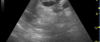
MRI examination of the pancreas: normal variant
MRI of the pancreas is a non-invasive method of visual diagnosis that allows you to accurately determine not only the presence of inflammatory processes, tumors and destructive lesions of the organ, but also the degree of their severity. The accuracy of the study is significantly higher than with computed tomography. MRI of the pancreas does not involve the use of ionizing radiation, so the procedure is practically safe for a person who does not have metal objects in the body.
Magnetic resonance scanning is a highly informative method in determining the cause of a number of urgent conditions associated with the biliary system: an inflammatory process, a gallstone blocking the duct, post-traumatic consequences, etc., but the main drawback of diagnosis is the duration of the procedure, which can take about an hour. In emergency cases, when there is no time to delay, a CT scan is performed, and to monitor the condition over time, depending on the clinical picture, the doctor may recommend an MRI or ultrasound. The greatest value is complex diagnostics, including MRI simultaneously with cholangiopancreatography and magnetic resonance angiography.
Advantages of the technique
Magnetic resonance today is the most highly accurate method for diagnosing lesions. The technique allows you to separate vascular elements and areas with fluid. This makes diagnosis much easier. An important advantage of MRI is the ability to obtain images in many projections. The procedure is harmless. Tomography is more informative than CT and ultrasound. The advantage is that MRI does not use harmful radiation.
Content:
- Brief information
- Advantages of the technique
- The essence of the technique
- Indications for the procedure
- Preparation and technique
- Application of contrast agent
- Contraindications to the procedure
- Safety of the procedure
- Price features
Features of magnetic resonance scanning
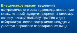
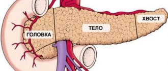
The MRI method is based on the ability of an electromagnetic field to influence hydrogen protons, causing magnetic resonance in them. As a result, the scanner visualizes gland tissue in the form of clear layer-by-layer images. Depending on the purpose of the study, the doctor selects the scanning plane and the frequency of sections (projections). The tomograph “photographs” the area being examined from different angles and transmits the data to a computer for subsequent processing.
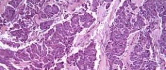
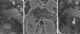
In the presence of cystic and malignant neoplasms, fibrous changes, inflammatory processes, acute forms of pancreatitis, deviations from the norm in the size, contours and structure of the organ are clearly visible on the images. To better visualize small tumors, MRI with contrast is usually performed. The data obtained during a magnetic resonance examination allows us to identify all abnormalities and make a correct diagnosis.
Preparation and technique
No special preparation is needed. You are allowed to eat and drink before the diagnosis.
If additional contrast will be used, then preparation is necessary. It is important to prepare for its implementation accordingly:
- Before contrast diagnostics, it is necessary to exclude fatty foods, do not drink alcohol, or take medications containing alcohol.
- Drinking coffee and tea is prohibited.
- Before the examination, it is recommended to remove any metal jewelry.
- It is prohibited to take the study with credit cards or telephones.
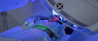
The table with the patient moves into the apparatus until the iron is under the magnet. There are innovative devices that eliminate the need to completely accommodate the patient.
Noise is generated during the study period. Therefore, it is possible to use headphones. The device is equipped with a speakerphone for two-way communication. It is allowed to have a loved one for support. To call a specialist, there is a button that is kept by the patient during the procedure.
Many more modern devices are additionally equipped with an internal camera to monitor the patient's condition. Therefore, during diagnosis, the patient is constantly under direct medical supervision for the entire duration of the scan (meaning from 20 minutes to a whole hour). Many cameras have special additional lighting and built-in air conditioners. Such solutions make the procedure even more enjoyable.
The procedure does not cause discomfort or pain to the patient.
It is extremely important to remain still - this is the key to high-quality photographs. At the doctor's signal, you will additionally need to hold your breath. Pictures are taken in series.
It is important to immediately report the first signs of pain or deterioration in health to your doctor.
The specialist receives images of the gland on the screen. Thanks to its versatility, the image is obtained in the desired projections. The doctor is deciphering. The patient receives the information in his hands.
MRI of the pancreas: preparation for examination
You should avoid certain foods before an MRI of the abdomen.
Preparation for an MRI of the pancreas involves switching 3 days before the diagnostic procedure to a gentle diet aimed at suppressing gas formation and stagnation of food in the intestines. The following are subject to exception:
- legumes (peas, beans, lentils);
- yeast containing baked goods;
- cabbage in any form;
- freshly squeezed fruit and vegetable juices;
- confectionery;
- carbonated drinks;
- strong tea or coffee;
- alcohol;
- milk, cream;
- fatty meat, smoked meats, spices.
The last meal should be 6-8 hours before the diagnostic procedure, so it is better to schedule an MRI of the abdominal cavity in the morning. Experts recommend coming to the examination on a lean stomach; you can take a sandwich and juice with you to have a snack immediately after the scan.
24-48 hours before magnetic resonance imaging of the pancreas, studies that involve contrasting the main pancreatic duct are prohibited. In a hospital setting or on the recommendation of a doctor, it is possible to have an intestinal enema, take special medications that reduce gas formation; sometimes, if motility is impaired, stagnant food can be removed through a tube, but these measures are not indicated for all patients.
Before diagnosis, you need to inform the doctor about any metal structures in the body, electronic devices and allergic reactions (the latter if an MRI of the pancreas with contrast is required). Before placing the tomograph on the table, make sure that all jewelry, cell phone, and plastic magnetic cards remain outside the diagnostic room. Choose loose clothing and underwear made from natural fabrics so that 45-60 minutes in a confined space will pass with greater comfort. Synthetics irritate the skin, causing itching, and unnecessary movements during the MRI procedure lead to blurred films.
To recreate a complete clinical picture, take the results of previous studies, tests, and extracts from the medical history.
Application of contrast agent
Diagnosis usually takes place without contrast. Difficult diagnostic cases are always accompanied by contrast, especially if the vessels are affected.
Contrast is a special iodine-containing substance. It is necessary for maximum clarity of visualization of the examined elements. It is administered parenterally a few minutes before diagnosis. It is neutral, low toxicity, and therefore does not have any effect on the patient’s condition.
By staining the vessels, further contrast of the soft tissues is improved. This significantly improves the quality of visualization, and the use of contrast is primarily justified for tumors. The dosage of the contrast element is always selected individually. It all depends on the age and weight of the patient. The substance is excreted over several days. After diagnosis, it is important to drink plenty of fluids. This is necessary to cleanse the body of contrast in a short time.
MRI diagnostics are performed by experienced doctors
Vlasov Evgeniy Alexandrovich, 10 years of experience.
Position held in the clinic: MRI doctor, Candidate of Medical Sciences.
Specializes in research, diagnosis of diseases and organ damage, description and diagnosis.
Professionally performs: MRI of joints, brain, internal organs, abdominal cavity and retroperitoneal space, pelvic organs in men and women, cancer search.
Make an appointment
Svetlana Sergeevna Koroleva, more than 8 years of experience.
Position held in the clinic: radiologist, MRI doctor.
Specializes in: MRI; radiology (CT, MRI).
Professionally performs: Conducting research and describing the results obtained using MRI
Make an appointment
Smolev Dmitry Mikhailovich, experience more than 14 years.
Position held in the clinic: MRI doctor, Candidate of Medical Sciences.
Specializes in: MRI; MRI diagnostics.
Professionally performs: MRI of joints, sports injury
Make an appointment
Contraindications to the procedure
There are a number of contraindications to the procedure. First of all, this is the presence of decompensation in the work of the heart. The procedure is not performed if the patient is in serious condition, if there is a metal-containing implant in the body (we are talking about pacemakers, pins, insulin pumps, hemostatic clips). Contraindications are also liver or kidney failure, epilepsy.
Closed-plan devices are used by patients up to 150 kg.
Tomography is strictly contraindicated in the first months of pregnancy. If the patient has tattoos, diagnosis is prohibited. It's all because of the paint with metallic elements.
For young patients, the procedure is performed for safety reasons. If necessary, children are given sedation before the procedure.
Indications for the procedure
Typically, examination of the pancreas using MRI is prescribed as the final stage of research if it is necessary to confirm the presumptive diagnosis. There are a number of indications for MTR of the pancreas.
The presence of any formations in the epigastric region identified by ultrasound
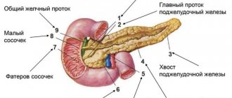
Since the MRI machine covers the entire abdominal cavity, it shows changes not only in the pancreas, but also in related organs: liver, ducts, which allows a more detailed picture of the disease.
Suspicion of calculous pancreatitis
This is a disease of the deposition of calcium salts in the pancreas and its ducts. It occurs due to changes in the composition of pancreatic juice, due to constant errors in nutrition or diseases of the liver and kidneys.
Pancreatic tumors
MRI of the pancreas
determines not only the presence of neoplasms at the earliest stages, but also their exact size and area of distribution. This is very important in cancer treatment. No other diagnostic method detects tumors with the same accuracy.
Chronic pancreatitis to assess organ structure, especially the pseudotumorous form
A description of the pancreas with MRI shows the structure of the tissues, changes in them, inflammatory processes, difficulty in the outflow of gland secretions, its deficiency, as well as an increase in the organ itself. All this, along with pain and possible jaundice, are symptoms of pseudotumor pancreatitis - an advanced stage of a chronic inflammatory process.
Pancreatic cyst
Cysts are also visible on ultrasound. MRI of the pancreas will help determine their exact size, quantity (down to the smallest), and location.
.
Prescription restrictions
An absolute contraindication is the presence of a pacemaker or insulin pump. Their work in a magnetic field will be disrupted. Also, when a metal object enters the scanning area, it quickly heats up, which leads to tissue burns. Therefore, this method is not prescribed in the presence of any metal structures - stents, clips installed during surgery on vessels, fragments remaining in the body, implants.
During pregnancy, MRI is not used in the first months, and then it is prescribed for health reasons.
Relative limitations are the fear of confined spaces and a weight of 130 kg or more. For these categories of patients, it is possible to use an open-type device designed for significant excess weight. In cases of severe motor agitation and the inability to remain still, sedatives may be additionally administered.