Any malignant tumor can develop metastases. In the presence of secondary tumor foci, the patient’s chances of recovery are reduced. Many patients who turn to oncologists are interested in what peritoneal carcinomatosis is and whether it can develop in oncology.
Peritoneal carcinomatosis is a secondary oncological disease, the prognosis of which in most cases is unfavorable. Patients with this diagnosis are prescribed palliative treatment, the methods of which are aimed at maintaining vital functions. Treatment of abdominal cavity carcinomatosis is carried out at the Yusupov Hospital, whose experienced specialists accept even those patients who were rejected in other clinics.
Causes of peritoneal carcinomatosis
Not every cell that breaks away from the mother’s cancerous tumor is capable of becoming a metastasis; the lion’s share of circulating malignant cells die in the bloodstream. To gain the ability to become a metastasis, a cancer cell must change internally - learn to produce substances that allow it to live independently and invade another place, suppressing normal cells.
Cells detached from the node migrate over long distances, pushing apart normal cells, are implanted into the abdominal mucosa, and can even penetrate inside other cells. After establishing itself in the area, reproduction and the formation of an entire cell colony begin.
In addition to the transfer of metastatic cells through the blood and lymph, the spread also occurs inside the abdominal cavity - transcoelomically. It is not entirely clear why malignant cells linger in the peritoneum; a beneficial effect of the microclimate is assumed. Most metastases are found in places with a calmer environment and weak peristalsis of organs, or where intra-abdominal fluid is actively absorbed.
Cells are often “scattered” during surgery and during laparoscopic surgery the likelihood of contamination is half as high as during classical surgery. During surgery, it is necessary to prevent cancer dissemination through repeated treatment with special solutions, but the most effective way to cleanse dissemination is intracavitary chemotherapy with hyperthermia (HIPEC).
Book a consultation 24 hours a day
+7+7+78
Metastases caused by surgery
On the surface of an organ damaged by a malignant neoplasm, there are many cancer cells. They can cause carcinomatosis of the peritoneal cavity. The second option for the appearance of abdominal metastases occurs after surgery, when malignant cells are easily introduced and seed the surrounding tissues and organs; it is often simply not possible to completely remove the tumor; the remaining at least one cancer cell can cause a relapse.
It takes a short period of time for metastatic cells to become fixed on the surface of the peritoneal cavity. The insidiousness also lies in the fact that traditional lavage of the abdominal cavity with saline solution is not effective in this case. Modern research in the field of oncology has revealed an interesting pattern. It has been established that the risk of contamination with malignant cells increases by 50-60% during classical abdominal surgery, and when performing a laparoscopic version of the operation, the risk of contamination is significantly lower (3-28%).
What diseases does it develop in?
Peritoneal carcinomatosis is diagnosed in every third patient with a neoplasm of the gastrointestinal tract. Metastases to the peritoneum are characteristic of carcinomas of the stomach and pancreas; up to 40% of patients are affected. In intestinal cancer, carcinomatosis is detected only in a tenth of patients. The highest percentage is due to malignant processes of the ovaries - at the time of diagnosis of the disease, two out of three patients already have tumor nodes on the peritoneum.
The likelihood of carcinomatosis depends on the degree of aggressiveness of cancer cells and the size of the primary tumor, so with total infiltrative gastric cancer it is detected more often than with a local process that has not destroyed the outer serous membrane of the organ.
However, in none of the malignant processes of any localization, be it breast or prostate cancer, lung or nasopharynx, intraperitoneal metastasis is excluded. Posthumously, carcinomatous changes are detected in every third person who died from the progression of the disease.
For sarcomas, such localization of metastases is atypical; peritoneal sarcomatosis is detected in hardly three out of a hundred patients. In the rarest cases, mucinous adenoma of the appendix and ovarian cystadenoma, completely benign in histology, can also lead to contamination of the peritoneum with the production of a gel-like secretion.
One in a million, and much more often it will be a woman, is diagnosed with a mucinous adenoma of the appendix or a mucinous cystadenoma of the ovaries, which subsequently often leads to colonization of the peritoneum. The spread of adenomucinous cells in the abdominal cavity with the production of a gel-like secretion is already called “pseudomyxoma”; often at this stage of the disease it is not possible to determine the original source of the tumor.
Diagnosis of the disease
Diagnosing carcinomatosis is extremely difficult. In this case, much depends on the doctor who has previously observed the development of the tumor in the patient. Information comes gradually over several surveys. Moreover, examinations should be carried out by different specialists, which will allow us to understand the full picture of the pathology.
You can diagnose the disease and determine the stage of carcinomatosis at the Sofia Oncology Center, which is located in the central district of Moscow. This center uses the most modern equipment, with the help of which specialists can determine the degree of carcinomatosis in a patient.
Personal inspection
An experienced oncologist can identify the disease even during the initial examination of the patient. Using special techniques and applying pressure to the abdominal cavity, the doctor tries to understand where the fluid has accumulated and what size the tumor is.
Examination using equipment
Modern medical devices make it possible to diagnose cancer with high efficiency.
Such devices include:
- X-ray;
- Ultrasound of internal organs;
- CT;
- MSCT, MRI;
- biopsy;
- laparoscopy.
Laboratory research
With the help of tests carried out in the laboratory, doctors can with a high probability determine the condition of the human body, as well as understand how its internal organs work and whether there is a malfunction.
Treatment of the disease
Today, many methods of treating carcinomatosis are used. Thanks to new technologies and modern equipment, doctors can help even patients with advanced cancer.
The main treatment methods include chemotherapy and surgery. Treatment also includes postoperative rehabilitation
Now in the center of Moscow, everyone can undergo examination at the Sofia Oncology Center. If the disease is detected, the patient will be able to undergo treatment for carcinomatosis in the clinic.
Forecast
The prognosis for effective treatment of carcinomatosis is determined in each specific case. It all depends on the general condition of the patient and the stage of carcinomatosis. You can now get an accurate forecast at the Sofia Cancer Center, which is located in the central district.
Diagnosis of peritoneal carcinomatosis
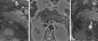
It is not difficult to identify tumor lesions of the peritoneum with ascites; in the absence of the production of pathological secretions, diagnosis is based on visualization - ultrasound and CT with contrast.
With ultrasound , on the inner layer adjacent to the muscles of the abdominal wall, which is normally very thin and invisible, you can see layers several centimeters thick, only small nodules are practically not visible.
Contrast-enhanced CT is much more informative than ultrasound and can detect centimeter-sized formations. The most accurate diagnostic method is laparoscopy. This examination is mandatory for gastric carcinoma; for ovarian cancer, surgery is preferable - diagnosis and treatment at the same time.
During laparoscopy or puncture, ascitic fluid is obtained to examine and determine the primary source of the malignant process. A precipitate is isolated from the exudate, which is examined under a microscope and specific reactions are carried out - PCR and IHC.
PET scan at the stage of primary diagnosis is not always informative, since not all malignant cells of the lung, liver, and kidneys are capable of accumulating isotopes.
Without a doubt, the most optimal diagnostic method is to obtain a piece of tumor tissue for examination. Biopsy is not appropriate if the source of metastases is known and after recent treatment of the primary cancer.
Modern diagnostics
In order for the diagnosis to be as accurate as possible, it is necessary to conduct modern research that is highly informative. The Top Assuta clinic has a diagnostic center where all types of tests can be carried out. The clinic's diagnostic doctors are high-class specialists with many years of experience. The entire examination takes no more than three days, after which the patient can immediately begin treatment. Everyone arriving from the CIS is provided with a personal Russian-speaking escort.
- Day 1
- Day 2
- Day 3
The treating oncologist conducts an examination: records symptoms and signs, studies the medical history, and specifies the list of studies.
All diagnostic measures are taken:
- Blood tests (general, biochemistry, hormones, tumor markers)
- CT
- Ultrasonography
- MRI
- Biopsy followed by histology
- Endoscopic studies
- PET-CT.
A treatment protocol is drawn up and the diagnosis is clarified. This happens at a medical consultation in the presence of specialized oncologists, chemotherapists, surgeons and radiologists.
Stages of abdominal carcinomatosis
Staging of peritoneal carcinomatosis cannot be called accurate; all classifications are approximate in determining the volume of damage and do not specify the location of the nodes. Often, staging gives a general idea of the prognosis for the effectiveness of treatment measures, rather than informing about the actual condition inside the abdominal cavity. The gradation of tumor spread into three degrees, developed by Japanese specialists, takes into account the total volume of the lesion, without the number and size of foci:
- P1 - limited;
- P2 - lesions separated by normal tissue;
- P3 is a set of nodes.
During surgery, surgeons determine the peritoneal carcinomatosis index (PCI), measuring nodules in 13 regions of the cavity; the total score affects treatment tactics, primarily the possibility of removing the peritoneum - peritonectomy and the advisability of intracavitary chemotherapy. In some malignant processes, complex formulas for calculating PCI are used. The best idea of the size of the cancerous lesion is given by staging by degree:
- 0 - the cavity is clean,
- I - nodules up to 5 mm in one anatomical zone,
- II - multiple nodules up to 5 mm,
- III - local lesion 0.5–2 cm,
- IV - 2 cm nodules.
The course of carcinomatosis is determined not so much by the size of the metastatic node as by the cellular potency for progression and production of ascitic fluid, the total area of tumor transformation and clinical manifestations.
Diagnosis of the disease
When visiting the hospital, the doctor prescribes a complete examination of the patient. This includes a detailed blood test, as well as hardware diagnostics using high-resolution imaging methods: CT, MRI, PET are used to detect peritoneal carcinomatosis . On the one hand, this helps to determine the location and size of the primary tumor, and on the other, the presence and number of metastases both in the peritoneum and throughout the body. To fully assess the tissue structure and the extent of primary or secondary infiltration of the abdominal space, laparoscopy, gastro- and/or colonoscopy is often required. During laparoscopy, the so-called peritoneal cancer index (PCI) is determined.
To do this, the abdominal cavity is conventionally divided into eight quadrants, each of which receives points from zero to three depending on the size of visible metastases. In addition, areas of the intestine, upper and lower jejunum, and ileum are assessed using the same criteria (see figure).
The sum of all points determines the degree (stage) of peritoneal carcinomatosis, on the basis of which prognoses are made and a decision is made on the choice of further therapeutic measures: surgical removal of the tumor (cytoreductive surgery) (CRS), radiation therapy, intraperitoneal chemotherapy (HIPEC).
To date, there is no gold standard for the treatment of peritoneal peritoneal carcinomatosis: this type of cancer is successfully diagnosed at the Nordwest clinic and is still the object of medical research to find the most effective way to combat it. In each individual case, the decision is made by the attending physician, based on the patient’s medical history, general condition, his preferences and possible risks of a particular type of therapy.
Symptoms of abdominal carcinomatosis
Peritoneal carcinomatosis of small extent may not manifest symptoms, especially in the absence of ascitic fluid production. On the other hand, fluid can be produced in the absence of visible metastases. As a rule, the symptoms are nonspecific, and a different set may include:
- painful sensations that change localization, and more often - incomprehensible discomfort in the abdominal cavity;
- increasing weakness to the point of loss of ability to work;
- weight loss with a stable diet regimen;
- progressive loss of appetite;
- functional disorders of the gastrointestinal tract.
Further growth of cancerous damage is accompanied by tumor intoxication, compression of the stomach by tumor nodes is complicated by nausea and vomiting, intestines - constipation and diarrhea with worsening partial obstruction. The disintegration of large nodes can cause pain and fever.
Ascites disrupts the breathing process and causes heart failure with constant edema, and frequent evacuation of pathological fluid leads to protein deficiency.
How is it carried out (HIPEC)
First, cytoreductive surgery is performed. Depending on the number of primary/secondary abdominal tumors, it can last from a few to many hours. After the surgeon has removed all visible parts of the tumor and the affected peritoneum, preparation for HIPEC begins.
To do this, special drains are installed in the lower and upper quadrants of the abdominal cavity. Some of them will deliver chemotherapy heated to 41-43 degrees, while others will install special monitor sensors that monitor the constant temperature of the chemotherapy drugs. The duration of this manipulation can be 45-60 minutes. The volume of chemotherapy infused into the abdominal cavity averages 3-3.5 liters. At the end of the procedure, the hot chemical is pumped out through the drains and the abdominal cavity is closed.
How is peritoneal carcinomatosis treated?
None of the modern methods of treating carcinomatosis guarantee radical removal of the tumor or can cure, but can improve the condition and significantly prolong life.
Surgical treatment of carcinomatosis is technically difficult for the operating team and difficult to tolerate by the patient, since it involves removal of the primary cancer, enlarged lymph nodes, omental bursae and all visible tumor formations along with the peritoneum.
Peritonectomy is a multi-stage intervention, including the removal of several organs and parts of the abdominal cavity. As a result of the operation, the patient may be left without a spleen, gallbladder, part of the intestines, uterus and appendages.
The standard treatment for carcinomatosis is systemic and local chemotherapy - intraperitoneal after removal of ascites or through a laparoport installed during surgery.
The effectiveness of drug therapy is low, with the exception of cases of primary ovarian cancer. Targeted and immuno-oncological drugs are only being studied in clinical trials.
Clinical picture
Symptoms of peritoneal carcinomatosis are observed in that part of patients who have already been diagnosed with a primary tumor of either the digestive system (more often it is localized in the intestines or stomach), or an invasive neoplasm of the ovaries in women. It should be noted that in patients with ovarian cancer, the likelihood of developing ovarian carcinomatosis is much higher (70%) than in patients with malignant processes of the digestive tract (only 40%).
Also, metastatic foci can form on the surface of surgical sutures, which seed the abdominal cavity. Neoplasm cells are surrounded by a fairly dense layer of fibrin (from the Latin fibra - fiber), which makes them practically invulnerable. It is this mechanism that explains the formation of relapse after surgery.
Which therapy methods give the best results?
The highest effect is demonstrated by a combination of three cancer treatment methods:
- An operation with the maximum possible removal of malignant tumors is cytoreduction.
- Local intraperitoneal hyperthermia.
- Intracavitary administration of chemotherapy drugs.
The use of intraperitoneal hyperthermic chemotherapy (IHCT or HIPEC) during surgery makes it possible to maintain a very high concentration of the cytostatic directly in the affected area for as long as possible and enhance the drug effect by heating the tissue. With very modest historical results for surgical intervention of cytotoxic-resistant pseudomyxoma, only HIPEC offers patients the prospect of a long life.
The IHCT technology is as follows: for an hour and a half, a heated chemotherapy drug is delivered under pressure into the abdominal cavity in a dose significantly exceeding the maximum allowed for intravenous administration. Due to local use, the spectrum of toxic reactions changes, life-threatening damage to hematopoiesis is excluded, but abdominal pain and temporary disruption of the functioning of the gastrointestinal tract are possible.
Intraoperative photodynamic therapy (PDT), when tumor foci identified using a photosensitizer are treated with a laser, is inferior to HIPEC in terms of effectiveness, since it is impossible for the laser to penetrate into all the “nooks and crannies” of the abdominal cavity. However, it is advisable to use photodynamic therapy for large and few cancerous nodes.
Signs and symptoms
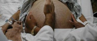
Peritoneal carcinomatosis, the treatment of which requires the patient to stay in the hospital and be monitored around the clock, is a secondary disease. The clinical picture for this diagnosis is determined by the signs of a primary malignancy. Peritoneal carcinomatosis in cancer is characterized by the formation of ascites - the accumulation of free fluid in the abdominal cavity.
The main signs of peritoneal carcinomatosis:
- body weight decreases quickly and the belly increases
- indigestion
- profuse sweating;
- dull, severe pain in the abdominal area
- Characterize carcinomatosis of the peritoneum; pain under the breast
- the patient shows signs of severe intoxication
- loose stools that may contain blood.
As the oncological process progresses, the patient may experience loss of consciousness and a state of delirium. Peritoneal carcinomatosis due to cancer requires immediate treatment, so the Yusupov Hospital accepts patients in serious condition every day, 24 hours a day.
Carcinomatosis and ascites can threaten the patient’s life, so people at risk should know the symptoms of the disease in order to promptly consult an oncologist. Specialists from the Yusupov Hospital answer questions from patients: abdominal carcinomatosis - what it is, what treatment methods exist and what is life expectancy.
Prognosis for carcinomatous lesions
The course of the process is influenced by the volume of the lesion at the time of initiation of therapy, the degree of malignancy of the tumor, which in turn determines the sensitivity to chemotherapy. The talent and experience of the surgeon, and undoubtedly the correct choice of treatment tactics, have a fundamental influence.
Only HIPEC showed clearly revolutionary results in clinical trials. After intraoperative hyperthermic chemotherapy, the five-year survival rate for gastric cancer carcinomatosis increased to a maximum of 20%; all other methods excluded such a long survival. With colon cancer with metastases to the peritoneum, every third patient lived more than 5 years, with carcinoma of the cecum and appendix - six out of ten entered the second five years of life.
Book a consultation 24 hours a day
+7+7+78
Bibliography
- Davydov M.I., Ter-Ovanesov M.D., Buydenok Yu.V. et al. / Hyperthermic intraoperative intraperitoneal chemotherapy for gastric cancer: is there a real opportunity to change the prognosis? // Bulletin of the Russian Cancer Research Center named after. N. N. Blokhin RAMS; 2010 T. 21; No. 1
- Stepanov I.V., Paderov Yu.M., Afanasyev S.G./ Peritoneal carcinomatosis // Siberian Journal of Oncology; 2014; No. 5
- Akiyama H., Yamaoka H., Tanaka K., et al./ Continuous hyperthermic peritoneal perfusion for peritoneal dissemination of gastric cancer // Hepatogastroenterology; 1998.
- Bozzetti F., Bonfanti G., Morabito A., et al/ A multifactorial approach for the prognosis of patients with carcinoma of the stomach after curative resection // Surg. Gynecol. Obstet.; 1986.
- Cotte E., Passot G., Gilly FN, Glehen O. /Selection of patients and staging of peritoneal surface malignancies // World J. Gastrointest. Oncol.; 2010; Vol. 2.
- Chua TC, Moran BJ, Sugarbaker PH, et al. /Early- and longterm outcome data of patients with pseudomyxoma peritonei from appendiceal origin treated by a strategy of cytoreductive surgery and hyperthermic intraperitoneal chemotherapy// J. Clin Oncol 2012.
- Deraco M., Santoro N., Carraro O., Inglese MG, et al./ Peritoneal carcinomatosis: feature of dissemination. A review // Tumori; 1999.
- Lansom J., Alzahrani N., Liauw W., Morris DL /Cytoreductive Surgery and Hyperthermic Intraperitoneal Chemotherapy for Pseudomyxoma Peritonei and Appendix Tumors // Indian J Surg Oncol. 2016 Jun.
- Sugarbaker PH/ Overview of peritoneal carcinomatosis // Cancerologia; 2008.
Cost of carcinomatosis treatment in Germany
To diagnose oncological diseases, the Nordwest clinic uses new generation equipment. The first signs of peritoneal carcinomatosis can be detected using CT and confirmed through other studies. A detailed examination and consultation with a doctor will help to identify the characteristic symptoms of the disease even at the stage of your visit to the hospital, carry out all diagnostic procedures in a timely manner and draw up an effective treatment regimen. Attentive attitude towards patients and many years of experience of the best oncologists in Germany are the reason for many positive reviews about the work of doctors, both in the treatment of peritoneal carcinomatosis and in the case of other diseases. The cost of an initial consultation, diagnosis and therapy at the Nordwest clinic can be clarified upon your first contact or by writing to
Making an appointment with an oncologist
Dear patients, We provide the opportunity to make an appointment directly with the doctor you want to see for a consultation. Call the number listed at the top of the site, you will get answers to all your questions. First, we recommend that you study the About Us .
How to make an appointment with a doctor?
1) Call the number 8-863-322-03-16.
1.1) Or use the call from the site:
1.2) Or use the contact form:
2) The doctor on duty will answer you.
3) Talk about what's bothering you. Be prepared that the doctor will ask you to tell you in as much detail as possible about your complaints in order to determine the specialist required for consultation. Keep all available tests at hand, especially those recently done!
4) You will be connected with your future attending physician (professor, doctor, candidate of medical sciences). Next, you will discuss the place and date of the consultation directly with him - with the person who will treat you.
OPT.MS.10.07.2016