Esophagogastroduodenoscopy (fibrogastroduodenoscopy, gastroscopy, FEGDS, FGDS, FGS, EGDS) is a method for examining the organs of the gastrointestinal system, with which you can examine the mucous membrane and structural features of the esophagus, stomach and duodenum from the inside.
Esophagogastroduodenoscopy allows not only to examine the digestive organs from the inside, but also to take a piece of tissue from the pathological area of the mucosa for cytological and histological studies, as well as to carry out therapeutic manipulations, if the need arises: to remove polyps, stop bleeding from an ulcer, etc.
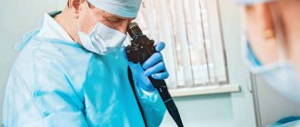
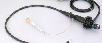
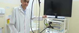
The manipulation is carried out using a special device - a gastroscope. This is a thin and flexible hose with a miniature optical camera at the end, which is inserted through the mouth into the esophagus, then into the stomach and duodenum.
In what cases is esophagogastroduodenoscopy (EGD) prescribed?
Indications for esophagogastroduodenoscopy are quite broad:
- differential diagnosis of diseases of the gastrointestinal tract (esophagitis, gastritis, peptic ulcer of the stomach and duodenum, tumor, diverticulum, pyloric stenosis, etc.);
- monitoring the effectiveness of treatment of gastrointestinal diseases;
- dynamic observation among people with chronic diseases of the gastrointestinal tract;
- suspicion of internal bleeding.
How is esophagogastroduodenoscopy (EGD) performed?
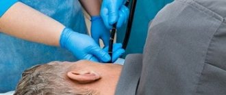
Before the examination, the person is placed on his side, and the oropharynx is treated with an anesthetic solution to avoid choking and the appearance of a gag reflex during insertion of the gastroscope into the esophagus. Then the gastroscope is carefully inserted inside. To examine the stomach and duodenum, air is pumped through it to straighten the walls of the organs.
The examination takes about 15–20 minutes and requires patience from the patient, but is usually not accompanied by pain. After the procedure, there may be discomfort associated with irritation of the esophagus and pharynx, as well as discomfort due to decreased sensitivity in the mouth, which disappears after a few hours. Until the effect of the anesthetic has completely worn off, you should not eat or drink, as you may choke.
How is gastroscopy performed? Gastroscopy in a dream
Gastroscopy can be performed under local anesthesia, as well as in a state of medicated sleep. Drug-induced sleep should be distinguished from general anesthesia.
If the procedure is performed under local anesthesia, gastroscopy lasts 5-10 minutes. The patient is asked to hold the mouthpiece through which the gastroscope is inserted.
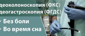
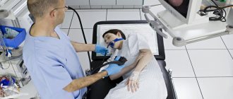
For those who are wary of this procedure, the doctors at Family Doctor recommend using the “sleep gastroscopy” service. This service is provided in a surgical hospital setting. Abroad (in Europe, Israel, the USA, Japan), gastroscopy during medicated sleep is the basic version of the procedure, which is abandoned only if there are contraindications.
To perform gastroscopy in your sleep, it is advisable to approach 30 minutes before the time for which the procedure is scheduled. In this case, you must have a fresh and deciphered electrocardiogram on hand. You will be examined by an anesthesiologist who will determine whether there are any contraindications to the study. To put you to sleep, the drug “Proviv” is used, which is administered under the supervision of an anesthesiologist. When the drug is administered, the patient falls asleep and wakes up almost immediately after the end of the administration. After 15-20 minutes, complete recovery occurs, however, the doctors at “Family Doctor” still recommend staying under the supervision of a doctor for another hour or two, which can be done in our comfortable hospital. It is considered acceptable to drive a car two hours after waking up from sleep, but it is better to drive no earlier than 6 hours later.
During the study, it may be necessary to carry out therapeutic and diagnostic manipulations (biopsy - taking a tissue element for laboratory analysis, removal of polyps, etc.). Similar manipulations are carried out during the study using a gastroscope. The biopsy material is sent to the Family Doctor’s own laboratory, which significantly speeds up the process of obtaining results.
Normal results of esophagogastroduodenoscopy
During the examination, the endoscopist describes in detail the structure of the esophagus, stomach, and duodenum and makes a conclusion based on these data.
Normally, each section of the gastrointestinal tract has a characteristic structure. Thus, the esophagus has longitudinal folds that are easily straightened by air, a uniform, light yellowish-pink mucosa, and a delicate network of blood vessels is visible. When moving into the stomach, the color of the mucous membrane becomes brighter, the walls become deeply folded. The initial sections of the duodenum have a reddish color, the distal section has a velvety appearance due to the different structure of the epithelium.
Gastroscopy
Gastroscopy (the full name of the study is esophagogastroduodenoscopy) is an examination of the upper gastrointestinal tract with an endoscope - a thin flexible probe with a high-resolution video camera and a channel for carrying out various instruments for therapeutic and diagnostic purposes. Gastroscopy examines the esophagus, stomach and duodenum.
Why is it worth undergoing a study at the Federal Scientific and Clinical Center of the Federal Medical and Biological Agency of Russia?
- modern approaches to diagnosis and treatment;
- doctors with extensive experience in endoscopy and oncology;
- consultation with a doctor before and after the study;
- video endoscopes with images of the examination on the screen;
- recording the study on the patient’s electronic media;
- the conclusion is sent to the patient by email;
- the possibility of outpatient examination and hospitalization in a hospital for preparation and examination;
- the ability to perform research “in your sleep”;
- the staff includes an anesthesiologist specializing in endoscopic interventions “during sleep”, as well as in gerontology;
- many years of experience in conducting two studies (colonoscopy + EGDS “during sleep”) in one day without hospitalization;
- no age restrictions for adult patients;
- comfortable recovery room for outpatients.
Along with and in combination with gastroscopy, if necessary, modern types of clarifying diagnostics are used, such as:
- collection of material for histological and cytological studies (biopsy) - to identify Helicobacter pylori, fungal lesions, pathology of the small intestine (celiac disease), diagnosis of inflammatory, pretumor and tumor lesions;
- chromoscopy – endoscopic examinations with vital dyes to identify precancerous changes and early forms of cancer;
- EUS is an ultrasound examination of the stomach with an endoscope, at the end of which there is an ultrasound sensor. The study allows you to evaluate the structure of the stomach wall, determine the nature of pathological formations of the mucous membrane and submucosal sections;
- examination of the mucous membrane in a narrow spectrum of light - chromoscopy using a certain length of light waves.
Possibilities of therapeutic endoscopy
Gastroscopy may include parallel performance of therapeutic procedures, such as:
- polypectomy – removal of pathological formations of the mucous membrane of the stomach, esophagus, duodenum;
- stop bleeding - endoscopic stop of bleeding that has complicated the course of a peptic ulcer, erosive processes in the stomach or duodenum;
- removal of foreign bodies of the esophagus, stomach, duodenum;
- ligation of varicose veins of the esophagus and stomach - “ligation” of dilated veins with special endoscopic rings to prevent bleeding;
- endoscopic installation of intraluminal prostheses (stents) in case of obstruction, usually caused by a tumor or non-tumor process - endoprosthetics of the esophagus and duodenum;
- bougienage, pneumodilation for achalasia cardia, various narrowings of the esophagus of a tumor and non-tumor nature.
Indications for the study
Gastroscopy is performed to carry out many diagnostic and therapeutic tasks, including:
- Determination of the etiological factors of vomiting with blood with the possibility of determining the source of bleeding;
- Determining the causes of pain in the epigastric region, bloating, dysfunction of the swallowing reflex, vomiting, unspecified causes of weight loss;
- Finding the cause of infection in the digestive system;
- Assessment of the mucous membranes of the upper digestive system after surgery;
- Study of the condition of the mucous membrane of the esophagus and stomach after acute poisoning with chemical poisons;
- Sampling of tissue materials (biopsy) for the purpose of further histological examination; usually used to exclude cancer;
- Removal of pathological formations - polyps from the lumen of the esophagus, stomach or duodenum;
- Removal of foreign bodies;
- Determination of the causes of chronic bleeding of unspecified etiology, contributing to the development of anemia;
- Conducting a search for pathologies and clarifying the diagnosis for:
- inflammatory phenomena on the mucous membrane of the esophagus - esophagitis;
- reflux disease of the stomach;
- focal narrowing of the esophagus;
- precancerous condition of the esophagus or Barrett's esophagus;
- hernial phenomena of the esophageal opening in the diaphragm (hiatal hernia);
- peptic ulcer of the stomach and duodenum;
- oncological pathologies of the upper digestive system.
Contraindications
Gastroscopy is strictly contraindicated if the patient has the following conditions and symptoms:
- general serious condition of the patient;
- elevated blood pressure at the time of the study;
- infarction processes in the myocardium in the acute period;
- acute period of cerebral stroke;
- a sharp decrease in blood clotting;
- psychopathological conditions and disorders.
How to prepare for research
- Last meal the night before at 7:00 pm if the test is performed in the morning or afternoon. Do not eat breakfast on the day on which your gastroscopy is scheduled. 6 hours before the test you can drink a glass of sweet tea.
- Come to the examination hall of the endoscopy department at the indicated time or a little earlier.
- Patients suffering from arterial hypertension, coronary artery disease, bronchial asthma, receiving hormone replacement therapy should take continuous medications in the morning 1 hour before the test, if necessary, with 1-2 sips of water.
- If you use a personal inhaler, take it with you.
Endoscopy diagnostics in the network of NEARMEDIC clinics
Diagnostic esophagogastroduodenoscopy is one of the most highly accurate methods for identifying pathologies, which is actively used in the gastroenterology departments of the multidisciplinary medical network NEARMEDIC.
Referrals for endoscopy can be received not only by patients of gastroenterologists, but also by oncologists, surgeons, and pediatricians in the children's departments of network clinics for the following symptoms and phenomena:
- signs of digestive disorders - belching, heartburn, nausea, vomiting;
- stomach pain after eating or not related to diet;
- constipation;
- diarrhea and associated dehydration;
- flatulence;
- pain in the upper digestive tract;
- chest pain during eating;
- rumbling in the stomach;
- heaviness in the stomach;
- feeling of a lump in the esophagus;
- appetite disorders;
- unpleasant taste in the mouth;
- bitterness in the mouth, including during regurgitation;
- swallowing problems;
- increased salivation;
- sensitivity of the esophagus to cold or hot food and drinks;
- change in body weight without changing diet and diet;
- foreign body in the esophagus;
- taking a biopsy sample;
- determination of surgical tactics.
- Contraindications for endoscopic examination of the stomach, esophagus and duodenum are diseases of the cardiovascular and respiratory systems in the decompensation stage, stroke, acute heart attack, cerebrovascular accident in the acute stage, blood diseases with bleeding disorders, some types and stages of neuropsychiatric disorders .
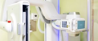
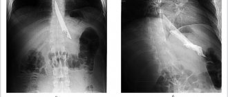
- The network's laboratories are equipped with all types of medical equipment for effective hardware examination of patients. But in some cases, esophagogastroduodenoscopy is the most preferable technique. If radiography reveals the presence of a pathological focus in the organs being examined, then endoscopy provides a comprehensive description of the location, dimensions, boundaries of the spread of the inflammatory process, and the presence of bleeding.
- Computed tomography, while highly accurate in detecting pathologies, does not allow visual assessment of the condition of the organ and the characteristics of the lesion, or sampling of tissues. Also, EGDS outperforms ultrasound diagnostics in terms of the possibility of developing a plan for surgical intervention. None of the methods, including MRI, make it possible to examine the digestive organs from the inside.
- Endoscopy is a self-sufficient method for studying the upper digestive tract. But if necessary, NEARMEDIC provides patients with all of the above listed diagnostic equipment, which will allow you to examine adjacent areas, which is sometimes necessary for tumors and perforations.
- The adult and pediatric gastroenterology departments employ experienced endoscopists who have basic medical education, many years of experience in their specialization and have been trained in the field of endoscopic diagnostics and have received the appropriate certificate. In this regard, they give an opinion after examination at a high expert level, with high accuracy of diagnosis, which allows you to prescribe effective treatment.
Esophagogastroduodenoscopy allows you to accurately detect:
- neoplasms (both benign polyps and neoplasia);
- foci of inflammation;
- disturbances in the structure of the organs being studied;
- narrowing, insufficiency of sphincters;
- processes of tissue deformation, erosion and ulcers;
- peptic ulcer of the stomach and duodenum;
- bleeding of the esophagus, stomach or intestines;
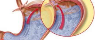
- gastroesophageal reflux;
- esophagitis;
- gastritis;
- duodenitis;
- gurgle;
- foreign bodies;
- mechanical, chemical injuries (burns from exposure to chemicals).
The method is used for highly accurate differentiation of pathologies that cause bleeding and intestinal obstruction, since these symptoms are characteristic of various diseases of the stomach, esophagus and duodenum.
During the diagnostic process, an experienced endoscopist can not only take tissue for histological and microscopic examination, but also carry out therapeutic manipulations.
EGDS as a medical procedure
Esophagogastroduodenoscopy is a therapeutic and diagnostic technique in which surgical and conservative therapy can be carried out. In gastroenterology departments, EGDS is used as a therapeutic procedure in the following cases:
- removal of polyps and other benign neoplasms (based on the results of histological and cytological examination);
- detection of a foreign body in the organs of the upper digestive tract;
- administration of medications for the treatment of ulcerative formations, inflammatory processes;
- blocking bleeding using mechanical and medicinal methods.