One of the most serious diseases of the esophagus is called Barrett's esophagus, a syndrome characterized by pathological changes in the mucous membrane. The disease develops against the background of gastroesophageal reflux, that is, due to the reflux of hydrochloric acid into the esophagus. This syndrome refers to conditions that can potentially cause the development of esophageal cancer. Diagnosis may require a number of instrumental studies, since the disease is manifested by belching, heartburn and other symptoms characteristic of various pathologies of the esophagus and stomach. In uncomplicated cases, conservative treatment is prescribed; in some cases, surgical intervention is required. If symptoms appear that are suspicious for the development of Barrett's esophagus, you should consult a gastroenterologist.
At CELT you can get advice from a gastroenterologist.
- Initial consultation – 4,200
- Repeated consultation – 3,000
Make an appointment
Features of the disease
The syndrome is named after the researcher who discovered this disease in the mid-20th century. At first, Barrett considered such pathological changes to be a variant of the norm. And only after years of research he was able to find out that the syndrome can be attributed to precancerous conditions. This disease is considered severe, since constant exposure to gastric juice, which has an extremely low pH, has a detrimental effect on the epithelium of the esophagus and provokes the development of oncology. The risk of developing a malignant tumor is reduced after successful treatment.
It is now common to associate GERD (gastroesophageal reflux disease) with Barrett's esophagus. There are studies showing that ~80% of patients with Barrett's esophagus previously had GERD. The pathology is associated with age and duration of GERD. Most often, the disease is diagnosed in men 50–60 years old. The risk of malignancy (malignancy) is 9 times higher for men than for women.
Treatment of Barrett's esophagus using minimally invasive technologies: laparoscopy and endoscopy.
My experience in treating patients with hiatal hernia and reflux esophagitis of varying severity is 24 years. During this time, I was able to successfully operate and treat more than 2,000 patients laparoscopically. In 10% of them, Barrett's esophagus was identified, which we successfully treated in 97% using our comprehensive approach of minimally invasive treatment.
It is worth noting that only as a result of a clear and complete examination can we obtain sufficient information about the patient. If Barrett's esophagus is detected on endoscopic examination and confirmed by histological tests, the question of mandatory treatment is raised, since the risk of degeneration into cancer is very high.
Puchkov K.V., Filimonov V.B. Hiatal hernia: monograph. - M.: MEDPRACTIKA - M., 2003. - 172 p.
At this stage, the results of cytological and histological studies help to accurately select the method of surgical treatment. We have already said that Barrett's esophagus is characterized by hyperkeratic, metaplastic and dysplastic changes in the esophageal mucosa. Consequently, when hyperkeratosis, gastric, small intestinal and colonic metaplasia, mild and moderate dysplasia are detected, we are talking only about a benign process. In the case of severe dysplasia or when squamous cell non-keratinizing cancer is detected, a diagnosis of cancer is made. This allows us to clearly distinguish between two types of treatment - in the first case, organ-preserving and in the second case, organ-sapping (Lewis operation).
Immunohistochemical examination, along with histological examination of biopsy specimens, allows us to identify early forms of esophageal adenocarcinoma. Esophageal cancer is characterized by an increase in the expression area of the Ki-67 marker and the anti-apoptotic factor bcl-2 compared to values in gastroesophageal reflux disease and Barrett's esophagus. Thus, the differential diagnosis of Barrett's esophagus and esophageal adenocarcinoma can be further achieved by studying the results of immunohistochemistry of esophageal cells producing nitric oxide synthase and endothelin-1.
If, as a result of endoscopic examination, histological and immunohistochemical studies, severe dysplasia and adenocarcinoma of the esophagus are not detected, we should only talk about organ-preserving treatment of Barrett's esophagus. The stages of this algorithm will be described by me below.
In the absence of a hiatal hernia, especially in young patients, we use radiofrequency ablation (RFA) of Barrett's esophagus. In the postoperative period, therapy with proton pump blockers (suppression of gastric secretion) and motilium (improvement of gastric motility) is prescribed for 6-8 weeks. Dynamic observation - gastroscopy - is carried out for several months. If a relapse of the disease occurs, this procedure can be repeated, since the effect is only within the mucous membrane, without causing damage to the entire thickness of the esophageal wall.
In the presence of a hiatal hernia, the first stage requires surgical intervention - laparoscopic Tope fundoplication (bilateral fundoplication at 270 degrees) and crurorrhaphy. This intervention eliminates the hernia and stops the pathological reflux of aggressive stomach contents into the esophagus. Without this stage, further treatment of Barrett's esophagus, as a rule, does not bring a positive result. In the postoperative period, for 3-4 months, therapy with proton pump blockers is prescribed.
This stage allows in 90% of cases to stop pathological reflux into the esophagus and thereby stop the pathological transformation of the mucous membrane and the reverse restructuring of the esophageal epithelium.
After surgery, after 2-3 months, it is necessary to perform radiofrequency ablation (RFA) of the affected mucosa, and then conduct follow-up examinations of the esophagus (FGS) after 3 and 6 months. In case of positive dynamics, further observation is carried out 1-2 times a year.
In a number of patients with long-term changes in the mucosa (long segment of Barrett's esophagus), an additional 1-2 sessions of RFA are necessary.
If, upon re-evaluation of the condition of the esophageal mucosa, according to FGS, the damage to the mucosa decreases, then we carry out further dynamic observation, performing FGS at intervals of 6 months. After 1 year, a final assessment is made for the presence or absence of Barrett's esophagus. In most patients, manifestations of Barrett's esophagus were reversed (especially in patients with hiatal hernia and acid reflux). If morphologically, during biopsy, we see persistence of changes in the mucosa (metaplasia), then we move on to the next stage - endoscopic (RFA) or argon plasma coagulation of the esophageal mucosa in the area of Barrett's esophagus.
Radiofrequency ablation and argon plasma coagulation are performed under endoscopic control, usually under general anesthesia (short-term intravenous sedation). For lesion segments up to 3 cm, one session is usually sufficient. If the lesion is more than 3 cm, a repeat procedure may be necessary. After radiofrequency ablation, the damaged tissue is replaced by squamous epithelium and heals without scarring. In our practice, we prefer to use RFA, which in principle is an improved version of the argon plasma coagulation technique, as it has fewer side effects.
It is worth noting that if areas of high-grade neoplasia were detected in the metaplasia zone, in which the likelihood of invasive growth increases significantly, radiofrequency ablation can also be performed. For deep lesions, endoscopic radical removal of a section of the esophageal mucosa is performed. If the area of changes is up to 2 cm2, endoscopic resection of the esophageal mucosa (EMR-C) is usually performed, and if the area of changes is more than 2 cm2, dissection of the neoplasm in the submucosal layer (ESD) is performed.
Next, lifelong clinical observation is mandatory - during the first year after 3 months, then FGS is performed once a year with a possible biopsy of the mucous membrane of suspicious areas.
It is extremely rare that after such complex and staged treatment, relapses of Barrett's esophagus occur (less than 5%), as a rule, this occurs with relapse of the hiatal hernia and remnants of altered mucosa after RFA.
A number of authors perform surgical treatment in the reverse order - at the beginning of RFA, and then laparoscopic fundoplication. It is worth noting here that the optimal period for performing laparoscopic surgery on the esophagus is 3-4 months after the first stage. Thus, according to ultrasound data of the esophagus, swelling of its wall persists for a very long time (against the background of RFA) and fundoplication performed at an earlier date can lead to the development of stenosis in the area of the esophagogastric junction. Naturally, the healing of the mucous membrane in the esophagus will take much longer, since the reflux of aggressive contents from the stomach remains uncorrected. That is why we use reasonable consistency in our work. First of all, the pathogenetic stage of stopping reflux and only after that is radiofrequency ablation of the altered mucosa. As I wrote earlier, in some patients the second stage is not required, since the mucous membrane returns to normal thanks to its own reparative processes.
Thus, laparoscopic fundoplication at the first stage of treatment makes it possible to reduce the size of the affected segment of the esophagus, which in the future may require only 1-2 sessions of RFA and, accordingly, a lower risk of developing esophageal stricture, and in some patients the second stage can be completely avoided.
Causes
Researchers point to a clear relationship between this disease and GERD. Therefore, the main etiological factor is considered to be the reflux of gastric juice into the esophagus, leading to metaplasia. In the future, degeneration of damaged cells is possible. It is believed that at least 4 years are required for the risk of malignancy to increase. Therefore, complications can only be prevented by timely treatment.
In addition to the immediate cause, the following risk factors are identified:
- Smoking. It is also the main risk factor for the development of GERD.
- Alcohol consumption.
- Taking certain classes of pharmaceutical drugs.
- Mature and old age.
- Chronic diseases of internal organs.
These risk factors do not directly cause the development of the disease, but they increase the risk of developing Barrett's esophagus in the presence of an underlying cause, and also contribute to a complicated course.
Prevention
Today there are no known methods to prevent the development of Barrett's esophagus, so the goal is to reduce the risk of the disease. Patients at risk are under constant surveillance and should regularly undergo endoscopy with targeted biopsy to determine the degree of degeneration of mucosal cells. Early recognition of the disease is important, so men over 60 years of age who suffer from symptoms of reflux, as well as those undergoing long-term and uncontrolled treatment, should undergo regular endoscopic examination. All patients should adhere to certain rules, for example, after eating it is recommended to walk slowly for 30-60 minutes, the last meal before bed no later than 2-3 hours, etc. A balanced diet and avoiding overeating are simple actions that can reduce the risk of developing the disease.
Symptoms
Complaints typical of GERD
This makes it particularly difficult to diagnose Barrett's esophagus. To make a diagnosis, it is necessary to conduct a histological examination of the pathologically altered tissue, because the symptoms are similar for this disease, for GERD, for gastritis.
Typical symptoms of Barrett's esophagus include:
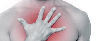
- Heartburn. Occurs most often. Develops after physical exertion, after eating, when bending over, and can intensify at night.
- Frequent belching of air, acid or even bile.
- Dysphagia is a swallowing disorder that makes it difficult for patients to eat. As a result, weight loss may occur.
- Vomiting and bleeding are characteristic of an advanced stage of the disease and may indicate the development of an oncological process.
Since it is impossible to determine the disease only by symptoms, instrumental and laboratory diagnostics are required. The scope of the examination is determined by the gastroenterologist after the examination.
Diagnostics
The sooner patients contact a specialist, the higher the effectiveness of treatment. If typical symptoms appear, you should go for examination to a gastroenterologist. If Barrett's esophagus is suspected, a standard test is prescribed - esophagoscopy. If the disease has been developing for a long time, an endoscopic biopsy of pathological epithelial tissue is required. Endoscopic examination clearly shows altered areas of the mucosa. In addition to esophagoscopy, other methods are used to clarify the diagnosis.
So, the diagnosis of Barrett's syndrome includes:
- Esophagogastroduodenoscopy.
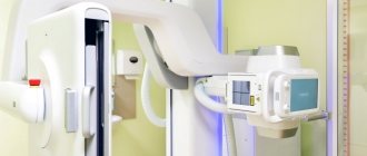
- X-ray of the esophagus.
- Electrocardiogram.
- Study of stomach acidity, pH-metry.
- Electrophysiological study of the functional state of the esophagus - impedance measurement with manometry.
- Laboratory examination of feces for occult blood. Allows detection of chronic bleeding.
- Histological examination of a biopsy specimen of the esophageal mucosa after esophagoscopy.
During morphological diagnosis, doctors assess the presence and level of metaplasia or dysplasia (pathological change) of epithelial tissue. If laboratory examination of the biopsy does not find altered cells, then the diagnosis of Barrett's esophagus is excluded.
Treatment
The treatment regimen depends primarily on the degree of damage to the mucosa, that is, on the degree of dysplasia. The presence of chronic diseases and general health are of great importance.
At the initial stage of the disease, treatment includes:
- Changing lifestyle and diet.
- Correction of gastroesophageal reflux disease. If you prevent the reflux of acid into the esophagus, you can stop the development of the disease.
- Symptomatic drug therapy.
- Surgical strengthening of the sphincter through which gastric juice flows into the esophagus.
It should be noted that treatment of GERD does not completely cure Barrett's syndrome, but it does help stop the development of this disease.
In addition, regular endoscopy is required to monitor changes in the esophageal mucosa. Treatment tactics largely depend on the rate of progression of pathological changes. If signs of cancer appear during endoscopy, you can immediately do a biopsy and diagnose cancer at an early stage.
In the presence of high-grade dysplasia and in advanced stages of the disease, more serious treatment is required. The developed syndrome with extensive damage to the mucous membrane is considered a precancerous condition that requires mandatory correction.
Treatment involves removal of pathologically changed tissue, which can be performed in several ways:
- Endoscopic surgery with submucosal resection of altered areas.
- Thermal radiofrequency exposure. Can be an addition to endoscopic surgery.
- Cryotherapy. With the help of cold, pathological cells can be removed.
- Laser surgery.
- Surgical resection. Used only in particularly severe cases.
After surgery, it is necessary to continue treatment for GERD and take medications that reduce the production of hydrochloric acid.
Diagnosis of Barrett's esophagus
Method for assessing the barrier function of valves of hollow organs in abdominal surgery
Barrett's esophagus is diagnosed by endoscopic examination of the esophagus and stomach. The study is carried out using a video esophagogastroscope with NBI or chromoscopy with taking a biopsy from at least 4 areas. The preparations are subjected to histological or immunohistochemical examination. In this case, visually in the lower third of the esophagus, changes in the mucous membrane with a bright red color are determined as “tongues of flame.”
The height of changes in the mucosa (the length of Barrett's esophagus) can be of three types: short - when the length of the affected area is less than 3 cm, medium - 3-5 cm and long - more than 5 cm. It is very important to understand the degree and type of dysplastic changes in the changed mucosa (classification P H. Riddell (1983)).
It is worth emphasizing that in order to examine such a delicate area as the lower third of the esophagus and the cardia of the stomach, assessing the condition of the gastroesophageal junction area and the function of the lower esophageal sphincter, the patient must be in a calm state and the absence of antiperistaltic contractions due to the gag reflex. As many authors say, complete “internal calm on the deck of a ship when there is a storm in the ocean” is necessary. Therefore, the use of short intravenous sedation makes it easier for the patient to tolerate the procedure and allows the doctor to make a more accurate diagnosis. In our clinic, we offer patients videofibroscopy using ambulatory anesthetics made in the USA.
In recent years, histologists have begun to note that material taken from one section of Barrett's esophagus may correspond to different types of dysplasia: intestinal metaplasia, gastric metaplasia, or cardiac type of metaplasia. Thus, two or three types of epithelium may occur in one section of the esophagus. This occurs as a result of pathological reflux of aggressive stomach contents into the esophagus (bile or acid) and the sequential restructuring of the esophageal epithelium, first into the cardiac one, and then into the intestinal one. Moreover, this situation is observed in 30% of patients. From a practical point of view, it is important to understand, this is based on large groups of studies, that the type of dysplasia does not affect the rate of degeneration into cancer. In this regard, indications for complex surgical treatment should be given in the presence of proven Barrett's esophagus, regardless of the type and stage of dysplasia.
It is worth noting that in 70% of cases, Barrett's esophagus develops against the background of a hiatal hernia (HHH) and gastroesophageal reflux disease (GERD) without a HHH. Therefore, in order to make an accurate diagnosis and, most importantly, understand the reasons for the development of this condition, we must have maximum information about the condition of the upper gastrointestinal tract. With this in mind, it is entirely necessary to perform esophagogastroscopy, in which to assess the condition of the mucous membrane of the esophagus and stomach, take a biopsy from several points (area of pronounced changes) and determine the extent of changes in the mucous membrane of the esophagus in centimeters, as well as detect the presence of bile in the stomach and assess the condition of the pylorus. Next, it is necessary to perform daily pH measurements of the esophagus and stomach to clearly understand the type of pathological reflux: acidic or alkaline. Then carry out an X-ray examination of the esophagus and stomach, during which it is possible to make or reject the diagnosis of hiatal hernia, as well as analyze the evacuation from the stomach and detect the phenomena and degree of duodenostasis.
This information forms the basis for choosing an algorithm for individual treatment of a particular patient.
To identify the hiatal hernia, determine the type of metaplasia and the degree of damage to the esophagus, as well as select the correct individual tactics for surgical treatment, you must send me a complete description of gastroscopy with a biopsy of the esophagus from at least 4 points, an x-ray of the esophagus and stomach with barium for the hiatus, preferably an ultrasound. abdominal organs, it is necessary to indicate age and main complaints. In rare cases, if there is a discrepancy between complaints, X-ray data and FGS, it is necessary to perform daily pH-metry and manometry of the esophagus. Then I will be able to give a more accurate answer to your situation.
Healthy lifestyle
The effectiveness of treatment largely depends on compliance with nutritional recommendations and maintaining a healthy lifestyle.
For patients with GERD and Barrett's syndrome, the following recommendations have been developed:
- Weight control. It is necessary to reduce weight in case of obesity and maintain it within the age norm.
- It is necessary to avoid wearing tight belts and tight clothing. There is evidence that excess pressure on the abdominal wall contributes to the worsening of GERD.
- You should avoid eating foods that cause heartburn. Traditionally, it includes spicy, fatty and fried foods. Each patient also has an individual list of foods and drinks that lead to discomfort.
- It is necessary to completely stop smoking and drinking alcohol.
- It is recommended to maintain a drinking regime.
- You should not go to bed immediately after eating. It is better to have dinner 3-4 hours before bedtime.
- Patients who suffer from heartburn at night are advised to raise the head of the bed. It is important that the entire upper half of the body is slightly elevated - pillows will not help.
You can undergo a full examination and begin timely treatment for Barrett's syndrome at the CELT multidisciplinary clinic.
How to cure Barrett's esophagus disease
Treatment of Barrett's esophagus can be carried out using methods:
- drug therapy;
- endoscopy;
- surgery.
Medications that can be used to treat Barrett's esophagus include:
- Proton pump inhibitors, which reduce the production of hydrochloric acid.
- Antacids that reduce the acidity of gastric juice and protect the esophageal mucosa from the irritating effects of acids.
- Selective non-steroidal anti-inflammatory drugs.
- Proton pump inhibitors in combination with prokinetics (stimulate the movement of food through the gastrointestinal tract) and ursodeoxycholic acid preparations (stimulate and accelerate digestive processes).
Endoscopic treatment of Barrett's esophagus disease involves the use of:
- photodynamic therapy (photosensitive substances are used, which, after being introduced into the patient’s body, accumulate at the site of the tumor/metaplasia/dysplasia and are irradiated with special light);
- laser therapy (changed areas of the esophagus are destroyed using a laser).
Surgical treatment of Barrett's esophagus is indicated when drug therapy is ineffective or serious complications develop. Two types of operations are possible:
- removal (resection) of the lower third of the esophagus,
- plastic surgery of the lower esophageal sphincter with part of the stomach to prevent acidic gastric contents from refluxing into the esophagus (Nissen fundoplication).
General recommendations for patients with pathology of Barrett's esophagus
Individuals undergoing treatment for Barrett's esophagus are advised to:
- sleep on a high pillow;
- do not wear belts or trousers that compress the stomach;
- after eating, walk for half an hour;
- do not eat before bedtime;
- eat a balanced diet, do not overeat;
- stop smoking and drinking alcohol;
- Avoid taking medications that reduce the tone of the quadrille sphincter (antidepressants, nitrates, calcium antagonists, birth control pills, non-steroidal anti-inflammatory drugs).
Treatment of Barrett's esophagus with folk remedies
It is important to understand that Barrett's esophagus is a chronic disease. It is completely impossible to cure it. You can only relieve the patient of pronounced symptoms and transfer the disease to the “dormant phase.”
Alternative treatment for Barrett's esophagus involves the use of recipes:
- Mix chamomile, calendula, St. John's wort, sage and elecampane in equal proportions. Add 1/2 part motherwort and 2 parts flaxseed. Mix and chop everything, put in a glass jar. Cover the top with a plastic lid. Store in a cool, dry place. Brew one spoon of the mixture with 400 ml of boiling water. Leave for 5 hours. Strain. Keep refrigerated. Drink 100 ml of decoction 3 times a day 30 minutes before meals or an hour and a half after.
- Mix 2 tbsp. small centaury herb, fennel fruits and trifoliate leaves, a spoonful of celandine and half a tablespoon of valerian roots. Pour 500 ml of boiling water over a spoonful of the mixture. Leave for 3 hours. Drink 150 ml half an hour before meals 3 times a day. The duration of the first course is 1.5 months. Then a 10-day break. Second course – 2 months. After a break of 1 month. Third course – 1.5 months.
Barrett's esophagus - what not to eat
The diet for Barrett's esophagus involves avoiding snacking late in the day and overeating. It is important to avoid using:
- cream, milk with a high percentage of fat, butter, spread;
- cakes;
- beef, pork, goose, duck, lamb;
- drinks containing caffeine (Coca-Cola, tea, coffee);
- citrus fruits;
- carbonated drinks;
- alcohol.
Take food 4 times a day in small portions. After eating, walk in the fresh air for 30 minutes. It is important that the diet is rich in proteins.
Our services
The administration of CELT JSC regularly updates the price list posted on the clinic’s website. However, in order to avoid possible misunderstandings, we ask you to clarify the cost of services by phone: +7
| Service name | Price in rubles |
| Gastroscopy (videoesophagogastroduodenoscopy) | 6 000 |
| Fluoroscopy and radiography of the esophagus | 2 600 |
| Ultrasound of the abdominal organs (liver, gall bladder, pancreas, spleen) | 3 800 |
| Histological examination of endoscopic material (simple - up to 3 pieces) of the esophagus, stomach, intestine, bronchus, larynx, trachea. | 2 500 |
All services
Make an appointment through the application or by calling +7 +7 We work every day:
- Monday—Friday: 8.00—20.00
- Saturday: 8.00–18.00
- Sunday is a day off
The nearest metro and MCC stations to the clinic:
- Highway of Enthusiasts or Perovo
- Partisan
- Enthusiast Highway
Driving directions