Ulcerative bleeding from gastric and duodenal ulcers treatment
It has already been noted that a gastric or duodenal ulcer complicated by bleeding is an indication for surgical treatment—gastric resection. The exception is isolated patients (see clinic of bleeding stomach ulcers). The difficulty in implementing this rule in emergency surgery is explained by the fact that it is not always possible to accurately differentiate ulcerative bleeding from gastric bleeding of another nature. Gastric bleeding of a non-ulcer nature is common.
In case of bleeding of a non-ulcer nature, surgery is unnecessary, and more often dangerous or disastrous (for example, in case of blood diseases). Differential diagnosis is complicated by the severe condition of patients and the inability to apply accurate diagnostic methods because of this. The second, important circumstance that must be taken into account when treating patients with massive gastroduodenal bleeding is a sharp decrease in their protective reactions, anemia and the doctor’s ignorance of the condition of the patient’s vital organs.
In combination with certain difficulties in emergency diagnosis of the nature of gastric bleeding, this circumstance very solidly argues for the need to begin treatment of all patients admitted with gastric bleeding using conservative methods. In this case, the doctor faces an important task that requires immediate resolution: to stop the bleeding. If a surgeon, when faced with massive bleeding, immediately sets the task of performing a radical operation to the fore, he risks making a serious mistake and, as a rule, makes it. In some patients with acute ulcer bleeding, the surgeon is forced to undergo a radical operation when he is convinced that intensive conservative therapy has no effect and the bleeding continues.
Here the indications for surgery become vital, which should be clearly justified and recorded in the medical history. A major operation on a patient weakened by bleeding is always a high risk. That’s why emergency operations for a bleeding ulcer are called vital—without surgery, the patient will die. It is impossible not to agree that it is better to achieve a complete stop of bleeding, examine the patient, make an accurate diagnosis, find out the condition of the liver, heart, respiratory system and, if indicated, after thoughtful preparation, perform the operation as planned. We consider this position to be basic in the treatment of patients with gastric bleeding.
Only the failure of conservative therapy forces the patient to be taken to the operating table at the height of bleeding. Of great importance in recognizing the intensity of bleeding, and therefore in choosing a treatment method, is the determination of the volume of circulating blood and its deficiency.
Conservative measures for massive gastric bleeding should be carried out quickly and thoughtfully. Every assignment, every order must be understood and, if necessary, fully explained to assistants. Immediately upon admission (in the emergency department), blood is taken from the patient's finger for a general analysis, determination of blood type and Rh status. Having begun to examine the patient, the surgeon gives instructions to prepare an ice pack and pieces of ice in a glass with a teaspoon. Immediately instructions are given on preparing the system for intravenous infusions and selecting the necessary medications (blood, epsilon-aminocaproic acid, calcium chloride, gelatin, proteinase inhibitors, etc.).
If the patient's condition causes concern, treatment measures begin in the emergency room. In seriously ill patients, the examination of the anamnesis should be concise, and the objective examination should be extremely specific. The surgeon must take measures to obtain large quantities of blood (1000-1500 ml). The patient is given complete rest. He should be placed on his back with a small pillow under his head or no pillow at all. An ice pack or a heating pad filled with cold water with pieces of ice is placed on the stomach, and small pieces of ice are given in a glass, which the patient swallows first every half a minute, and then after 2-3 minutes.
Immediately, 50-200 ml of a 5-6% solution of epsilonaminocaproic acid is administered intravenously, and then 20-50 thousand units of proteinase inhibitors (traziol, tsalol, contrical, etc.). Epsilonamiocaproic acid (fibrinolysis inhibitor) is a powerful hemostatic agent. Research in recent years has shown that protease inhibitors have a pronounced hemostatic effect and, therefore, find clinical use in the fight against bleeding.
Blood transfusions are performed with rare drops in fractional portions of 100-150-200 ml. If there is a collapse, you can start a jet transfusion. Blood is the best remedy for the treatment of gastric bleeding: it replenishes blood loss, eliminates hypoxia (especially detrimental to vital organs), hypoproteinemia and returns to the patient lost (inhibited) protective reactions while simultaneously correcting homeostasis.
Direct blood transfusion has a good effect. For direct blood transfusions, donors are called or blood is taken from volunteer employees in compliance with existing rules. Additional hemostatic agents can be a 10% solution of calcium chloride (5-10 ml intravenously), gelatin (5-10% solution orally, 1 tablespoon every hour; warmed under the skin, 10-50 ml or intravenously from calculation 0.1 - 1 ml of 10% solution per kilogram of patient weight). It is advisable to prescribe atropine (1 ml of 0.1% solution twice a day).
If the patient is bothered by regurgitation of blood or “coffee grounds” and there are signs of stomach fullness, it is useful to insert a gastric tube, pump out the contents, rinse the stomach with ice water and inject up to 100 ml of epsilonaminocaproic acid into it. It should be considered advisable (if the patient tolerates the probe well) to leave the thin probe permanent. This ensures the evacuation of spilled blood and acidic gastric contents, creates rest for the stomach and allows the use of hemostatic agents locally (epsilonaminocaproic acid, gelatin, ice water).
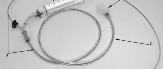
Emptying the stomach from blood is also necessary for reasons of pathogenetic effects on developing changes in the patient’s body (see “Pathogenesis”). In this regard, it becomes advisable to take measures to free the intestines from blood (in case of duodenal bleeding). The use of local hypothermia has a good hemostatic effect (Wangensteen, 1962; Miller et al., 1963; Turner et al., 1963; Mialaret et al., 1964; Crampton et al., 1964; Gross et al., 1965; B. A Petrov, N. N. Kornev, 1968; Rovati et al., 1968; etc.). A probe with an inflatable balloon shaped like a stomach is inserted into the stomach, filled with liquid cooled to 2°-20°, and this liquid is circulated for 2-3 or more hours.
The patient's nutrition is of certain importance. For the first day or two, it is better to keep the patient on hunger. This is advisable for reasons of giving the stomach rest. Eating causes motor and secretory activity of the stomach. Then they begin to feed, first with liquid and pureed food, and then gradually switch to the usual diet for an ulcer patient. Food should be high in calories (up to 2500 calories), rich in complete proteins and vitamins.
Once bleeding has stopped, treatment is carried out in accordance with the patient’s condition, the severity of anemia and changes in laboratory parameters indicating the condition of the liver, kidneys, heart, electrolyte levels, etc.
If intensive conservative treatment does not give the desired effect and bleeding continues, as evidenced by the patient’s serious condition, repeated collapse, low blood pressure, repeated vomiting of blood, and the diagnosis of ulcerative bleeding is beyond doubt, the patient should be taken to the operating room.
The failure of conservative therapy for ulcer bleeding is an indication for emergency surgery. In such cases, the operation cannot be delayed. The patient should be operated on on the first, or at least on the second, day after the onset of bleeding. As the period from the moment of bleeding increases, the patient’s condition worsens, the functions of vital organs decrease, the body’s defenses and the ability of tissues to reparative regeneration sharply decrease.
It is especially dangerous to operate on patients 5-10 days after bleeding. Many die from failure of the anastomotic sutures or duodenal stump, hepatic-renal failure, and from peritonitis, which can also develop when the sutures are tight.
Emergency surgery for ulcer bleeding requires at least a liter of blood. Without the presence of 500 ml of blood, the operation cannot be started. During the operation, at least another 300 ml of blood must be obtained. In difficult cases, employees, students or relatives of the patient become donors. Czeruski (1972) from Poland considers it possible, in the absence of blood, to operate on patients under the cover of dextran, physiological saline solution or 5% glucose solution in an amount of 1.5-3 liters.
The best operation is gastric resection using the methods that the surgeon knows. The operation involves removing a bleeding ulcer. If the ulcer cannot be removed, then the bleeding vessel is sutured from the side of the organ lumen (in this case, a free piece of omentum can be used). For intractable duodenal ulcers, in many cases the problem of stopping bleeding is solved by the methods of treating the stump of S. S. Yudin and B. S. Rozanov. In extremely severe patients who cannot undergo gastric resection, surgery is limited to excision of the ulcer or suturing of a bleeding vessel, followed by suturing of the gastric wound. In recent years, recommendations have appeared to excise the ulcer and perform selective vagotomy (Tenner et al., * 1972; Vogel, 1972, V. G. Borisov, 1973).
Introduction
Recently, there has been a trend towards a decrease in the incidence of peptic ulcer disease in the world [8, 12], in Russia this figure in 2008 was 1127.2, in 2009 - 1099.6 per 100,000 population. There is a widespread decrease in the level of surgical activity [10] due to a decrease in Helicobacter pylori
and as a result of preventive measures used when taking non-steroidal anti-inflammatory drugs (NSAIDs) [1, 8]. Modern principles of surgical intervention do not imply curing a patient from a peptic ulcer; its goal is to directly stop bleeding followed by drug therapy in situations where endoscopic hemostasis is impossible or ineffective [3], while the need for surgical intervention, according to summary data, arises in 13 % of observations [2]. Although surgical treatment is effective in a number of patients with uncontrolled bleeding, in most cases endoscopic hemostasis, both primary and for recurrent bleeding, is more justified and produces fewer complications [4, 5, 7]. The problem of recurrent ulcer bleeding, which worsens treatment results and directly leads to death, remains an urgent problem. The incidence of recurrent bleeding ranges from 1.8% (with the thermal method of hemostasis) to 21% (with clipping) and depends on a number of reasons [2]. Mortality from ulcerative bleeding continues to remain high - within 5-10% [6, 11]. Recent studies have shown that the majority (80%) of deaths from ulcerative bleeding are not associated with the fact of bleeding itself and depend on criteria such as decompensated concomitant diseases, generalized oncological diseases, and multiple organ failure [6].
The purpose of the work is to assess the structure of mortality in acute ulcerative gastroduodenal bleeding, its dependence on concomitant diseases, treatment results and the possibility of predicting death using the Rockall scale.
Material and methods
During the period from 2006 to 2010, 895 patients with ulcerative gastroduodenal bleeding were hospitalized; death occurred in 220 (24.6%) of them (main group). 45 (5%) patients died directly from ulcer bleeding. The control group randomly included 100 patients who survived an episode of ulcer bleeding and did not differ from the patients of the main group (Table 1).
All patients with signs of bleeding from the upper gastrointestinal tract upon admission underwent endoscopy with subsequent classification of ulcerative defects according to Forrest, conservative treatment using injectable forms of proton pump inhibitors for 3 days, followed by switching to oral forms. The objective of this study was to analyze 30-day mortality from the moment of ulcer bleeding. Concomitant drug therapy, including the use of NSAIDs and the use of antiplatelet drugs, was taken into account. Analysis of concomitant diseases was carried out on the basis of detailed processing of patients’ medical records and autopsy results. Severe concomitant diseases were considered to be sub- or decompensated cardiac, pulmonary, hepatic, renal, neurological and generalized oncological diseases. Criteria such as the severity of hemorrhagic shock upon admission, the nature of endoscopic hemostasis, recurrence of ulcer bleeding and emergency surgery were assessed. To predict mortality, the Rockall et al. scale, developed in 1996, was used [9].
Statistical analysis was performed using the Biostat program, qualitative analysis was performed using the &khgr;2 test and Fisher's exact test, and Student's t-test was used to analyze parametric independent variables.
Results and discussion
When analyzing the indicators, it was revealed that in the group of those who died, the average age of patients was 69.2±13.4 years and exceeded this indicator in the group of survivors - 55.8±18.2 years (p=0.017). The distribution depending on gender was homogeneous, men were more common in both groups - in 64 and 57.7%, respectively. In both groups, more than half of the patients (52 and 73.2%, respectively) had an ulcer history. When analyzing the localization of ulcerative defects, it was revealed that in the group of patients who died, the source of bleeding in 51.4% of patients was a gastric ulcer versus 34% in the group of survivors (p=0.006); on the contrary, duodenal ulcer was more common in the group of survivors - 56%, in the group of those who died - 37.3% (p=0.003). The number of patients taking NSAID drugs was the same in both groups (27 and 26.8%, respectively), which casts doubt on the significance of the effect of this group of drugs on the mucous membrane of the digestive tract. Signs of hemorrhagic shock were detected in 60% of those who died versus 18% in the group of survivors (p=0.001). The state of ulcerative defects and bleeding status, assessed using the Forrest classification, indicated a higher frequency of FIIA-FIIB type bleeding in the survivor group - 53% versus 40.5% in the deceased group (p = 0.049), as well as the insufficient prognostic significance of these indicators , since clinical parameters are not taken into account. Recurrent bleeding was observed in 28.6% of the group who died and in 11% of those who survived (p=0.001). Analysis of concomitant diseases revealed the following patterns. Thus, only 3 (1.36%) of the deceased patients had no concomitant diseases, while in the group of survivors this figure was 38% (p = 0.001). The use of the Rockall scale confirmed its diagnostic significance: in the group of surviving patients, the average value was 4.3±2.12 points, in the group of deceased patients - 7.16±2.35 points (p=0.001).
Death within 30 days occurred in 204 (92.7%) of 220 patients. Of the 204 patients, in 40 (19.6%) the death was classified as associated with ulcer bleeding and in 164 (80.4%) it was classified as unrelated to ulcer bleeding (Table 2).
Only 4 (10%) patients died as a result of uncontrolled bleeding, 18 (45%) died after emergency surgery, 10 (25%) patients died within 48 hours from the onset of bleeding due to NC.
Of the 164 deaths not related to bleeding, the most common cause was multiple organ failure - 59 (36%), diseases of the cardiovascular system - 40 (24.4%) and generalized oncological diseases - 26 (15.9%) .
All deaths were classified as follows: death associated with ulcer bleeding occurred in 45 patients, and death not associated with ulcer bleeding occurred in 175 patients (Table 3).
When analyzing the results obtained, no differences were revealed depending on the age and gender of the deceased patients. An ulcer history was more common in the group of those who died directly from bleeding than in the group of those who died from other causes (33.3% versus 18.3%; p = 0.046). The difference in the use of NSAIDs and aspirin in both groups was not significantly significant (p = 0.187), since the use of NSAIDs, in our opinion, is a marker indicating the presence of a concomitant disease, and not the cause of ulcer bleeding. The anatomical localization of ulcerative defects made it possible to identify some patterns. Thus, in the group of those who died from ulcerative bleeding, duodenal ulcers were most common (40%), in the group of those who died from other causes - gastric ulcers (51.4%), a combination of ulcerative defects in the duodenum and stomach was noted in 35.6 and 19 .4% of observations, respectively.
In the presence of signs of hemorrhagic shock, the mortality rate was significantly higher in the group of those who died from ulcerative bleeding. The profuse nature of bleeding and the rapid initiation of the pathogenetic mechanism of hemorrhagic shock, despite adequate therapy, lead to unfavorable results. When analyzing concomitant diseases, a difference was revealed in the prevalence of kidney diseases (8.9% versus 1.1%; p = 0.002) in the group of those who died from ulcerative bleeding, regarded as a consequence of hemorrhagic shock. Recurrent bleeding was more common in the group of those who died directly from ulcer bleeding (53.3% versus 24.4%; p = 0.001), and a third of patients had repeated relapses, which indicates the significance of relapse as a prognostic factor. The use of the Rockall prognostic scale in both groups did not reveal any differences, which is natural, since the scale was designed to predict death, which occurred in both groups.
Therefore, hemorrhagic shock, recurrent bleeding, and Rockall scale scores can be used as risk factors and criteria for predicting death directly related to bleeding.
Thus, in most countries there has been a decrease in the number of patients with ulcer bleeding, which has led to a decrease in mortality in this disease. This situation is also explained by the improvement of methods of endoscopic hemostasis and pharmacotherapy. The question remains controversial about the need to stop taking non-steroidal anti-inflammatory drugs and antiplatelet drugs in patients who have suffered an episode of ulcer bleeding, which can contribute to decompensation of the condition and complicate treatment.
Up to 80% of patients die from causes not directly related to the bleeding episode [11], while mortality depends on the severity of concomitant diseases and in most cases is caused by multiple organ failure, decompensation of oncological and cardiovascular diseases, which often leads to incorrect formulation of final diagnoses and determining the immediate causes of death. In the structure of deaths directly related to ulcer bleeding, the leading place is occupied by hemorrhagic shock and the recurrent nature of bleeding. The significance of the Rockall score for predicting lethal outcome in gastroduodenal bleeding was confirmed.
The results obtained in this work indicate the importance of concomitant diseases, the need to monitor and correct the function of the main organ systems, which will improve the results of treatment for ulcerative gastroduodenal bleeding.
