When is the procedure indicated?
The examination is carried out if diseases and pathologies are suspected that may pose a serious danger to the patient. In most cases, barium X-ray of the stomach allows you to confirm or clarify the diagnosis and prescribe the correct treatment.
Suspicion of the formation of gastric and duodenal ulcers
Peptic ulcer of the stomach and duodenum (DPC) leads to the periodic appearance of ulcers. It can take years to develop and cause serious harm to health. Using a barium X-ray of the stomach, the specialist finds the ulcer and evaluates its size, shape and other parameters, as well as establishes the stage of the disease and detects accompanying symptoms.
Detection of a malignant process in an organ
If the development of a malignant process is suspected, an X-ray of the stomach with barium is recommended without fail, since the consequences of the disease can be deadly for the patient. In the photographs, the cancerous process looks like a defect in the mucous membrane. Often, after a barium X-ray of the stomach, an endoscopic examination is performed and a sample of the tumor is taken for laboratory testing.
Diverticulosis and other deformities of the gastric walls
To diagnose wall protrusion (diverticulosis), a barium X-ray of the stomach requires additional preparation. After administration of the contrast agent, the patient alternately lifts the upper and lower parts of the body so that the contrast fills the entire organ cavity. On a barium X-ray of the stomach, small diverticulosis looks like an ulcer, but has a horizontal level and a neck.
Any inflammation of the stomach
Foci of inflammation in the stomach can appear as a result of various processes. Including peptic ulcers or oncology. An X-ray of the stomach with barium allows you to find the source of inflammation and, based on indirect signs, determine its cause.
Swallowing dysfunctions
Dysphagia, or swallowing dysfunction, is often expressed by the patient complaining of the inability to swallow food and its accumulation in the esophagus. In this case, the patient cannot always accurately indicate the location of the obstacle. Therefore, barium radiography of the stomach is used to accurately localize the pathology.
Pain in the abdominal area or navel area
Pain in the navel and abdominal area may indicate the development of malignant processes in the stomach and duodenum. Therefore, with such symptoms, the specialist often refers the patient to an X-ray of the stomach and duodenum with barium.
Everything for work: SOP X-ray examination of the stomach
Full text of the article:
SOP X-ray examination of the stomach
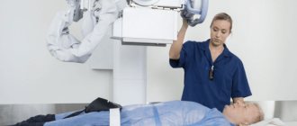
X-ray examination
The stomach involves the introduction of a contrast agent (barium suspension) into the organ. According to the technique, barium and air are injected into the stomach, which straightens the folds of the organ so that the doctor can more carefully examine the relief of the mucous membrane and evaluate the elasticity of the muscle layer. X-ray examination of the stomach includes fluoroscopy (monitoring the progress of contrast through the esophagus, stomach, small intestine in real time) and radiography in different positions.
Target:
standardization of the procedure for performing x-ray examination of the stomach
Application area
Where:
X-ray diagnostic room of outpatient service (APS) and 24-hour hospital (CSS)
When:
as prescribed by a doctor
Responsibility:
The responsible person for carrying out the manipulation in accordance with the requirements of the SOP is the radiographer of the OLD. Monitoring compliance with SOPs is carried out by the senior nurse of the OPD
Regulatory and reference documentation
· Federal Law No. 3-FZ dated 01/09/1996 (as amended on July 19, 2011) “On Radiation Safety of the Population” · Federal Law No. 323-FZ dated November 21, 2011 “On the Fundamentals of Protecting the Health of Citizens in the Russian Federation” · Order of the Ministry of Health RSFSR from 02.08.1991 No. 132 “On improving the radiology diagnostic service” SanPin 2.6.1.1192-03 “Hygienic requirements for the design and operation of X-ray rooms, devices and conducting x-ray examinations” SanPin 2.6.1.2523-09 “Radiation safety standards” (NRB-99/2009 ) · SP 2.6.1.2612-10 “Basic sanitary rules for ensuring radiation safety” (OSPORB 99/2010) · SanPiN 2.1.3.2630-10 “Sanitary and epidemiological requirements for organizations engaged in medical activities” · SanPiN 2.1.7.2790-10 “Sanitary -epidemiological requirements for the management of medical waste" · Atlas of placement during X-ray studies // edited by A.N. Kishkovsky.
Resources:
1 X-ray machines: — Complex X-ray diagnostic device RUM-20 — X-ray diagnostic complex Medix-R “Amiko” — X-ray diagnostic complex “SpektrAp” KRD-SM 50/125-1 2 2 Sterlix OPTIMA developing machine. 3 X-ray cassettes 4 X-ray film 5 Protective plates for working with cassettes during the production of images 6 Radiation protection equipment for personnel and patients 7 Solutions for the developing machine (developer and fixer) 8 Non-standard lights in the darkroom 9 X-ray viewer 10 Disinfectant solution, disposable diapers, gauze wipes , gloves, mask 11 Bar-Vips 12. Citric acid, soda (or 0.5 liters of carbonated unsweetened water) or carbonated mineral water 13 Glasses, teaspoon.
Main part of the SOP
1. Introduce yourself, identify the patient based on medical documentation (ask for full name, date of birth). 2. Register the patient in the journal (according to the medical history or as directed by the outpatient service doctor) 3. Inform the patient about the study, check for informed consent for the procedure. 4. Take the patient into the treatment room, place the patient behind the device screen, warning the patient not to move and follow the doctor’s commands. 5. Give a glass of barium suspension solution to your left hand and explain that you need to drink from it at the doctor’s command. 6. Go to the control room, set the technical parameters on the control panel, and at the doctor’s command, change the technical parameters on the control panel. 7. Return to the treatment room, take the cassettes filmed by the doctor, go to the darkroom, turn off the lights, turn on the non-actinic lights, open the cassettes sequentially, remove the X-ray film from the cassette and insert it into the developing machine that was turned on in advance. Load the cassettes with new film and return the cassettes back to the doctor. 8. After completing the procedure, take the glass from the patient and place the glass in the disinfectant solution, help the patient get out from behind the device. 9. We prepare the device for the next patient: treatment with a napkin moistened with a disinfectant solution. 10. Take the developed photographs to the doctor, having signed them first. 11. Obtain the result of the study from the doctor, register it in the journal, indicating the dose of radiation exposure in the journal and on the conclusion form. 12. Give the patient a conclusion form and inform that the procedure is completed. 13. Invite the next patient to the office
Preparation for the procedure
— The study is performed strictly on an empty stomach; before this, you cannot eat or drink for 8-10 hours. — To obtain objective data, it is recommended to follow a diet for 3 days before the procedure. Foods that cause flatulence (legumes, brown bread, fatty, fried, smoked foods, fruits, vegetables) should be excluded. Preference is given to lean boiled meat (chicken, beef), lean fish, white stale bread, water-based porridge, and eggs. — If the patient has pyloric obstruction, then before performing an X-ray with barium, the stomach is washed with a probe. — Immediately before the procedure, you must remove any jewelry.
Parameters for assessing and quality control of the methodology:
- compliance with the technology of performing the manipulation, - timely execution of the procedure, - ensuring radiation and infectious safety of the procedure, - availability of a record of the appointment in the medical documentation, - patient satisfaction with the quality of the procedure, - doctor satisfaction with the quality of the manipulation.
Distribution of this SOP
Copy Department Original Chief nurse Copy 2 Head nurse OLD
Responsible executors are familiar with and undertake to perform:
| No. | Surname | Signature | date |
What can be detected with radiography?
If a barium stomach x-ray is performed correctly and with proper patient preparation, the examination can provide enough comprehensive information to make a diagnosis in most cases. At the same time, to detect a particular disease, a specialist evaluates a number of parameters.
Gastric lumen disorders
Disturbances in the lumen of the stomach may indicate the development of a malignant process or diverticulosis. If the lumen of the stomach is narrowed on a barium X-ray, diffuse fibroplastic cancer is possible. With diverticulosis, on a barium X-ray of the stomach, the lumen appears larger than it should be normally.
Abnormal placement of the stomach
Gastroptosis (prolapse of the stomach) is often diagnosed with an abdominal wall hernia or diaphragmatic hernia. The disease is clearly visible using barium X-ray of the stomach.
Niche symptom
A niche is a shadow of a contrasting mass that fills a defect during an ulcer or the development of a malignant process. Depending on the location of the defect, an X-ray of the stomach with barium distinguishes between a contour and a relief niche. In the first case, the silhouette of the defect is visible from the full face, in the second - in profile.
The lack of filling is visible in the image as a dark area
A dark area on a barium x-ray of the stomach indicates that there is a place where the contrast could not reach. As a rule, this happens with atrophic gastritis or tumor.
Carrying out
An X-ray of the esophagus and stomach begins with a general image of the abdominal cavity. After this, the patient is asked to drink prepared barium sulfate. The initial targeted photograph is taken after two sips of the drug. At this moment, the relief of the walls of the esophagus with barium is determined. Then the patient is allowed to finish the rest of the drug. During the examination, the doctor may press on the patient's abdomen to promote better distribution of the contrast.
During the process, various pictures are taken in different positions - lying on your back as standard or with the pelvis raised at an angle of 45°, lying on your side, standing. At the same time, at the command of the radiologist, the patient must hold his breath. Modern X-ray rooms are equipped with a special table that rotates while taking pictures.
As a rule, to examine the upper part of the gastrointestinal tract, it is enough for an adult to take 250-300 ml of dissolved barium sulfate (in some cases the dose may be increased). If radiography is prescribed for a child, then the required amount of suspension is calculated based on the age category. For children, as a rule, 100 ml of barium paste is sufficient.
Bowel examination
X-ray of the small intestine with barium is performed in stages. The procedure begins with the patient drinking 0.5 liters of barium suspension. If the study is carried out with double contrast, the drug enters the body through a special tube that is inserted into the patient’s mouth. Along with the contrast, air or inert gas is supplied.
After this, wait at least 2 hours - during this time the barium has time to reach the small intestine. As contrast fills the small intestine, the radiologist takes a series of pictures, asking the patient to assume different body positions. And after relieving themselves, they take the last control shot. After filling them with contrast, the diagnostician examines the various segments of the small intestine in detail on the monitor for half an hour.
The contrast procedure allows you to evaluate intestinal motility and its mucous membrane. While the contrast agent is still present in small quantities, the relief of the inner wall of the intestines is examined, and when there is a lot of contrast, the shape, size, contours and functionality of the intestines are assessed. With maximum barium filling, it is possible to identify inflamed segments, ulcerative processes and identify neoplasms.
If the passage of barium is disrupted, the radiologist gently presses on the anterior wall of the peritoneum to distribute it evenly. During a procedure with contrast, as the substance is distributed in the intestinal lumen, an experienced radiologist can draw conclusions about the presence of pathological processes. If the barium suspension is distributed in the form of flakes, then this is a clear sign of impaired absorption. And if the contrast fills the lumen unevenly, this may indicate oncopathology.
How should a patient prepare for the procedure?
Since the patient needs time to prepare for a barium stomach x-ray, it is best to start a few days before the examination. The first step is to exclude from the diet foods that cause increased peristalsis and gas formation. 8 hours before the examination you should completely abstain from food. To relax the stomach before the x-ray and prepare it for filling with barium, the doctor may recommend taking a special drug half an hour before the examination. Contrast is administered orally a few minutes before the procedure. When a double contrast examination is required, after the patient has prepared for a stomach x-ray and taken barium, he will need to drink a gas-forming mixture.
When is x-ray prohibited?
An X-ray of the stomach with barium is a safe examination, but since it is done using ionizing radiation and a contrast agent, the method has a number of contraindications.
Hematopoietic disorders caused by pathological conditions of the bone marrow
Ionizing radiation can worsen the disease. Therefore, if hematopoiesis is disrupted, an X-ray of the stomach with barium is not performed.
Cataract
When X-raying the stomach with barium, the radiation dose does not exceed permissible limits. However, with cataracts, even a limited amount of ionizing radiation may be enough to have a negative effect on the condition of the lens.
Malignant neoplasms of the bronchi and lungs
The examination is contraindicated if the patient is undergoing radiation or chemotherapy treatment.
Pregnancy
During pregnancy, even minor radiation can have a detrimental effect on the development of the fetus. Therefore, X-rays of the stomach with barium are contraindicated for expectant mothers.
Childhood (before the onset of puberty)
In childhood, the body reacts poorly to exposure to even small doses of ionizing radiation. Therefore, doctors refer children for an X-ray of the stomach with barium only if absolutely necessary.
results
- abnormalities in the structure of the digestive tract;
- acute expansion or narrowing of the lumens of the stomach/esophagus;
- malformations of certain organs of the gastrointestinal tract;
- hypertonicity/hypotonicity of the muscular wall of the stomach;
- tumors, papillomas, foreign bodies;
- reduction or radial arrangement of shell folding;
- cicatricial changes at the site of tumors, ulcers, chemical burns.
- pathological narrowing;
- saccular protrusions and elasticity of the walls;
- the introduction of one section of the intestine into another with the possible development of gastrointestinal obstruction;
- intestinal motor function;
- the presence of ulcerative and inflammatory processes;
- tumors, polyposis.
An examination can show how the bauhinium valve functions. This is the structure that separates the small and large intestines and is responsible for passing food between them. If it has pathological changes, then the food gets back access, and this poses a danger to the patient’s life.