Causes of the disease
Esophageal achalasia can occur equally often in both women and men.
The disease can manifest itself at any age, but most often occurs between 30 and 50 years of age. According to statistics, the disease occurs in 0.001-0.002% of people. The causes of achalasia cardia have not been fully established. It is believed that achalasia of the cardia occurs due to a mismatch in the nervous regulatory mechanisms responsible for the peristaltic movements of the esophagus and the reflex opening of its lower sphincter when a bolus of food approaches it. Some researchers believe that nutritional disorders, especially insufficient dietary intake of B vitamins, play a large role in the occurrence of the disease.
Symptoms and course of the disease
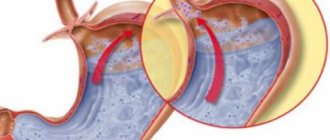
Cardiac achalasia occurs in patients of any age and develops gradually. The patient is bothered by regurgitation of food he has just eaten without signs of digestion. Pain appears. Pain may be the first sign of illness. Their nature is very diverse: from diffuse pain in the chest to a place precisely indicated to the patient. Pain can radiate to the shoulder, neck, shoulder blade, ear, etc. Painful sensations appear after eating, but quite often the pain may not depend on food intake. Over time, the pain syndrome decreases. The patient may complain of a feeling that food is not entering the stomach on time. Some patients accurately indicate the place of food retention on the anterior surface of the chest.
Patients often use various methods to facilitate the penetration of food into the stomach: drink a large amount of liquid in one gulp, tilt or tilt the body, hold the neck or sternum with their hands. Belching or regurgitation first appears immediately after eating, then gradually, due to the expansion of the lower esophagus, an increasing amount of food accumulates there. This food is regurgitated later. But in this case, a large volume of food is regurgitated at once. This condition is sometimes called esophageal vomiting. Unlike normal vomiting, regurgitated food masses are chewed food without signs of digestion in the stomach. Regurgitation of food can cause part of the bolus to be thrown into the trachea and further into the lungs. Inflammatory lung diseases and aspiration pneumonia occur. Sometimes the patient complains of persistent hiccups. Cardiac achalasia is often accompanied by constipation.
There are four stages of achalasia cardia:
- The initial stage or stage of functional intermittent spasm. There is no narrowing of the lower esophageal sphincter (cardia) and expansion of the esophagus itself above the cardia. Difficulties in swallowing food occur periodically.
- This stage is called stable. In this case, a constant (stable) spasm of the cardia occurs. The esophagus above the cardia is slightly dilated. The patient's complaints become constant.
- In the third stage, cicatricial changes occur in the tissue of the lower esophageal sphincter. The sphincter undergoes sclerosis, loses its elasticity and cannot fully open. The esophagus above the sphincter expands sharply.
- The fourth stage is the stage of complications. Stenosis (narrowing) of the cardia is pronounced. The esophagus above the cardia is significantly dilated. Inflammatory phenomena occur in the wall of the esophagus (esophagitis), necrotic ulcers on the walls of the esophagus. Inflammatory phenomena can spread to surrounding tissues. Inflammation of the mediastinum occurs - mediastinitis.
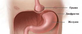
The most common complication of achalasia is congestive esophagitis (inflammation of the esophagus), which can lead to the development of esophageal cancer. Also, the dilated esophagus can compress the recurrent and vagus nerves, the right bronchus, and the superior vena cava. Achalasia can provoke chronic nonspecific lung diseases due to aspiration of food into the respiratory tract, leading to the formation of esophageal diverticula and hiatal hernia.
Treatment of the disease
Treatment for achalasia cardia involves eliminating cardiospasm and can be carried out using conservative or surgical methods, and sometimes drug therapy.
A conservative method for eliminating cardial achalasia is pneumocardiodilation - balloon expansion of the cardiac sphincter, which is carried out in stages, using balloons of different diameters with a consistent increase in pressure. With the help of cardiodilatation, overstretching of the esophageal sphincter and a decrease in its tone are achieved. Complications of balloon dilatation can include cracks and ruptures of the esophagus, the development of reflux esophagitis and cicatricial strictures of the cardiac sphincter.
A lasting result in the treatment of cardia achalasia is achieved after surgical intervention - esophagocardiomyotomy - dissection of the cardia followed by plastic surgery (fundoplication). The operation is indicated for the combination of achalasia cardia with a hiatal hernia, diverticula of the esophagus, cancer of the cardia of the stomach, failure of instrumental dilatation of the esophagus, or its ruptures.
If achalasia cardia is combined with duodenal ulcer, selective proximal vagotomy is additionally indicated. In the presence of severe peptic erosive-ulcerative reflux esophagitis and severe atony of the esophagus, proximal resection of the stomach and abdominal part of the esophagus is performed with the application of intussusception esophagogastroanastomosis and pyloroplasty.
Drug therapy for achalasia cardia plays a supporting role and is aimed at prolonging remission. For this purpose, it is advisable to prescribe antidopaminergic drugs (cerucal, raglan), antispasmodics, minor tranquilizers, calcium antagonists, nitrates. In recent years, botulinum toxin administration has been used to treat achalasia cardia.
Important points for achalasia cardia are adherence to a gentle diet and diet, normalization of the emotional background, and avoidance of overexertion.
Until now, treatment for achalasia cardia has been carried out using surgery, in which an incision in the muscles of the lower esophageal sphincter is performed laparoscopically or using a robot. The new POEM (Per Oral Endoscopic Myotomy) method does not require surgery, its solution is more “elegant”: the patient is inserted through the oral cavity into the esophagus, where an incision is made in the mucous membrane to create a passage between the mucous membrane and the muscles of the esophagus to the stomach, and then a muscle incision is made.
This operation is considered innovative and is performed endoscopically - a muscle incision is made while preserving the integrity of the mucous membranes of the esophagus and stomach.
Achalasia cardia
Modern treatment of achalasia cardia is aimed at eliminating the functional barrier in the form of an unrelaxed lower esophageal sphincter and can be carried out conservatively or surgically, and in some cases with the help of medications. And if pharmacotherapy of the present disease has a purely auxiliary value, then the question of the advantage of the first two methods has not yet been resolved in the literature. A number of authors prefer surgical intervention.
Proponents of cardiomyotomy and its modifications cite the following arguments: 1) relatively high efficiency of operations (the average percentage of “good” long-term results is 85; however, it should be noted that in the literature there are no sufficiently developed objective criteria to reliably assess the effectiveness of the surgical method of treating achalasia) ; 2) the possibility of auditing the distal esophagus to identify a malignant neoplasm. The mortality rate from surgery, according to some authors, is 1.2%.
The main arguments of supporters of cardiodilation are the completely satisfactory tolerability of the procedure by the vast majority of patients, its cost-effectiveness and relative safety. The percentage of good long-term results is quite high.
Thus, it is advisable to begin treatment of almost all patients with achalasia cardia with cardiodilation. In other words, the indications for its implementation are both newly diagnosed achalasia cardia of the first and second types, and relapses after primary treatment with this method. Recurrence of the disease after cardiomyotomy requires, first of all, the exclusion of the development of peptic stricture as a complication of the operation, which is preferably carried out in close contact with surgeons. Attempts to compensate for postoperative relapse of achalasia through cardiodilation are also acceptable. And only the ineffectiveness of conservative therapy and pronounced cicatricial changes in the cardia can be the basis for cardiomyotomy.
- Conservative treatment and pneumocardiodilation
Before pneumocardiodilation, it is necessary to conduct a thorough examination of the patient. During the examination, special attention is paid to an objective assessment of his general condition, as well as the condition of the esophagus through x-ray examination and esophagomanometry. In this case, the main parameters are the diameter of the expanded part of the esophagus, its shape, the level of fluid and food retention in it, as well as expiratory pressure in the lower esophageal sphincter, reflecting its tone. Changing these parameters is the basis for assessing the effectiveness of treatment. It is best to begin preparing the patient for cardiodilation by rinsing the esophagus through a probe with a warm 4% solution of sodium bicarbonate or a weak solution of potassium permanganate. Evacuation of stagnant contents significantly improves the patient’s well-being: the feeling of heaviness behind the sternum and regurgitation at night disappear, and the manifestations of congestive esophagitis decrease. The use of a thick gastric tube is a kind of “rehearsal” before cardiodilation and facilitates the introduction of a pneumocardiodilator at the beginning of treatment.
In most cases, intramuscular injections of 0.1% atropine sulfate solution (1 ml), 50 or 25% analgin solution (2 ml) and 1% diphenhydramine solution (2 ml) are used for premedication. The combination of drugs significantly reduces the severity of the gag reflex and salivation, as well as the intensity of chest pain caused by stretching of the cardiac segment of the esophagus during the procedure. In special cases, additional superficial anesthesia of the pharyngeal mucosa with solutions of dicaine and xylestezin is permissible. In case of concomitant diseases of the cardiovascular system, which occur in approximately 15% of all patients with achalasia cardia, premedication should be supplemented with antianginal or antihypertensive drugs according to the situation. Complete anesthesia with narcotic analgesics is impractical, since this increases the risk of not noticing in time the possibility (albeit very rare with qualified cardiodilation) of such a formidable complication of the procedure as rupture of the esophageal wall.
The requirement for maximum safety for patients is met by gradual expansion of the cardia, first with a balloon with a diameter of 30 mm at a pressure of 180 - 200 mm Hg. Art. with a gradual increase in the diameter of the balloon and the pressure in it to 40 - 50 mm and 340 - 360 mm Hg. Art. respectively. The exposure is 1 - 2 minutes. The first procedure is the most important, since the risk of esophageal rupture is greatest. The fact is that in 36.5% of patients during the first cardiodilation there is a sharp increase in pressure in the system, caused by unexpected gagging movements. Such phenomena are caused by a reflex reaction of the cardia in response to its stretching, which can be spontaneously leveled out during subsequent procedures.
Control of the position of the pneumocardiodilator balloon both during its installation and during the procedure itself is carried out radiographically. However, pneumocardiodilation is quite acceptable without X-ray control. In addition, there are situations in which X-ray irradiation of the patient is contraindicated (for example, during pregnancy).
The most important point when carrying out extensions without x-ray control is determining the distance at which the probe should be inserted. For this purpose, an original technique was developed based on the use of esophagomanometry. When removing an esophagomanometric probe from the stomach into the esophagus, in accordance with the nature of the change in the curve, three marks are made on its outer side: the beginning of the cardia from the stomach, the place of greatest narrowing and the transition of the cardia to the thoracic esophagus (length of the cardia). The interval between the second mark (the place of greatest narrowing) and the esophagotonograph balloon is the required distance, which is then laid directly from the middle of the expanding part of the pneumocardiodilator. We have successfully used this method to treat more than 250 patients with achalasia cardia. Excellent and good immediate results were achieved in 95.2% of patients, excellent and good long-term results - in 86.4%.
An important advantage of this technique is the reduction in radiation exposure to the patient by 30.7% of the total dose. This is especially valuable considering that many patients additionally have to undergo cholecystography, irrigoscopy, etc. during the examination.
Pneumocardiodilation without direct x-ray control of the probe position is indicated for type 1 achalasia cardia and in most cases of type 2 achalasia. Severe curvature and atony of the esophagus in advanced cases significantly complicate the introduction of a cardiodilator into the esophagus and the installation of its balloon in the cardia. In such rather rare situations, it is advisable to use the pneumocardiodilation technique under endoscopic control, which allows not only to successfully carry out the procedure, but also to avoid serious complications.
The interval between repeated procedures should be at least 3 - 4 days. If, when removing the dilator, traces of blood are found on it, then this period is increased to 5 - 8 days. In 50% of cases, longitudinal tears were found, disappearing after 6-10 days. This period determines the frequency of procedures.
Under the influence of cardiodilation, a decrease in the tone of the lower esophageal sphincter occurs due to overstretching, and possibly tears, of its circular muscle fibers. As a result, patients note a significant reduction in the symptoms of achalasia. However, the ultimate goal of treatment is not only to achieve an immediate effect, but also to ensure remission for the longest possible period. It has been empirically established that this task is feasible in the case of reduction of expiratory pressure in the lower esophageal sphincter in conventional units to 12-15 mm Hg. Art. or up to 3 - 5 mm Hg. Art. The volume of the balloon sensor significantly affects the tone of the cardiac sphincter. The given figures were obtained under different recording conditions: the first - when using a pressure gauge probe with a volume of 2 cm3 canisters, the second - when using a probe with a 1 cm3 canister volume; they are virtually identical. The lower limit of these intervals is limited by the possibility of developing cardia failure and reflux esophagitis. Maintaining residual pressure in the cardia above the specified values creates the prerequisites for the rapid occurrence of relapse of achalasia.
The effectiveness of treating this disease with cardiodilation depends not only on the degree of reduction of the tone of the lower esophageal sphincter, but also on the condition of the smooth muscles of the thoracic esophagus. As mentioned above, the function of the latter is disrupted under the influence of the immediate cause of the disease, as well as secondarily due to overload with the residual volume of food and excessive work against the cardiac sphincter that does not relax when swallowing. Thanks to cardiodilation, this functional barrier to the food bolus is eliminated, which indirectly helps restore the tone of the smooth muscles of the thoracic esophagus. Radiologically, a decrease in the diameter of the suprastenotic expansion and the appearance of a gas bubble of the stomach are noted. An esophagotonogram demonstrates an increase in the amplitude of contractions in the thoracic esophagus. However, in a number of cases, esophageal atony is not eliminated even as a result of adequately performed cardiodilatations, which is apparently due to the severity of the initial damage to the smooth muscles of the esophagus.
The immediate results of treatment of patients with achalasia cardia type 1 (according to the subjective feelings of patients) are somewhat worse than patients with achalasia cardia type 2, which is explained by the persistence of spastic contractions of the lower part of the esophagus with completely satisfactory patency of the cardia. However, long-term results are better: after a course of treatment, excellent and good long-term results were obtained in 74.7 and 50.7% of patients with achalasia cardia types 1 and 2, and after repeated courses - in 93.1 and 74%, respectively. When comparing the duration of remission with the age of the patients and the duration of the disease, it was found that after 45 years, good and excellent results decrease to 56.5%. The duration of the disease for more than 15 years reduces this figure to 28.6%, which can be explained by cicatricial degeneration of the cardia.
The percentage of good and excellent long-term results of treatment of achalasia cardia using pneumocardiodilation ranges, according to the literature, from 50-80. Our many years of experience indicate that methodically correctly performed pneumocardiodilation, including without direct x-ray control of the probe position, allows us to achieve excellent and good results in 84.4-86.4% of cases. In this case, good and excellent results mean the following: complete disappearance or significant reduction in complaints of dysphagia, reduction in the tone of the lower esophageal sphincter to the above optimal values, good passage of barium sulfate suspension from the esophagus into the stomach during X-ray examination (a short delay of a column of radiopaque suspension with a height of no more than 2-4 cm above the cardia, since its stream entry into the stomach indicates the possibility of gastroesophageal reflux), the presence of a gas bubble of the stomach.
Complications during pneumocardiodilation occur in 2.3% of cases. The mortality rate from them or during the procedure itself is 0-0.1%. Such complications include perforation or rupture of the esophageal wall with the development of mediastinitis, reflux esophagitis, peptic stenosis of the esophagus, bleeding, trauma to the pyriform sinus of the larynx, and fainting. The integrity of the esophagus also depends not only on the absolute value of the pressure in the balloon, but also on the rate of its increase. This provision has great practical significance.
The recurrence rate of severe dysphagia in achalasia cardia ranges from 5.6 to 18%. According to summary statistics, these figures vary over a much wider range - from 3 to 30%.
There are three types of such relapses. In the genesis of relapses of the first type, spastic contractions of the lower third of the esophagus are of primary importance. The second (true) type is caused by a repeated increase in pressure in the lower esophageal sphincter and is most common. Relapse of the third type is observed only in isolated cases and is associated with a progressive disturbance of peristalsis of the esophagus due to atony of its thoracic part, although the pressure in the cardia does not go beyond the limits.
According to various authors, the return of symptoms of the disease may depend on a wide variety of reasons: restoration of cardiac tone due to “depression of the automatism of myoneural connections”; iatrogenic reflux esophagitis and scars in the cardia area; an increase in serum gastrin after a course of treatment with cardiodilation due to periesophagitis and mechanical irritation of the vagus nerves; incompleteness of treatment, i.e. maintaining residual pressure in the cardia above 3-15 mm Hg. Art.
Thus, some authors explain the occurrence of relapses of achalasia by achalasia itself, others by non-compliance with certain therapeutic recommendations. In other words, if the appropriate empirically compiled instructions are followed, a relapse of the disease may not occur. It is difficult to agree with this opinion. Cardiodilation in all cases, even with excellent clinical results, is purely symptomatic. This means that this procedure does not provide a complete cure. It is incompetent to even talk about “cure with a defect.” The symptoms of the disease are eliminated for a certain period of time, depending on strict adherence to methodological recommendations (“adequate treatment”).
In other words, the reason for the resumption of dysphagia after treatment of achalasia cardia with pneumocardiodilation lies in the disease itself and is still unknown to us. Hence the question logically follows: how to deal with these relapses?! In our opinion, the approach to solving this problem should be based on two principles: firstly, on the individualization of treatment tactics when correcting relapses and, secondly, on the organization of dispensary observation of such patients.
The first position is essentially the result of dividing relapses of achalasia cardia into three types, which have practical significance when choosing the optimal method for their correction. Relapses of type 1 dysphagia (spasmodic contractions of the distal esophagus) are an indication for the use of nitrates and calcium antagonists. In some cases, repeated courses of pneumocardiodilation have a significant effect.
Patients with type 2 relapses require repeated pneumocardiodilation. However, in this case, the risk of complications after the procedure increases due to the development of cicatricial changes in the lower esophageal sphincter in a number of patients. In addition, the pressure in the cardia during relapses almost never reaches such a high level as was noted before the initial treatment. It is this circumstance that determines the tactical features of the correction of relapses of the second type: lower pressure in the dilator balloon during the first procedure (on average 140 mm Hg) and a smaller increase in pressure during subsequent dilations (on average 40 mm Hg) . The final pressure in the cylinder should not exceed 300 mmHg. Art.
For recurrent dysphagia of the third type, treatment with pneumocardiodilation is less effective. Therapy has not yet been sufficiently developed. You can use antidopaminergic drugs (metoclopramide, Raglan, Cerucal), which tonic the smooth muscles of the thoracic esophagus.
The duration of remission of achalasia cardia increases as a result of patients regularly taking medications such as minor tranquilizers, antipsychotics, myotropic antispasmodics, and anticholinergics. However, these drugs do not actively influence the tone of the lower esophageal sphincter and thoracic esophagus in achalasia cardia, since their effect is to one degree or another mediated by the autonomic nervous system. α-adrenolytics and fh-stimulants are not widely used.
Currently, the most popular drugs are from the group of nitrates and calcium antagonists. The beneficial effect of nitroglycerin on dysphagia caused by achalasia of the cardia has been known for a long time: nitroglycerin and amyl nitrite significantly reduce pressure in the cardia, but its function is not normalized. The creation of long-acting nitrates has opened up new prospects for the conservative correction of achalasia. Isosorbide dinitrate (nitrosorbide) at a dose of 5 mg significantly reduces pressure in the lower esophageal sphincter in such patients with a maximum decrease in the 10th minute. Unfortunately, the clinical value of nitrates is significantly reduced due to their side effects (dizziness, headache, tachycardia).
It is also known that fluctuations in the concentration of this ion in the blood plasma cause changes in esophageal motility. Acute hypercalcemia increases thoracic peristalsis and tones the lower esophageal sphincter. However, with achalasia cardia, no electrolyte shifts were observed.
Intravenous administration of verapamil to patients with cardial achalasia leads to a significant decrease in pressure in the cardia by an average of 31.4%, especially at the 12th minute. Nifedipine is a more active drug. When taken orally 10-20 mg (1 - 2 tablets) the degree of reduction in cardiac pressure is 35-50% with a maximum at the 60th minute. The effect of the drug depends on its dose. There was also a decrease in dysphagia and cramping pain behind the sternum when swallowing. Hemodynamic side effects are minor.
The effect of nifedipine on motility of the entire esophagus has been less studied. Scintigraphy demonstrates acceleration of food passage through the esophagus, but normalization of peristalsis does not occur. From this point of view, this drug is inferior to nitrosorbide.
In our opinion, the clinical effect of any drug, in particular Corinfar (nifedipine), for achalasia cardia depends on at least three parameters: the tone of the lower esophageal sphincter, the amplitude of contractions in the thoracic esophagus and the number of extra-swallowing contractions. The passage of food through the esophagus is carried out due to peristalsis of the thoracic part of the organ and synchronous opening of the cardia. If the reduction of the tone of the latter in this case is a positive factor, then the inhibition of contractility of the thoracic esophagus is negative. Therefore, against the background of the use of Corinfar, in some cases one can expect an increase in dysphagia, caused precisely by drug-induced atony of the thoracic esophagus. In this regard, we studied the effect of 20 mg of sublingually administered Corinfar on expiratory pressure in the cardia, the number of extraswallowing contractions in the lower third of the esophagus, reflecting its excitability, as well as on the amplitude of contractions in this area. Corinfar significantly reduces pressure in the lower esophageal sphincter and the number of extra-swallowing contractions in its lower third. The amplitude of contractions in the distal part of the esophagus does not change. It should be noted, however, that such effects of the drug are very selective and do not occur in every patient. Therefore, in order to avoid unreasonable prescription of the drug, a manometric study or at least a clinical assessment should be carried out every time.
Indications for the use of Corinfar for achalasia cardia:
- relapses of dysphagia after preliminary treatment with pneumo-cardiodilation (a means for secondary prevention);
- combination of this disease with coronary heart disease and hypertension;
- correction of esophageal dyskinesia in achalasia. It is advisable to take the drug sublingually until it is completely dissolved 60 minutes before meals.
The second most important measure aimed at reducing the number of relapses of achalasia and prolonging the period of remission is medical examination. All patients are subject to dispensary registration. In most cases, it should be carried out 2 times a year: examination by a therapist, chest X-ray, esophagomanometry, as well as other diagnostic procedures necessary at the moment.
Taking into account the peculiarities of the neuropsychic status of patients with achalasia cardia, care should be taken to organize a therapeutic and protective regime. It is necessary to recommend them a clear daily routine, an optimal combination of work and rest, daily walks in the fresh air and the use of Sundays exclusively for relaxation. Morning exercises are very useful in this regard, but without exercises with the torso tilted forward or in a lying position and without tension in the abdominal muscles, since regurgitation of stagnant contents of the esophagus and gastroesophageal reflux are possible. Gymnastics should be completed with water procedures. To prevent nocturnal regurgitation, patients with achalasia cardia are recommended to sleep with the head of the bed elevated and not to go to bed immediately after eating.
The employment of these patients also has some peculiarities. They are not recommended to work associated with frequent neuropsychic overload, on night shifts, on a rotating schedule, long business trips, as well as all types of work accompanied by bending the body or lifting significant weights.
It is advisable to pay attention to patients’ adherence to a strict diet with four or five meals a day, and more often if necessary. Frequent and small meals help reduce dysphagia and regurgitation, normalize the function of the stomach and intestines. It is necessary to eat food as slowly as possible, chewing it thoroughly. Food should not be too cold or hot: the muscles of the esophagus react to the low temperature of the contents of the esophagus with an additional spasm, while hot food can intensify the inflammatory process in the wall of the esophagus. Removal of stagnant contents is facilitated by drinking a glass of warm water after meals, which to a certain extent is a prevention of stagnant esophagitis.
The diet should be mechanically and chemically gentle. The consumption of hot seasonings, sauces, spicy foods, salty and heavily fried, as well as smoked foods is prohibited. It is advisable to limit the consumption of canned food, especially fish and vegetables, as they contain large amounts of thermally processed vegetable oil or tomato sauce.
There are also a number of foods that increase dysphagia in patients with achalasia cardia - apples, persimmons, peaches, kefir, soft bread, meat, boiled potatoes, which should not be consumed.
It is necessary, however, to note that in some cases the diet of patients with achalasia cardia is determined by their individual characteristics and habits. For example, tasty and favorite foods cause less dysphagia than those recommended by a doctor, but perceived with indifference or even disgust. In this regard, sometimes you can deviate from a strict diet and recommend foods that evoke positive emotions.
For the period of stay of patients with achalasia cardia in the hospital, a diet 1 or 5a according to Pevzner or the esophageal table type is recommended.
- Surgery
Indications for surgical treatment are the combination of achalasia with a hiatal hernia or esophageal diverticulum complicated by diverticulitis, the inability to exclude cancer of the cardiac part of the stomach, the failure of instrumental dilatation of the esophagus, as well as fresh ruptures that occurred during this manipulation. Severe diseases of internal organs with decompensation of vital functions are contraindications to surgical treatment.
The most common operation currently used for achalasia cardia is extramucosal esophagocardiomyotomy using the Heller technique. The surgical approach used is left-sided thoracotomy in the seventh-eighth intercostal space or upper median laparotomy. The first approach is preferable, as it provides a wider view of the surgical area. After opening the mediastinal pleura and mobilizing the distal esophagus from the surrounding tissues, its muscular layer is dissected longitudinally to the mucosa. The length of the incision should be at least 8-10 cm, and in the distal direction it must be extended to the muscular layer of the cardiac part of the stomach.
Mortality with this type of operation averages 1.5-3%. In 65-85% of patients it is possible to achieve excellent and good results. The disadvantage of Geller's operation is the development of reflux esophagitis in a significant part of the operated patients due to a decrease in intraluminal pressure in the area of the lower esophageal sphincter. In addition, some patients experience the development of esophageal diverticula in the area of the dissected muscles. With incomplete intersection of the muscle bundles, the frequency of relapses of achalasia cardia is very high.
To prevent the above undesirable consequences of this type of intervention, a number of surgical techniques have been proposed: closing the defect in the muscular lining of the esophagus with a strand of the greater omentum on a pedicel, a flap cut from the diaphragm, suturing the dissected muscular lining in the transverse direction, suturing a flap from the esophagus to the defect in the muscular lining of the esophagus anterior wall of the proximal stomach. It should be noted that their effectiveness in preventing the development of gastroesophageal reflux after esophagocardio-myotomy was not high enough.
A very effective method of surgical treatment of achalasia cardia is a combination of Heller's operation with Nissen fundoplication. This allows not only to eliminate achalasia, but also to reliably prevent reflux esophagitis and the formation of esophageal diverticulum. When achalasia cardia is combined with duodenal ulcer, selective proximal vagotomy is performed along with this surgical intervention. However, during this operation, a number of patients experience the development of dysphagia, which may be associated with “hyperfunction” of the fundoplication valve. In these situations, cardiodilation is indicated.
Particularly great difficulties are encountered when achalasia cardia is combined with severe peptic erosive-ulcerative reflux esophagitis and severe atony of the esophagus. In these cases, economical proximal resection of the stomach and abdominal part of the esophagus is indicated, followed by intussusception (if possible, areflux) esophagogastroanastomosis in combination with pyloroplasty. Other types of surgical interventions in such a situation are usually ineffective. It should be borne in mind that proximal gastrectomy is accompanied by a fairly high postoperative mortality due to failure of the sutures of the esophagogastric anastomosis. In the presence of a pronounced inflammatory process in the wall of the esophagus, the frequency of this complication can reach 15-25%. In this regard, the use of this type of operation should be limited to strict indications.
Esophagofundostomy, which was used for a long time for advanced forms of achalasia cardia using the Heyrovsky method, is now almost completely abandoned due to the high incidence of severe reflux esophagitis.
More favorable consequences of surgical treatment are observed in patients with type I esophageal achalasia. It should be emphasized that there is a close relationship between the effectiveness of a particular type of intervention and the degree of morphological and functional disorders of the esophagus present at the time of surgery. The best long-term results are observed in patients in the absence of pronounced irreversible changes in the motor function of the esophagus. In this regard, it is advisable to resort to surgical treatment at earlier stages of the disease, if one or two courses of drug treatment in combination with pneumocardiodilation are unsuccessful. Indications for surgical treatment for type II achalasia cardia should be expanded.
Diagnosis of the disease
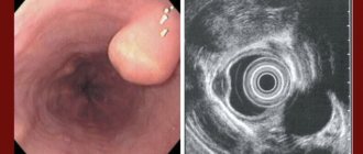
The disease can be diagnosed based on fluoroscopic data (an overview image of the chest organs and more informative fluoroscopy with contrasting the esophagus with a suspension of barium sulfate) or endoscopic examination. Esophagomanometry is of great value for diagnosing achalasia. As an additional method, pharmacological tests with intramuscular injection of solutions of carbacholine or acetylcholine are used.
Differential diagnosis for achalasia cardia is carried out with benign tumors of the esophagus, esophageal diverticula, cardioesophageal cancer, and esophageal strictures.
Classification
Taking into account the morphological signs and clinical picture, the following stages of development of this pathological process are distinguished:
| First stage | the esophagus does not expand, disturbances in the passage of food are periodic; |
| Second stage | moderate dilatation of the esophagus, dysphagia and stable tone of the cardiac sphincter appear; |
| Third stage | the esophagus is dilated at least twice, which is due to scar changes and significant narrowing of the esophagus |
| Fourth stage | inflammation of nearby tissues and deformation of the esophagus. |
It should be noted that these stages of the pathological process can develop either within a month or several years. It all depends on the patient’s history and general health.
Treatment with conservative methods is possible only up to the third stage - until scar changes begin. Starting from the third stage, treatment is only surgical with drug therapy and diet.
Prices
| Disease | Approximate price, $ |
| Prices for examinations for stomach cancer | 5 730 |
| Prices for diagnosing Crohn's disease | 3 560 — 4 120 |
| Prices for diagnosing gastrointestinal cancer | 4 700 — 6 200 |
| Prices for hepatitis C diagnostics | 5 700 — 6 300 |
| Prices for treatment of Vater's nipple cancer | 81 600 — 84 620 |
| Prices for colorectal cancer treatment | 66 990 — 75 790 |
| Prices for treatment of pancreatic cancer | 53 890 — 72 590 |
| Prices for treatment of esophageal cancer | 61 010 — 81 010 |
| Prices for treatment of gallbladder cancer | 7 920 — 26 820 |
| Prices for treatment of nonspecific ulcerative colitis | 5 670 |
| Prices for treatment of stomach cancer | 58 820 |
| Prices for diagnosis and treatment of gallstone disease | 9 000 — 11 950 |
| Prices for the treatment of gastroenterological diseases | 4 990 — 8 490 |
| Prices for diagnosing Crohn's disease | 5 730 — 9 590 |
| Prices for treatment of viral hepatitis C and B | 5 380 — 7 580 |
| Prices for treatment of gastrointestinal cancer | 4 700 — 6 200 |
Causes
There are a huge number of theories trying to establish the prerequisites for the development of the disease.
- Some scientists associate the pathology with a defect in the nerve plexuses of the esophagus, secondary damage to nerve fibers, infectious diseases, and a lack of vitamin B in the body.
- There is also a theory according to which the development of the disease is associated with a violation of the central regulation of the functions of the esophagus. In this case, the disease is considered as a neuropsychic injury, which led to a disorder of cortical neurodynamics and other pathological changes.
- It is believed that at the very beginning the process is reversible, but over time it develops into a chronic disease.
There is another opinion according to which the development of the disease is associated with chronic inflammatory diseases that affect the lungs, hilar lymph nodes, and vagal neuritis.
Symptoms of esophageal achalasia
The following symptoms are characteristic of achalasia cardia:
- dysphagia,
- regurgitation,
- chest pain,
- weight loss.
Difficulty swallowing food (dysphagia) occurs as a result of slower evacuation of food into the stomach. With cardiospasm, this symptom has characteristic features:
- the passage of food is not disrupted immediately, but 3-4 seconds after the start of swallowing;
- subjectively, the feeling of obstruction occurs not in the neck or throat, but in the chest area;
- paradoxicality of dysphagia - liquid food passes into the stomach worse than solid and dense food.
As a result of impaired swallowing, food masses can enter the trachea, bronchi or nasopharynx. This causes hoarseness, hoarseness and a sore throat.
Chest pain is bursting or spastic in nature. They are caused by stretching of the walls of the esophagus, pressure on surrounding organs and irregular violent contractions of the muscle layer. Because of the pain, patients are afraid of eating, so they gradually lose weight. Weight loss is also associated with insufficient supply of nutrients through a spasmodic esophageal sphincter.
Another sign of achalasia cardia, regurgitation, is the passive (involuntary) flow of mucus or undigested food through the mouth. Regurgitation can occur after eating a large amount of food, when bending the body and in a lying position, during sleep.
This disease occurs in waves: periods of exacerbation and severe pain can be replaced by times when the state of health is satisfactory.
Diagnostics
The most common methods for diagnosing the disease are the following:
- diagnostics using a chest x-ray;
- use of contrast radiography;
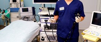
- examination of the esophagus using an esophagoscope;
- esophageal manometry (this study is indispensable in making an accurate diagnosis). Helps determine the ability of the esophagus to contract.
However, this disease significantly complicates diagnosis, since such symptoms may be characteristic of esophageal cancer and other formations in it. Therefore, if any defects are detected in the gastrointestinal tract, it is necessary to perform a biopsy.
Forecast
The course of achalasia cardia is slowly progressive. Untimely treatment of the pathology is fraught with bleeding, perforation of the esophageal wall, the development of mediastinitis, and general exhaustion. Achalasia cardia increases the risk of developing esophageal cancer.
After pneumocardiodilation, a recurrence of achalasia cardia in 6-12 months is not excluded. The best prognostic results are associated with the absence of irreversible changes in esophageal motility and early surgical treatment. Patients with achalasia cardia are advised to undergo clinical observation by a gastroenterologist with the necessary diagnostic procedures performed.