Causes of the disease
The exact causes of the formation of esophageal leiomyoma have not been established to date.
Experts put forward only a number of factors that predispose to tumor formation. The following can serve as an impetus for the development of this disease of the esophagus:
- bad habits;
- living in unfavorable environmental conditions;
- genetic predisposition;
- poor nutrition (excessive passion for salted and smoked foods, poor diet, lack of vitamins).
Symptoms and course of the disease
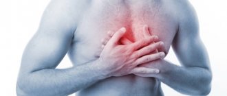
In most cases, leiomyoma does not show any symptoms. However, sometimes the tumor begins to grow and then a number of specific symptoms may appear, such as dysphagia, chest pain, gastrointestinal reflux.
Treatment of the disease
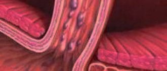
For esophageal leiomyoma, only surgical treatment is performed. These neoplasms are characterized by slow growth, so their removal is indicated only if the functions of the esophagus are impaired. For small tumors, long-term observation with mandatory periodic examination is possible, the purpose of which is to timely establish the indications for surgery. However, early removal of esophageal leiomyomas saves the patient from more complex and extensive surgery in the future.
Most often, surgical removal of leiomyoma is performed by enucleation (husking) of the tumor during thoracotomy. After removal of the tumor formation, the defect in the muscular wall of the esophagus is sutured; for large defects, plastic surgery with a flap of the diaphragm or parietal pleura is possible. Extensive surgery, the purpose of which is to remove part of the esophagus (resection), is performed extremely rarely - in case of large multiple nodes and there is no way to exclude malignancy.
In the postoperative period, the patient is prescribed a diet (mechanically, thermally and chemically gentle dishes). For a long period, drugs from the group of proton pump inhibitors are prescribed, which prevent damage to the mucous membrane of the esophagus by acidic gastric contents. This is especially important if esophageal leiomyoma is accompanied by symptoms of esophagitis.
Diagnosis of esophageal leiomyoma in Israel
At the Top Ikhilov Clinic, diagnostics are carried out using modern equipment, which makes it possible to determine the presence of a tumor at the earliest stages. It is especially important to undergo preventive studies for those who have a history of benign tumors of the esophagus or have a hereditary predisposition to them. Diagnosis of esophageal leiomyoma in Israel is carried out in the most convenient environment for those coming to Israel, and takes a minimum of time.
- Day 1
- Day 2
- Day 3
First day. Inspection
The patient, accompanied by the clinic's case manager, goes to see the attending physician. An initial consultation takes place, the patient finds out what tests he needs to undergo, and asks questions that interest him. The case manager acts as a translator if necessary.
Second day. Survey
The task of diagnosis is to determine the exact location of the tumor and determine which treatment tactics will be optimal. If leiomyoma is suspected, the following studies are prescribed:
- General blood analysis.
- X-ray.
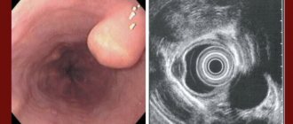
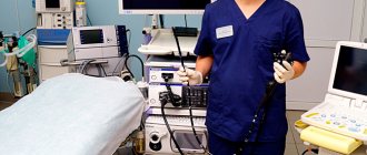
- Endoscopy of the esophagus (fibroesophagogastroduodenoscopy).
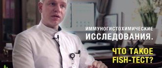
- Histological analysis of tumor cells.
- Coprogram.
- Ultrasound of the abdominal cavity.
- MRI of the abdominal cavity.
The third day. Collegial decision
After all the research has been completed, a council of specialized doctors determines the most effective therapy. At the Top Ikhilov Clinic, treatment of esophageal leiomyoma is carried out according to international protocols; in each specific case, a program is selected that will be the most effective.
Prices
| Disease | Approximate price, $ |
| Prices for examinations for stomach cancer | 5 730 |
| Prices for diagnosing Crohn's disease | 3 560 — 4 120 |
| Prices for diagnosing gastrointestinal cancer | 4 700 — 6 200 |
| Prices for hepatitis C diagnostics | 5 700 — 6 300 |
| Prices for treatment of Vater's nipple cancer | 81 600 — 84 620 |
| Prices for colorectal cancer treatment | 66 990 — 75 790 |
| Prices for treatment of pancreatic cancer | 53 890 — 72 590 |
| Prices for treatment of esophageal cancer | 61 010 — 81 010 |
| Prices for treatment of gallbladder cancer | 7 920 — 26 820 |
| Prices for treatment of nonspecific ulcerative colitis | 5 670 |
| Prices for treatment of stomach cancer | 58 820 |
| Prices for diagnosis and treatment of gallstone disease | 9 000 — 11 950 |
| Prices for the treatment of gastroenterological diseases | 4 990 — 8 490 |
| Prices for diagnosing Crohn's disease | 5 730 — 9 590 |
| Prices for treatment of viral hepatitis C and B | 5 380 — 7 580 |
| Prices for treatment of gastrointestinal cancer | 4 700 — 6 200 |
Advantages of treatment at the Top Ichilov Clinic
- Accurate diagnosis.
The center eliminates the possibility of making an erroneous diagnosis: ultra-precise instruments used by highly professional diagnosticians guarantee a 100% correct diagnosis. - High level of training of doctors.
Only those specialists who have sufficient experience and have the skills necessary to perform complex operations are allowed to treat leiomyomas. - Minimally invasive interventions.
When choosing a method of performing an operation, doctors give preference to those that are least traumatic for the patient.
- 5
- 4
- 3
- 2
- 1
(0 votes, average: 5 out of 5)
Double blow to the disease
To combat the disease, a combination of surgical and hormonal treatment is often used. Hormonal therapy for submucous fibroids is not used as a monomethod; it is used as an auxiliary method before or after surgical treatment. If the size of the submucosal node is less than 5 cm, and the entire tumor is located in the uterine cavity, then such a node can be removed simultaneously during hysteroresectoscopy.
If the diameter of the node is more than 5 cm, half or more of the node is located in the muscle of the uterus, then two ways to solve the problem are possible: either the accessible part is first resected and after hormone therapy the remaining part of the node is removed, or after hormone therapy and a reduction in the size of the node, it is carried out resection.
In the presence of concomitant small intermuscular nodes that are inaccessible for removal, our department also uses hormonal therapy, which in some cases allows us to avoid repeated operations.
Removal of a submucosal node
- Cost: 62,000 - 95,000 rubles.
- Duration: 20-40 minutes
- Hospitalization: 1 day in hospital
More details
BENIGN TUMORS AND TUMOR-LIKE DISEASES OF SYNOVIAL TISSUE (JOINTS)
Benign synovioma without giant cells
The existence of benign synoviomas is debated. Most authors are inclined to believe that all synoviomas are malignant, regardless of the degree of maturity. The tumor mainly affects the knee joint, in the form of small dense nodes. Treatment is surgical, but patients should be observed for 5-9 years. The disease can cause relapses and metastases.
Benign giant cell synovioma (nodular tenosynovitis)
Pseudotumor process occurs quite often. In 15% the process occurs in the area of the synovial membrane of the joints, in 80% in the tendon sheaths, in 5% in the mucous bursae. It is a nodular formation, most often localized on the fingers, less often on the feet, and even more rarely in the area of large joints. Favorite localization is the interphalangeal joints. It is more common in women 30-60 years old. If it persists for a long time, it can cause atrophy of surrounding tissues, including bones. The process often recurs, most of the relapses are associated with incomplete removal. Does not give metastases.
Pigmented villonodular synovitis
It is located inside the shell of the joints, most often in the area of the knee, elbow and shoulder joints. Occurs in middle age. The etiology is not clear.
Treat and no nails!
Submucous fibroids cannot always be removed with hysteroresectoscopy. If the size of the nodes is more than 5 centimeters, the operation is considered inappropriate due to possible complications during and after the operation. If there is a large intramuscular part of the node, it is impossible to completely remove it in one operation. In this case, hormone therapy comes to the rescue, with drugs such as Zoladex and Decapeptyl-Depot. These drugs create a hormonal background corresponding to postmenopause. Thus, under the influence of low estrogen levels during treatment, the size of the nodes decreases and they become accessible for hysteroresectoscopy. The minimum course of treatment with such drugs is usually 3-4 injections (one injection every 28 days).
Discussion
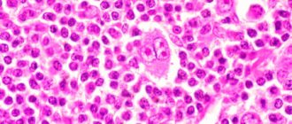
The size of the removed tumors ranged from 0.5 cm in diameter to 8.0×10.0 cm, with an average of 2.8±0.6×2.6±0.5 cm (Fig. 3). In 1 case, 2 adjacent tumors with a diameter of 0.5 and 1.5 cm were removed, creating a single filling defect of 2.0×2.0 cm on the radiograph.
Rice. 3. Leiomyoma of the esophagus (macro specimen).
Histological examination of macroscopic specimens in all operated patients confirmed a benign tumor of smooth muscle tissue (leiomyoma).
An intraoperative complication (damage to the esophagus) occurred in one patient, which was diagnosed on the operating table. Defect S.O. sewn up with an intracorporeal continuous suture with absorbent thread to restore the integrity of the muscular membrane. On the 2nd day after the operation, the sutures of the lower third of the esophagus failed, which was manifested by the X-ray picture of left-sided hydrothorax. After video-laparoscopic suturing of the perforated opening of the esophagus, video-laparotranshiatal drainage of the posterior mediastinum, video-assisted jejunostomy according to Meidl, and drainage of the left pleural cavity, scarring of the perforated opening was achieved.
In the second patient, perforation of the lower third of the thoracic esophagus occurred on the 7th day after right-sided video-assisted thoracoscopic enucleation of esophageal leiomyoma. To eliminate the complication, staged sanitary video-thoracoscopy on the right with drainage of the posterior mediastinum was performed, followed by video-laparotranshiatal drainage of the posterior mediastinum and video-assisted jejunostomy according to Meidl.
There were no deaths. The average duration of postoperative rehabilitation of patients was 18.2±7.8 days, the duration of inpatient treatment was 26.4±8 days. In 1 case, 7 months after surgery, cicatricial stenosis of the lower third of the esophagus developed. After excluding recurrence of the disease, the patency of the esophagus was restored after a single orthograde bougienage of the esophagus along the guide. No relapse of the disease was observed in any of the patients from 1 to 15 years after surgery.
"Final Analysis"
Hysteroscopy (diagnostic, therapeutic)
- Cost: 47,500 rub.
- Duration: 20 to 60 minutes
- Hospitalization: 1 day in hospital
More details
The most common symptom of submucous uterine fibroids is uterine bleeding. They can occur both during menstruation, which becomes very heavy and prolonged, and during the intermenstrual period. The next most common manifestation is cramping pain during menstruation. Very rarely, such fibroids remain asymptomatic. The likelihood and severity of bleeding does not depend on the size of the node. Uterine bleeding, as a rule, leads to anemia, which dictates the need for surgical treatment, since medications are powerless in this situation. When diagnosing such symptoms, you must consult a qualified doctor who will conduct the necessary research. The final diagnosis of uterine fibroids is made after ultrasound and hysteroscopy.
The authors presented a case report of esophageal leiomyosarcoma in a 57-year-old man. X-ray and endoscopic examination revealed an exophytic dense tumor with ulceration. The operation was performed: laparothoracocervicotomy, extirpation of the esophagus, esophagogastroplasty with anastomosis on the neck. Histological conclusion: leiomyosarcoma of the esophagus.
The Case of Leiomyosarcoma esophageal
The authors present a description of the case of leiomyosarcoma of the esophagus in men 57 years old. X-ray and endoscopy study revealed exophytic research dense tumor with ulceration. An operation: laparotoracocervicotomy, extirpation of the esophagus, esophagogastroplasty with anastomosis in the neck. Histological conclusion: leiomyosarcoma of the esophagus.
Malignant non-epithelial tumors - sarcomas of the esophagus - are rare and account for no more than 0.5-1% of all malignant neoplasms of this organ [3, 6]. According to V.Kh. Vasilenko et al. (1971), they are almost 200 times less common than cancer. Sarcomas develop mainly in older people, more often in men, and much less often in women. This group of tumors is characterized by the same clinical symptoms (local and general) as with esophageal cancer. Esophageal sarcomas are divided into polypoid and diffuse infiltrating.
Leiomyosarcoma usually develops from the tissue of the walls of large vessels (superior vena cava) containing smooth muscle tissue; most often localized in the lower half of the esophagus. Leiomyosarcoma of the esophagus is extremely rare [2], accounting for about 0.3% of malignant neoplasms of the esophagus [7]. Only isolated observations of this disease have been described in the literature [4, 5, 8]. The surgical approach remains the main one in the absence of signs of dissemination of the process.
We observed a case of esophageal leiomyosarcoma in a 57-year-old man. The patient was admitted with complaints of dysphagia. FGDS revealed a dense infiltrative tumor with ulceration in the esophagus from the mouth to 35 cm along the postero-right wall. X-ray: the esophagus from the tracheal to the supradiaphragmatic segment is sharply dilated, in its lumen there is a tuberous “defect” of filling measuring about 13 by 2.5 cm, with a barium “depot” in the center. The elasticity of the walls of the esophagus in the affected section is limited, and in some areas the walls are rigid. There were no folds of mucous membrane in the affected area.
Rice. 1. X-ray of the esophagus of patient T., 57 years old. The esophagus is dilated and deformed due to a tuberous “defect” of filling, with a barium “depot” in the center. The elasticity of the walls is sharply limited
Rice. 2. X-ray of the esophagus of patient T., 57 years old. In the lumen of the esophagus there are multiple nodes with fairly clear contours. “Niche” on the contour due to ulceration
An operation was performed: laparothoracocervicotomy, extirpation of the esophagus, esophagogastroplasty with anastomosis on the neck. With thoracotomy in the 5th intercostal space on the right, the esophagus is dilated to 8-10 cm. A large exophytic tumor is palpated in the lumen of the esophagus, occupying the upper and middle thirds of the esophagus from the upper thoracic aperture to the supradiaphragmatic region. The case was found resectable. The mediastinal pleura was dissected along the anterior and posterior surfaces of the esophagus, the azygos vein was ligated and resected. The esophagus is mobilized from the esophageal opening of the diaphragm along with paraesophageal tissue and the bifurcation group of lymph nodes to the upper thoracic outlet.
Upper-median laparotomy. Correction of access with four retractors RSK-10. Mobilization of the stomach with ligation of the left gastroepiploic artery, short branches of the splenic artery and right gastric artery, left gastric artery at the origin. Using UO-60 devices, a graft was formed from the greater curvature of the stomach with peritonization of a tantalum suture strip using a continuous suture. Through the posterior mediastinum, the graft was brought into the cervical wound with satisfactory blood flow at the apex of the graft. An end-to-end anastomosis was performed between the proximal part of the graft and the esophageal stump in the cervical wound. A double-lumen nasogastric tube was inserted into the graft.
Macro specimen: a soft tissue tumor measuring 12 x 2.5 cm in the shape of an hourglass with clear contours. Histologically: leiomyosarcoma of the esophagus against the background of leiomyoma. There are no metastases along the resection lines and in the lymph nodes and omentum.
Immunohistochemistry: tumor cells are positive for L-smooth muscle actin, about 20% for Ki67, negative for S100, CO 117, CO 34, desmin. Calponin is a uniform positive reaction. Diagnosis: leiomyosarcoma of the esophagus.
R.M. Taziev, D.A. Abdulkhakova, N.V. Balatenko
Kazan State Medical Academy
Republican Clinical Oncology Dispensary of the Ministry of Health of the Republic of Tajikistan
Taziev Ravkat Mingazovich – Doctor of Medical Sciences, Professor of the Department of Oncology and Surgery
Literature:
1. Vasilenko V.Kh., Grebenev A.L., Salman M.M. Diseases of the esophagus. - M.: Medicine, 1971. - 402 p.
2. Davydov M.I., Machaladze Z.O., Polotsky B.E. and others. Mesenchymal tumors of the mediastinum (literature review) // Siberian Journal of Oncology. - 2008. - No. 1 (25). — P. 64-72.
3. Choh JH, Khazei AH, Ihm HJ Leiomyosarcoma of the esophagus: report of a case and review of the literature // J Surg Oncol. - 1986. - No. 32. - P. 223-226.
4. Farkas E., Renyi-Vamos F., Matrai Z. et al. Leiomyosarcoma of the oesophagus: a case report // Magy Seb. — 2006 Dec. — No. 59 (6). - P. 441-4.
5. Hou Y., Wang J., Zhu X et. al. A comparative study of esophageal stromal tumors and smooth muscle tumors // Zhonghua Bing Li Xue Za Zhi. — 2002 Apr. — No. 31 (2). - P. 116-9.
6. Jutley RS, Gray RD, MacKenzie JM, Cockburn JS // A leiomyosarcoma of the oesophagus presenting incidentally without dysphagia. — Eur J Cardiothorac Surg. — 2002 Jan. — No. 21 (1). - P. 127-9.
7. Rocco G., Trastek VF, Deschamps C. et al. Leiomyosarcoma of the esophagus: results of surgical treatment // Ann Thorac Surg/ - 1998 Sep. No. 66 (3). - P. 894-6.
8. Visioli A., Daniel FJ Leiomyosarcoma of the oesophagus: a case report and literature review of leiomyosarcoma // Australas Radiol. — 1997 May. — No. 41 (2). - P. 160-5.