Gastric fistula
To make a correct diagnosis, an abdominal surgeon can use various techniques, but all of them are aimed at studying the fistulous tract, the condition of the gastric and intestinal mucosa, the relationship between the stomach and its surrounding organs, and passage through the digestive tract. The examination of the patient begins with clinical and biochemical blood tests. An unformed fistula is characterized by pronounced inflammatory changes in the general blood test: leukocytosis, increased ESR, shift of the leukocyte formula to the left.
Formed fistulas cause biochemical changes: disturbances in the water-electrolyte state and protein metabolism, metabolic alkalosis with a significant decrease in chlorine and potassium levels, hypoprothrombinemia. To assess the possibility of closing the tract, the albumin level is determined - an albumin level above 3.5 mg/dl is considered sufficient; levels below 2.5 mg/dL are associated with high mortality in 40% of cases.
X-ray techniques are of great importance in diagnosis. To assess the state of gastrointestinal motility and the movement of food masses through the small intestine, radiography of the stomach with contrast and barium passage radiography are performed. For the same purposes, dynamic scintigraphy of the esophagus and stomach and static scintigraphy of the intestine are used.
The main method of studying a fistula is fistulography - the introduction of a contrast agent into the fistula tract to determine its direction, pockets and leaks. After fistulography, digital examination of the wide fistulous tract may be necessary. The introduction of contrast through the mouth can show how food masses are removed from the stomach - mainly through a gastric fistula or through the intestines.
A consultation with an endoscopist is mandatory: he must assess the condition of the gastric mucosa during esophagogastroduodenoscopy. During the examination, it is necessary to find the fistula tract, determine its size, and the condition of the mucous membrane. It is also necessary to examine the duodenal bulb to identify its kinks and deformations that create difficulties for the passage of food and provoke the persistence of the fistula. To determine the condition of the organs surrounding the stomach, a multislice spiral computed tomography (MSCT) of the abdominal cavity may be required.
Differential diagnosis of external tracts usually does not present any difficulties if there is a history of peptic ulcer, stomach cancer, or surgery on the abdominal organs. Difficulties in diagnosis may arise in the presence of internal anastomosis. Internal gastric fistula should be differentiated from acute cholecystitis, pancreatitis, peritonitis with the presence of interintestinal abscesses, peptic ulcer of the anastomosis, phlegmon of the retroperitoneal tissue, fistulas of the intestines and other parts of the digestive tract.
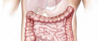
Intestinal fistulas
Intestinal fistula is a connection between the intestinal lumen and the surface of the body or with the lumen of another hollow organ.
Classification of intestinal fistulas
- I. By time of occurrence: congenital, acquired.
- II. By etiology: traumatic, applied for therapeutic purposes, arising from diseases of the small intestine.
- III. By function: complete, incomplete.
- IV. The nature of the fistula: labiform, tubular.
- V. According to the level of location on the intestine: high, low, mixed.
- VI. According to the presence of complications: uncomplicated, complicated.
- VII. By quantity: single and multiple.
Fistulas of the small intestine can be congenital (for example, when the vitelline duct is not closed) or acquired. Acquired fistulas arise as a result of injury, disease (colon diverticulosis, Crohn's disease), operations in which the fistula is applied for therapeutic purposes (jejunostomy for inoperable total gastric cancer, colostomy for acute obstructive intestinal obstruction due to sigmoid colon cancer).
Fistulas can develop as a result of prolonged standing of tampons and drainages in the abdominal cavity, or failure of the sutures of the small or large intestine.
A fistula connecting the intestinal lumen with the surface of the body is called external, one organ with another is called internal. With a complete fistula, all intestinal contents pour out; with an incomplete fistula, part of it passes into the efferent loop of the intestine. If the fistula opens directly on the surface of the body and the intestinal mucosa is fused to the skin, this condition is called a labiform fistula. When there is a passage between the intestine and the surface of the body, it is a tubular fistula.
Fistulas located on the duodenum and jejunum are classified as high, while those located on the ileum and colon are classified as low.
The changes that occur in the body are associated with the loss of proteins, fats, carbohydrates, vitamins, water, electrolytes and enzymes through the fistula. The more oral the fistula is located, the greater these losses and the more pronounced metabolic and water-electrolyte balance disorders are. Maceration (dermatitis) may occur on the skin around the fistula. Local complications include abscesses, phlegmon of the abdominal wall, purulent or fecal leaks. In addition, intestinal prolapse, parastomal hernia, bleeding from the fistula, and enteritis (colitis) may occur. Common complications include cachexia, renal and liver failure.
Clinical picture and diagnosis. External small intestinal fistulas are manifested by the release of liquid small intestinal contents. Fistulas of the colon release feces and gases. The localization of the fistula, its function and level of location are clarified during an X-ray examination. For small intestinal fistulas, a contrast agent is administered through the mouth and its passage is monitored; for large intestinal fistulas, a contrast agent is administered through the rectum (irrigoscopy). An important diagnostic method is fistulography, in which a water-soluble contrast agent is injected into the external opening of the fistula.
For tubular fistulas of the duodenum, jejunum and ileum, conservative treatment is usually carried out: high-calorie parenteral and enteral (tube) nutrition, correction of metabolic disorders and water-electrolyte disorders, occlusion of the fistula using various devices (pellets, obturators), skin care around the fistula . In a number of patients, positive results are achieved with total parenteral nutrition. In general, a course of conservative therapy is effective in 30-40% of patients with a duration of 1-1.5 months.
The ineffectiveness of conservative treatment of duodenal fistula after gastrectomy may be due to duodenostasis, afferent loop syndrome, which requires reconstructive surgery. In case of fistulas of the descending branch of the duodenum due to failure of the biliodigestive anastomosis or trauma, accompanied by significant losses of bile and intestinal contents, surgery to disconnect the duodenum is indicated, but the prognosis for this category of patients, especially with infrapapillary fistulas, is questionable.
For labiform and long-term non-healing fistulas of the small intestine, surgical treatment is indicated. For incomplete tubular and labial fistulas, it is advisable to use extraperitoneal methods of closing them; for other types of fistulas, the method of choice is laparotomy with intraperitoneal resection of the section of intestine bearing the fistula, and the imposition of an end-to-end anastomosis between the afferent and efferent loops.
For labiform fistulas of the colon, operations are performed, the type of which depends on the type of fistula (complete or incomplete). For small incomplete labial fistulas, extraperitoneal methods of closing them are used. To do this, the intestinal wall in the fistula area is isolated and the defect is sutured with a double-row suture. For large incomplete and complete labiform fistulas, the use of intra-abdominal closure methods is indicated. For this purpose, the intestine is isolated along the entire perimeter of the fistula, brought into the wound and the fistula opening is sutured (for incomplete fistulas) or an anastomosis is performed (for complete fistulas). In case of multiple fistulas located on one intestinal loop, it is advisable to resect it and perform an anastomosis.
Rectal fistula - symptoms and treatment
Fistula dissection, fistulotomy
Dissection of the fistula (fistulotomy) is used for 85-95% of primary fistulas (subcutaneous, intersphincteric and low transphincteric).
The probe is passed into the fistula tract through the external and internal openings. Using a scalpel or an electrocoagulator, the skin, subcutaneous tissue and internal sphincter are dissected, thus opening the entire fistula tract.
When the fistula is located low, the internal sphincter and the subcutaneous part of the external sphincter can be divided at right angles to the main fibers. Curettage is performed to remove granulation tissue at the bottom of the wound. The wound is left open and not sutured.
Fistula excision (fistulectomy) is the complete removal of the fistula tract and surrounding tissue, which leaves wound defects that require more time to heal and does not provide any advantages over fistulotomy.
Carrying out a ligature (seton, seton)
The seton can be placed alone, in combination with a fistulotomy, or in stages.
Indications:
- complex fistulas (high transphincteric, suprasphincteric, extrasphincteric) or multiple fistulas;
- recurrent fistulas after previous fistulotomy;
- anterior fistulas in women;
- anal sphincter insufficiency;
- patients with Crohn's disease or patients suffering from immunosuppression.
In addition to visually determining the amount of sphincter muscle involved, ligatures drain the fistula, stimulate fibrosis, and gradually cut through the fistula. Setons can be made from non-absorbable sutures or latex.
One-step technique (cutting)
The ligature is passed through the fistula tract and tightened from the outside. Over time, the fistula tract gradually erupts, and fibrosis occurs above the ligature. Treatment time is 6-8 weeks.
Recurrence and fecal incontinence are important factors to consider when using this method. Success rates for cutting setons range from 82-100%; however, long-term incontinence rates may exceed 30%.
Two-stage technique (drainage/fibrosis)
The ligature is placed around the deep part of the external sphincter after incision of the skin, subcutaneous tissue, internal sphincter muscle and subcutaneous portion of the external sphincter.
Unlike the cutting seton, with this option the ligature remains loosely tied to drain the intersphincteric space and stimulate fibrosis in the deep part of the sphincter. Once the superficial wound has completely healed (2-3 months), the sphincter muscle bound by the ligature is separated.
Moving a mucosal flap (FLAP technique, Advancement Rectal Flap)
Mucosal relocation is indicated in patients with a chronic high fistula, but the indications may be the same as for ligation.[19] Advantages: one-step technique, without additional damage to the sphincter. Disadvantages: low effectiveness in patients with Crohn's disease or acute infection.
This procedure involves a complete fistulotomy with removal of the primary and secondary tracts and complete removal of the internal orifice.
A muco-submucosal flap with a wide proximal base (twice the width of the apex) is exposed. The internal muscle defect is closed with absorbable sutures, and the flap is sutured over the internal opening so that its suture line does not overlap the sphincter sutures.
Plugs and adhesives (fibrin glue and collagen plug)
Advances in biotechnology have led to the development of new tissue adhesives and biomaterials formed into fistula plugs. Due to their less invasive nature, these treatments result in reduced postoperative complications and the risk of incontinence, but long-term results, especially in complex fistulas, have a high recurrence rate.
Registered formulations containing fibrin glue for the treatment of anal fistulas have yearly recurrence rates of 40 to 80%.
There is evidence of success with newer materials such as acellular dermal matrix and Gore Bio-A bioabsorbable plug in low fistulae. Evaluation of long-term success rates with plug technologies for complex disease will be based on additional data from randomized trials.
In a randomized controlled trial designed to evaluate the effectiveness and safety of a plug in patients with perianal fistulas due to Crohn's disease, Senéjoux et al. did not find that the plug was superior to the seton for fistula closure, regardless of whether the fistula was simple or complex.
Combined sphincter-sparing treatment has also been proposed, which includes both fistulous plug and rectal flap reduction to treat transfincteric fistulas.
LIFT procedure (Ligation of Intersphincteric Fistula Tract)
Ligation of the intersphincteric fistula tract (LIFT) is a sphincter-sparing procedure for complex transphincteric fistulas first described in 2007. It is performed by accessing the intersphincteric space to ensure safe closure of the internal opening and removal of diseased cryptoglandular tissue.
The intersphincteric tract is identified and divided by making a careful dissection through the intersphincteric space after making a small incision over the probe connecting the external and internal openings. After isolation, the intersphincteric tract is ligated near the internal sphincter and then brought out distal to the ligation point. Hydrogen peroxide is injected through the external opening to confirm proper division of the fistula tract. Curettage of the remaining part of the fistula is performed. The intersphincteric incision is sutured with absorbable material. The wound in the area of the external opening is left open for dressings.
Due to its relative novelty, the LIFT method has not been widely studied. The randomized trial involved 39 patients with complex fistula who had failed previous operations and were treated with the LIFT technique, with success rates comparable to those observed with the FLAP technique. The probability of recurrence within 19 months was 8% for the LIFT technique versus 7% for the FLAP technique. Recovery time to performance was shorter in the LIFT group (1 vs. 2 weeks), but there was no difference in incontinence rates.
Laser treatment of fistulas (FiLaC – Fistula laser closure)
FiLaC - treatment of rectal fistula using an invented radial emitting laser probe. It is a new, insufficiently studied method of treating chronic paraproctitis using a special laser probe, which eliminates the fistulous epithelium and simultaneously destroys the remaining fistulous tracts. In this case, the anorectal fistula is removed gently, without trauma to the sphincter, while maintaining the function of the anal sphincter. Recent studies have shown very encouraging results with this new type of fistula treatment.
Stoma
In rare cases, a diverting colostomy may be indicated to facilitate treatment of a complex, recurrent anal fistula. The most common indications:
- necrotizing fasciitis of the perineum;
- severe anorectal Crohn's disease;
- recurrent rectavaginal fistulas;
- radiation-induced fistulas.
Postoperative care
After surgery, most patients can be treated on an outpatient basis, with discharge recommendations followed and close monitoring. Sitz baths, analgesics, and stool softeners (eg, psyllium bran and preparations) are used in aftercare.