June 19, 2021 Types and places of papillomas
Among all the types of benign neoplasms that arise in the digestive system, esophageal papilloma is a very rare occurrence. Only 0.5% of such cases of all gastrointestinal pathologies are diagnosed by medicine.
Previously, they were found mainly in older people, but now cases of this disease at a young age have become more frequent. This is explained by the living conditions characteristic of modern humans, under which immunity decreases. Bad habits, a polluted environment, stress, and an incorrect and unbalanced diet also have a negative impact. Among the patients there are more males than females.
Etiology
Etiology
in most cases viral. Thus, human skin papilloma is intertwined with cell-free filtrates into the lab. animals, and the virus is detected in the keratinizing papilloma cells using an electron microscope. Moreover, as the papilloma “aging”, the virus disappears from these cells. During the development of papilloma, large acidophilic inclusions are detected in the nuclei of epithelial cells located above the basal layer of the epithelium; such inclusions are found in the cytoplasm; are much less common.
The causative agent of papilloma is human papillomavirus - a virus of the genus Papillomavirus (Papillomavirus) family. papovaviruses (see).
Human papillomavirus is highly host specific. Its cytopathic effect has been established in kidney cell cultures of monkeys, humans and mice. There is no reliable system for titrating the infectivity of this virus in vitro. In cell culture, papillomavirus transforms human cells at a very low frequency.
Papillomavirus can normally be detected in urine, which suggests that in humans this virus, in addition to skin cells and mucous membranes, can multiply in kidney cells, from where it enters the urine.
There are no special laboratory diagnostic methods aimed at identifying human papillomaviruses.
Causes of papilloma in the esophagus
The causative agent of growths is papillomavirus, which has many strains. In this case it is the 52nd type. Every carrier of the virus is at risk of developing cancer, but HPV may never appear, remaining in the inactive stage in the human body until the end of life.
A connection has been established between decreased immunity and the appearance of tumors. Some factors also influence their growth:
- frequent consumption of alcoholic beverages;
- smoking;
- abuse of spicy and fatty foods;
- chronic heartburn;
- recurrent gastritis.
All this creates microtraumas of the esophagus, which creates a favorable environment for the development of tumors.
Pathological anatomy
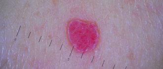
Macroscopically, papilloma is usually delimited, dia. up to 1-2 cm, dense or soft to the touch tumor on a thin long or short stalk, less often - on a wide base. In rare cases, papillomas reach large sizes. With papillomatosis, the surface of the skin or mucous membrane is covered over a significant area with papillary growths. The surface of the papilloma is uneven, fine- or coarse-grained, reminiscent of cauliflower or cockscombs. Skin papilloma can have a different color - from white to dirty brown (depending on the blood supply to the stromal vessels and the pigment content in the basal layer of the epithelium); Papilloma of the mucous membrane is often colorless or pearly white, but sometimes acquires a purple or black color due to hemorrhages into its tissue. Bladder papilloma can be hardened due to deposits of calcium salts.
Rice. 4. Microscopic specimen of papilloma of the oral mucosa: the surface of the papilloma is covered with a layer of stratified squamous keratinizing epithelium (1) with deep acanthotic cords plunging into the connective tissue stroma rich in thin-walled vessels (2); hematoxylin-eosin staining; x 20. Fig. 5. Microscopic specimen of papilloma of the esophageal mucosa: numerous papillary growths of stratified squamous epithelium and connective tissue stroma are visible; hematoxylin-eosin staining; x 20.
Microscopically, papilloma (color Fig. 4) consists of two tissue components - connective tissue stroma and epithelium. Based on the nature of the epithelium, they distinguish between squamous cell (covered with stratified squamous epithelium) and transitional cell (covered with transitional epithelium) papillomas. The connective tissue of the tumor stroma can be loose or dense, often edematous, sometimes with mucus phenomena, a greater or lesser number of vessels and, as a rule, with signs of inflammation. In cases where the papilloma stroma is significantly developed and sclerotic, we speak of fibropapilloma. In the epithelium covering the papilloma, the number of cell layers is usually increased, and the epithelial cells are larger than normal. At the same time, noticeable hyperkeratosis is observed in the skin papilloma; in papillomas that arise on the mucous membranes, keratinization is usually less pronounced (tsvetn. Fig. 5), the cells of the superficial layer of the epithelium are pale colored. Sometimes there are papillomas of the mucous membranes, covered with stratified squamous keratinizing epithelium, which developed as a result of metaplasia (see).
In some papillomas, the phenomena of acanthosis (see), usually accompanied by high mitotic activity of the cells of the basal layer of the epithelium, may be expressed.
Various skin papillomas may differ from each other in their histological structure. Thus, ordinary skin papillomas are characterized by the presence of vacuolated epithelial cells in the basal layer and areas of parakeratosis; with senile keratosis, papillomas with atypia and polymorphism of epithelial cells appear; basal cell papilloma is characterized by the presence of the same type of dark cells and horny cysts.
Esophageal papilloma - what is it?
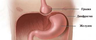
Neoplasms that are found on the walls of the digestive organ are a cluster of benign cells. They form small mastoid processes of the same color as the healthy surface of the mucosa. Their sizes vary from a few millimeters to 3 centimeters.
Most often, single growths occur, but there are cases of papillomatosis, when they take on bush-like forms. Neoplasms are located on the lower or middle sections of the food tube.
So far, this disease has been little studied by specialists. It may not have a clear pattern of symptoms for many years, so it is rarely detected at an early stage. In itself, such a tumor is not dangerous, but it can transform into a malignant one at any time and lead to a serious condition and death.
Clinical picture
The clinical picture is characterized, as a rule, by a long course and depends on the hl. arr. on the location of the lesion. For example, papillomas of the skin of the face and neck can cause a cosmetic defect, papillomas of the mucous membrane of the larynx - cause disturbances in phonation and breathing, papilloma of the ureter - narrow or obstruct its lumen with impaired outflow of urine, papilloma of the bladder is often subject to ulceration, with separation of individual papillae and bleeding . Transitional cell papilloma of the paranasal sinuses can, while remaining morphologically benign, have infiltrative growth and grow into surrounding tissues. Sometimes malignancy is observed, which usually occurs due to the epithelial component of the tumor.
Diagnosis and treatment of esophageal papillomas
The International Classification of Diseases, 10th revision, includes only malignant neoplasms in the alimentary canal. Esophageal papilloma ICD 10 belongs to class C15 if it has oncological activity. Subclasses are also distinguished according to the anatomical description and location of growths in thirds of the organ.
To identify a neoplasm, determine its characteristics and options, and how to treat esophageal papilloma, a number of diagnostic examinations are performed:
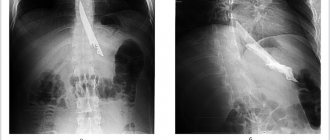
- contrast radiography – establishes the location of the tumor and its diameter;
- endoscopic, histological, cytological studies - help to clarify the type of growth cells, the presence of oncology, the tendency of cells to malignancy;
- blood test - informs about the state of health, the presence of any other diseases;
- MRI or CT also clarify the picture of the disease;
- in rare cases, chromoscopy and esophagoscopy are performed.
When esophageal papilloma is discovered, treatment will include a set of measures aimed at increasing immunity, suppressing the activity of the virus, and saturating the body with vitamins. In cases of detection of neoplasms that have prerequisites for oncology, they are removed surgically, even if they are benign, since there is a high probability of their degeneration into cancer.
The doctor prescribes medications:
- antiviral drugs;
- immunomodulators;
- vitamin and mineral complexes.
Removal is carried out in three different ways:
- endoscopic method - is carried out using an endoscope, which penetrates the papilloma through natural openings: the nose and mouth. In this case, a special video camera is used that allows you to see the internal cavities;
- Laparoscopy is a method in which the surgeon makes a centimeter incision and removes the growth through it. This option is the most acceptable, since during the operation the organs are not injured, and the small incision heals quickly without leaving a scar;
- if the growth is attached to the wall of the food tube with a wide leg, experts give preference to esophagotomy removal.
The rehabilitation period does not last long. These operations are usually easily tolerated and do not cause complications.
Treatment
Treatment Ch. arr. surgery; methods of cryodestruction and sclerosing treatment are also used. Indications for surgical treatment include both cosmetic defects and localization of papilloma, which causes significant functional disorders, causing frequent trauma with repeated bleeding and inflammatory processes, as well as the risk of malignancy. Removed papillomas are subjected to histological examination. If the papilloma is localized in the area of the ureteric orifice, resection of the bladder and transplantation of the ureter are performed.
Surgical treatment for papillomatosis consists of excision of the largest number of papillomas, electrocoagulation of small papillomas and the surrounding mucous membrane. In this case, care should be taken to remove all elements of the papilloma, since implantation of its individual fragments can lead to tumor recurrence.
Forecast
, as a rule, favorable. However, in some cases, relapses are possible, sometimes multiple, or malignancy of the tumor occurs.
Bibliography:
Apatenko A.K. Epithelial tumors and skin malformations, p. 40, 44, M., 1973; Golovin D.I. Atlas of human tumors, p. 63, M., 1975; Guide to pathological diagnosis of human tumors, ed. N. A. Kraevsky and A. V. Smolyannikov, p. 407, M., 1976; Strukov A.I. and Serov V.V. Pathological anatomy, p. 112, 171, M., 1979; Fenner F. et al. Biology of animal viruses, trans. from English, vol. 1-2, M., 1977; Lever WF a. Schaumburg-Lever G. Histopathology of the skin, Philadelphia—Toronto, 1975.
I. G. Balandin, M. N. Lanzman.
Symptoms and diagnosis of neoplasms in the digestive tract
For many years a person may not feel that there is a growth in his food tract. The disease occurs without obvious symptoms for a very long time. In some, such tumors were discovered only after death from other causes.
If the size of the tumors increases or the cells turn into cancer-active ones, then specific symptoms appear:
- discomfort or the presence of a foreign body is felt;
- becomes difficult to swallow;
- belching is often bothersome;
- nausea occurs for no reason;
- saliva is released more abundantly;
- constantly feeling tired and powerless;
- pain in the upper chest.
Such manifestations are signs of tumor development, which leads to dangerous consequences, so if they occur, you should consult a doctor.
Complications
Treatment does not always go without leaving a trace, and surgical intervention may have complications during the rehabilitation period. The most common complications:
- divergence of fabrics, seams;
- rejection of the created connection between tissues;
- inflammation of the fiber;
- the lumen becomes scarred, causing narrowing.
The latter is considered the most harmless complication; a greater danger arises when inflammation or necrosis occurs at the surgical site. The rehabilitation process largely depends on the complexity of the operation and how severely the papilloma has affected the esophagus.
Reasons for the development of papillomatosis
In most cases, papillomas have a viral etiology. The causative agent of the disease is a DNA-containing human papillomavirus (HPV) of the papovavirus family. To date, about 100 types of this virus have been identified, many of which have oncogenic potential of varying degrees of risk.
The virus can be transmitted from person to person through contact (including sexual contact); It has been established that one of the ways of infection with the virus is transmission from mother to fetus during pregnancy or during childbirth, which causes the appearance of papillomatosis in young children.
Most often, HPV infection leads to asymptomatic carriage, but in situations where immunity decreases (after a long illness, in stressful situations, during vitamin deficiency, during pregnancy, when taking certain medications, for example, glucocorticosteroids), an immediate clinical manifestation occurs in the form of papillomas.
In addition to a viral cause, papillomatosis can be a consequence of chronic inflammatory processes. It is interesting to note that in some situations, when papillomatosis is detected, no signs of viral infection are detected, which prompts the search for other causes of the development of this disease.
Preventive measures
It is always better to prevent a disease than to treat it, and this also applies to papillomas of the esophagus. The most effective measure would be to consult a doctor in a timely manner, since if the virus is diagnosed early, treatment does not last so long and surgery can be avoided. There are preventive measures that need to be followed daily. A healthy lifestyle, quitting smoking and alcohol will reduce the likelihood of developing the disease. Also, a lot depends on the foods a person eats, so the diet should be balanced and meals should be regular.
Esophageal papilloma: treatment with folk remedies
Traditional medicine does not have in its arsenal a way to completely cure tumors. But it can counteract viral infections, preventing the development of tumors. Herbs with antiviral properties, propolis .
A mixture of the following components is very useful for strengthening the immune system and all body systems:
- raisin;
- dried apricots;
- walnuts;
- lemon;
- honey.
Grind nuts and dried fruits, grind the lemon in a meat grinder, mix everything and add honey. This medicine is taken daily for a month, 1 tbsp. spoon on an empty stomach.
This recipe is good for preventing many diseases, but does not guarantee complete health. In order to detect dangerous diseases in time, you need to be regularly examined and be attentive to various symptoms.
HPV papilloma biopsy: diagnosis of genital warts
Previous post
Papillomas throughout the body: why growths appear and how to remove them
Next entry
Symptoms
When the virus just begins to manifest itself, that is, it is small in size, there are no symptoms as such. As it grows, the lumen of the esophagus narrows and this affects a person’s normal life activities.
Squamous cell papilloma
Symptoms of esophageal papilloma:
- food has difficulty passing through the esophagus;
- there is a feeling that something is stuck to the wall of the esophagus;
- epigastric pain;
- nausea, which is accompanied by vomiting;
- burping becomes more frequent;
- salivation is much higher than usual;
- general weakness.
Rarely do patients go to the doctor with these symptoms, and papilloma is usually discovered by chance when a person is examined for another reason. This is because symptoms may not appear for years after the virus has entered the body. And also for the reason that no one perceives excessive salivation and frequent belching as a real symptom of the disease.
Certain types of papillomatosis
Papillomas can affect the skin and mucous membrane of almost any internal organ. Below are descriptions of individual types of papillomatosis depending on what part of the body is affected.
Laryngeal papillomatosis
It is caused predominantly by HPV types 6 and 11, types 16 and 18 are less common. The incidence of laryngeal papillomatosis in the population is 2 per 100,000 among adults and 4 per 100,000 among children.
Characteristic symptoms are hoarseness and breathing problems. Despite the fact that laryngeal papillomatosis is a benign disease, serious complications are possible, such as stenosis of the larynx, spread of papillomas to the trachea and bronchi with subsequent development of pulmonary failure, as well as degeneration into a malignant tumor, especially if papillomatosis is caused by the HPV virus type 16 or 18, which have a high level of potential oncogenicity.
In addition, laryngeal papillomatosis can recur; distinguish between “aggressive” and “non-aggressive” types of relapse. The aggressive type includes papillomatosis, for which 10 or more operations have been performed, or more than three operations in a year, or when the process has spread to the subvocal part of the larynx.
Vestibular papillomatosis
This term is used to refer to small papilloma-like formations of the vaginal vestibule. It occurs quite often in women under the age of 30 and becomes an indication for examination of the mucous membranes of the vagina and cervix, including for the presence of human papillomavirus, as it can be combined with damage to the cervix, although vestibular papillomatosis itself may not have a viral nature.
Vestibular papillomatosis can often develop without clinical symptoms and is detected during examination by a gynecologist for prophylactic purposes or for other complaints, but it can also manifest as leucorrhoea, pain and burning in the vulva, symptoms of dyspareunia, and be combined with a local inflammatory process.
Vestibular papillomatosis, unlike lesions of the cervix, is prone to recurrence - according to the literature, the relapse rate is up to 17%. Typically, relapses are characteristic of infection with papillomavirus of low oncogenic risk.
Skin papillomatosis
Skin papillomatosis is the growth of numerous skin tumors in a limited area. Papilloma is formed when the upper layer of skin, the epidermis, grows. Typically, skin papillomas are 5–7 millimeters in size, less often up to 2 centimeters. The shape varies from a dotted or slightly hanging outgrowth of skin to a pea. The color of the papilloma is often indistinguishable from the color of the surrounding skin, but it can also be white or brown. Ordinary papillomas are most often localized on the skin of the back, palms and fingers, soles and feet, filamentous papillomas - in places with thin skin (eyelids, neck, axillary and groin areas).
It usually has an asymptomatic course and is predominantly a cosmetic defect, however, skin papillomas can often be injured, which can lead to the development of inflammation or be a risk factor for their malignancy.
Gottron's carcinoid papillomatosis of the skin deserves special attention - a rare precancerous disease characterized by specific growths of the epidermis. The main factor in its occurrence is a predisposition to the development of multiple skin lesions by papillomas; Among other risk factors, regular trauma to the skin, chronic skin diseases, and circulatory disorders should be mentioned. In this disease, the lesions are located symmetrically on the legs, often on the front surface, against the background of long-existing skin lesions, they look like papillomatous warty growths and vegetations in the form of plaques of significant size protruding above the surface by 1–1.5 cm. The grooves between the lesions are filled with yellowish-white sticky masses with an unpleasant odor; in some areas they shrink into yellowish-gray crusts. Sometimes erosions, superficial ulcers, and easily bleeding granulations occur. Due to the high risk of malignancy (it is worth noting that a number of authors equate carcinoid papillomatosis with well-differentiated squamous cell skin cancer), this disease requires consultation and treatment with an oncologist.
Papillomatosis of the esophagus
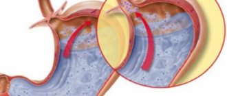
It is a rare pathology - according to the literature, its frequency is only 4.5%. Papillomas have the appearance of a polypoid or leaf-shaped formation of a whitish color, usually up to 1 cm in size. The lesions are typically localized in the distal parts of the esophagus. Although the main reason for the development of this pathology is considered to be the presence of human papillomavirus infection, according to a number of authors, esophageal papillomatosis also occurs in the absence of viral contamination. In addition, there is a connection between the development of papillomatosis and gastroesophageal reflux, which, due to prolonged exposure to hydrochloric acid contained in gastric juice, leads to damage and chronic inflammation of the esophageal mucosa.
Papillomas of the esophagus may not show any symptoms for a long time, but as they grow, the following complaints may appear: difficulty swallowing, mild or moderate chest pain, belching, nausea.
Currently, it is believed that esophageal papillomas are a precancerous disease, and it is papillomatosis, due to the prevalence of damage to the esophageal mucosa, that has the greatest risk of malignancy, therefore the identification of esophageal papillomas is an indication for surgical treatment.
Intraductal papillomatosis
This term refers to papillomas located in the milk ducts of the mammary gland. Papillomas can occur both in the peripheral ducts of any quadrant of the mammary gland, and in the ducts located immediately behind the nipple. Peripheral papillomatosis is usually represented by small, up to 1 cm, formations. In some cases, intraductal papillomas can have a gigantic (more than 5 cm) size, in which case they significantly deform the mammary gland. As the papilloma grows in the lumen, a mechanical expansion of the duct occurs, which may cause the development of pain.
Intraductal papillomas can develop at any age, but most often occur between the ages of 35 and 55 years. Risk factors for their formation are considered to be the use of oral contraceptives and hormone replacement therapy, family history, endocrinological disorders, and the presence of chronic inflammatory processes in the appendages. The most common symptom is the appearance of discharge from the nipple, not associated with lactation, amber in color or mixed with blood. In addition, due to the growth of papillomas, pain may appear, and it becomes possible to palpate lumps in the mammary gland.
Intraductal papillomatosis requires surgical treatment involving resection of the affected quadrant or, in the case of a significant prevalence of the process, radical mastectomy, since it has a significant potential for degeneration into ductal cancer.


