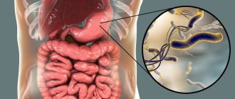
What is Meckel's diverticulum?
Meckel's diverticulum in children is a true diverticulum, or a protrusion from the small intestine at a distance of 20–70 cm from the ileocical angle in the form of a cylindrical or cone-shaped sac, 3–8 cm long and up to 2 cm in diameter. The inner layer of the diverticulum can consist of 2 types ectopic cells, which in their structure will be similar to the mucous surface of the stomach and pancreatic cells.
Pathology of Meckel's diverticulum
Bibliographic description:
Burlakova, E. S. Pathology of Meckel's diverticulum / E. S. Burlakova. — Text: direct // Research of young scientists: materials of the IX International. scientific conf. (Kazan, April 2020). — Kazan: Young scientist, 2021. — pp. 17-19. — URL: https://moluch.ru/conf/stud/archive/368/15726/ (access date: 05.26.2021).
This article discusses the pathology of Meckel's diverticulum and analyzes the complications caused by this disease. There is a relationship between the features of the anatomical structure and the options for surgical removal of Meckel's diverticulum.
Particular attention is paid to the clinical case of surgical treatment of complicated Meckel's diverticulum with strangulating small bowel obstruction.
Key words: diverticulum, obstruction, complication, perforation, laparotomy.
Meckel's diverticulum (MD) was first described by the German anatomist J. F. Meckel (1781–1833). However, this pathology was known earlier - in 1598, an unusual intestinal extension was discovered by Fabricus Hildanus. But it was Meckel who studied it in detail. The scientist publishes several works (1808–1820), in which he describes the appendage in detail and proves that it appeared in the process of abnormal development of the embryo. DM is one of the most common congenital anomalies of the gastrointestinal tract and the cause of a number of severe pathological conditions. From the point of view of embryology, the DM is a remnant of the vitelline-intestinal duct, which connects the yolk sac of the embryo with the primary intestine and normally undergoes reverse development after 6–8 weeks of the prenatal period. DM is one of the earliest formations in ontogenetic terms. From the point of view of anatomical and topographical features, it should be noted that DM is a protrusion of a section of the ileum of various shapes and lengths, which resembles a vermiform appendix located at a distance of 70–100 cm from the ileocecal angle with the same structure of the intestinal wall and its blood supply being preserved. The frequency of pathology ranges from 2–3%, with clinical manifestations and complications of the disease observed in 25% of cases. The length of the process varies from a few millimeters to 15 cm, and the diameter - from 0.5 cm to 5 cm. Its shape is varied: cylindrical, spherical, worm-shaped. In the abdominal cavity, the DM lies freely or is fixed by a fibrous cord to the navel, intestine, mesentery, etc. [1, p. 104]. Histologically, its wall is built in the same way as the wall of the small intestine, but the muscular layer is less pronounced [3, p. 488].
Often, complications develop in children under 10 years of age and in adults over 30 years of age. Diverticulitis is one of the most common complications, observed in approximately 20% of patients with DM. Basically, the complication develops in diverticula with a narrow neck, where food debris accumulates, and later a bacterial infection occurs. There is a possibility of developing diverticulitis as a result of torsion of the diverticulum itself and disruption of its blood supply. [2, p. 74]
Quite often there are ulcerations of the DM, which can cause perforation into the abdominal cavity, thereby causing peritonitis.
Diverticula can contribute to the formation of umbilical pathologies (about 12.5% of cases) - fistulas, cysts, fibrous cords between the diverticulum and the navel. [5, p. 146]
Another dangerous complication of Meckel's diverticulum is intestinal obstruction, which develops due to compression of the intestinal lumen by a diverticulum attached to it. Sometimes the DM fixed to the abdominal wall or to the mesentery forms something like a ring-shaped hernial orifice. As a result of the penetration of intestinal loops through them, internal strangulated hernias can occur.
Intussusception, as a complication, can occur when a diverticulum with a wide base penetrates into the intestinal lumen and, under the influence of peristalsis, moves along with the intestinal chyme, carrying with it the intestine from which it originates.
For patients with intestinal obstruction caused by Meckel's diverticulum, emergency surgery should be considered.
Options for surgical removal of Meckel's diverticulum:
- like a vermiform appendix - with a narrow base of the diverticulum;
– cutting off using a clamp followed by suturing of the ileum with a double-row suture in the transverse direction - in case of a narrow base or inflammation of the diverticulum;
– wedge-shaped excision of the diverticulum between two clamps, followed by suturing of the ileum with a double-row suture - in case of a wide base or inflammation of the diverticulum;
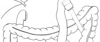
– resection of the ileum with a diverticulum followed by an end-to-end anastomosis - if the intestine is involved in the inflammatory process [4, p. 152].
Patient A., 37 years old, underwent surgery - mid-median laparotomy: elimination of intestinal obstruction, excision of Meckel's diverticulum, nasogastric intubation, sanitation and drainage of the abdominal cavity.
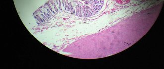
The loops of the small intestine are moderately swollen to 2 diameters, 40–50 cm from the ileocecal angle there is a diverticulum of the small intestine (Meckel), 7 cm long, 3 cm in diameter with a rod pressing on the wall of the ileum; below the rod the intestine is not thickened. The strangulation is cut, the intestinal obstruction is eliminated, the intestine in the area of strangulation is viable. Meckel's diverticulum was excised using clamps (10cm x 3cm), the intestine was sutured with a double-row suture. Sanitation of the abdominal cavity was performed. The patient was discharged after 9 days.
Postoperative care is similar to that for an appendectomy or small bowel anastomosis. Fluid and electrolyte balance is maintained intravenously until the intestines regain their ability to contract. The nasogastric tube is then removed and progressive feeding begins. Any residual inflammation, peritonitis, or drained abscess is treated with appropriate general antibiotics and blood and plasma replacement.
The above clinical case proves that Meckel's diverticulum may be one of the causes of acute intestinal obstruction. Consequently, knowledge of such anatomical anomalies as DM and the principles of its surgical treatment makes it possible to eliminate the complications caused by it.
Literature:
- Noskov A. A., Lazarev S. M., Efimov A. L., Ershova N. B., Chepak D. A. A rare observation of a giant Meckel’s diverticulum // Bulletin of Surgery named after. I. I. Grekova. 2021. No. 1. - pp. 104–105.
- Pimenov I. A., Oganesyan M. V., Merenkova I. V. Meckel’s diverticulum: anatomical features and localization options [Electronic resource] // International Student Scientific Bulletin. 2017. - No. 5. Access mode URL: https://eduherald.ru/pdf/2017/5/17335.pdf (02/14/2020)
- Littman I. Operative surgery: atlas. - Budapest: Publishing House of the Hungarian Academy of Sciences. - 1985. - 1143 p.
- Zhuk I. G. Topographic anatomy and operative surgery: textbook. — Grodno: Grodno State Medical University. - 2012. - 269 p.
- Klimov A. E., Cherepanov D. E. Cases from practice: two cases of complications of Meckel’s diverticulum // Bulletin of RUDN University, Medicine series. — 2003. — No. 3. — P. 146
Key terms
(automatically generated)
: ileum, diverticulum, intestinal obstruction, abdominal cavity, double-row suture, small intestine, subsequent suturing, surgical treatment, appendix, wide base.
Statistics
According to the frequency of congenital diseases in children, Meckel's diverticulum occurs in 2% of cases. The incidence of complications with this developmental anomaly is 5–7% of cases. In half of the cases in children under 2 years of age, diverticulum is symptomatic. When comparing data, the frequency of this developmental anomaly in boys is almost 3 times greater than in girls.
Statistics show that Meckel's diverticulum most often forms in parallel with other developmental anomalies and genetic diseases of the gastrointestinal tract:
- omphalocele – remnants of the vitelline duct (10 times);
- esophageal atresia – lack of connection between the stomach and esophagus (6 times);
- anorectal anomalies (5 times);
- Crohn's disease (3 times);
- various neurological diseases (3 times).
Reasons for the development of Meckel's diverticulum in children
The cause of such a diverticulum is pathological embryonic development. During the 1st trimester of pregnancy, the fetus has a vitelline-intestinal duct passing through the umbilical cord. The main task of this duct is to transfer nutrients from mother to fetus.
Starting from the 3rd trimester of pregnancy, this duct forms into the middle ligament, which is located on the inner surface of the abdominal wall. If this formation is disrupted, the vitelline duct does not completely close, which is why a diverticulum (protrusion) appears, which can connect to the navel with a fibrous cord.
Meckel's diverticulum in children
Meckel's diverticulum in children is an intermittently encountered diverticulum of the lower third of the ileum, a remnant of an incompletely reduced vitelline stalk, in other words, it is a congenital anomaly of the ileum, resulting from a violation of obliteration of the proximal part of the vitelline duct. Meckel's diverticulum in children is dangerous due to its complications: bleeding, intestinal obstruction, inflammation, perforation, strangulation, and tumor processes. To diagnose Meckel's diverticulum in children, radiography of the small intestine with a barium suspension, scintigraphy, ultrasound and CT of the abdominal organs, and laparoscopy are performed. Complicated cases of Meckel's diverticulum in children require surgical tactics - resection of the diverticulum or section of the intestine. Symptoms of Meckel's diverticulum in children
Symptoms of Meckel's diverticulum always serve as signs of its complications. The following complications of Meckel's diverticulum are observed in children: bleeding, intestinal obstruction, inflammation, tumors.
Bleeding is the most common clinical manifestation of Meckel's diverticulum in children under 4 years of age, accounting for up to 22% of all children with this intestinal anomaly.
Painless rectal bleeding is typical of Meckel's diverticulum, which appears as melena and is characterized by tarry, black stools. The color of the bloody discharge can vary from bright red to black, tarry or in the form of “currant jelly”. Hidden bleeding is very rare; on the contrary, it can be massive, accompanied by clinical and laboratory signs of anemia.
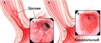
The mechanism of bleeding from Meckel's diverticulum is associated with its peptic ulceration due to the secretion of hydrochloric acid by the cells of the ectopic gastric mucosa. The ileum, unlike the stomach, is not able to weaken the effect of hydrochloric acid, which leads to ulceration. The typical localization of such ulcers is at the base of the diverticulum at the border of the ectopic gastric mucosa and the normal ileal mucosa.
Intestinal obstruction is the second most common complication of Meckel's diverticulum.
Uncomplicated Meckel's diverticulum in children is asymptomatic and may be an incidental finding during laparotomy for another disease or may not be recognized. The clinical manifestation of Meckel's diverticulum in children is usually associated with the development of complications: intestinal bleeding, inflammation (diverticulitis), intestinal obstruction (intussusception, strangulation), tumors. Bleeding from a peptic ulcer, as the most common complication of Meckel's diverticulum in children, can be acute, profuse or chronic, hidden. A sign of intestinal bleeding with Meckel's diverticulum in children is tarry, black stool. Bleeding is accompanied by general weakness, dizziness, tachycardia, pallor, and clinical and laboratory signs of posthemorrhagic anemia. Unlike bleeding from esophageal varices or gastric and duodenal ulcers, children with complicated Meckel's diverticulum never experience "coffee-ground" vomiting. The clinical picture of acute diverticulitis, complicating Meckel's diverticulum in children, resembles the symptoms of acute appendicitis. The child has abdominal pain (near the navel or in the right iliac region), nausea, fever, leukocytosis, and a positive Shchetkin-Blumberg sign. Usually, the correct diagnosis is established intraoperatively, when an intact appendix is identified, and inspection of the ileum reveals the presence of an inflamed Meckel's diverticulum in the child. Inflammation and ulceration of Meckel's diverticulum in children can cause its perforation into the free abdominal cavity with the development of peritonitis. Intestinal obstruction caused by Meckel's diverticulum in children is accompanied by nausea, vomiting, cramping abdominal pain, and increasing intoxication. Obstruction can be caused by intussusception, torsion of ileal loops around a diverticulum, or strangulation of intestinal loops. Sometimes in children, Meckel's diverticulum becomes strangulated in the hernial sac of an inguinal or femoral hernia (Litre's hernia). If a hernia is strangulated, there is a sharp pain, tension and uncontrollability of the hernial protrusion, and the absence of a symptom of a cough impulse. Less often than other complications, tumors of Meckel's diverticulum, both benign (hamartomas, fibroids, lipomas) and malignant (adenocarcinomas, carcinoids), occur in children. Clinical manifestations of Meckel's diverticulum tumors in children may be associated with intestinal obstruction, wall perforation, and bleeding.
Treatment of Meckel's diverticulum in children
There is no consensus regarding asymptomatic Meckel's diverticula in children. Some pediatric surgeons believe that an unchanged diverticulum accidentally discovered during surgery should not be removed, and that asymptomatic diverticula should not be removed; others insist on its mandatory removal in a favorable surgical situation, that Meckel's diverticulum must be removed in case of diverticulitis, diverticulum ulcers, intestinal obstruction caused by a diverticulum, umbilical fistulas, and also, according to a number of experts, if it is accidentally discovered during surgery: the diverticulum is resected with suturing of the intestinal wall (resectio diverticuli Meckelii).
Meckel's diverticulum in children, complicated by inflammation, perforation, bleeding, intestinal obstruction, strangulation, clearly requires urgent surgical intervention. In this case, the child can undergo excision of the diverticulum of the small intestine (diverticulectomy) or segmental resection of the small intestine with end-to-end enteroenteroanastomosis. In pediatric surgery, preference is given to endoscopic resection of the small intestine.
The method of choice for treating diverticulitis in children is conservative drug therapy: antibiotic infusions, injections of anti-inflammatory drugs. With the recurrent nature of inflammation of Meckel's diverticulum in children, the issue of resection of the diverticulum is resolved. With the development of peritonitis, in addition to resection of the small intestine, it is necessary to carry out drainage and sanitation of the abdominal cavity, prescribe massive antibiotic therapy, infusion and detoxification therapy.
Diagnostics
Scintigraphy , a radionuclide isotope method, is the primary method for diagnosing Meckel's diverticulum in children complicated by bleeding. This method is based on radioactive isotopes that are attracted to and collected from ectopic cells located in the gastric mucosa. If scintigraphy results are negative and bleeding continues, diagnostic laparoscopy should be performed.
With this research method, the results can be false positive and false negative. The presence of a duodenal ulcer, intestinal duplication and vascular tumor means the result is false positive. The minimum content of ectopic gastric mucosa in the diverticulum means the result is false negative.
X-ray is the best way to detect intestinal obstruction and diverticulum perforation. This method is performed using a contrast agent (barium suspension).
Treatment methods and prevention
There are not many methods for treating Meckel's diverticulum; one can say that this is surgical removal of the diverticulum. But there are many contradictions in the opinion of doctors regarding the removal of uncomplicated diverticulum. Such a diverticulum can be detected during various operations on the abdominal organs. And then the question arises: “To delete or not?”
There are many opinions and studies that Meckel's diverticulum is removed in all cases. A comparative analysis was carried out, comparing patients with complicated and uncomplicated diverticulum. This analysis found that children with complicated diverticula had much larger diverticulums. Using this analysis, it can be determined that the lifetime risk of developing complications is 6% for all ages.
There are 2 types of surgical treatment for Meckel's diverticulum.
- A median laparotomy is performed. This approach is used when the length of the diverticulum is less than 3 cm and it has a wide base. The diverticulum is resected along with part of the small intestine, followed by enteroanastomosis. This method avoids leaving part of the ectopic gastric mucosa, which can cause postoperative complications.
- Laparoscopy. Less invasive method for removing diverticulum. Indications for this method are a diverticulum length of more than 5 cm and a narrow base.
In surgical practice, there are 2 methods of resection of Meckel's Diverticulum using the laparoscopic method.
- Place a ligature at the base of the diverticulum if there are no changes (tumor) there.
- Use the endoscopic stapler ENDO-GIA 30 with a 12 mm trocar.
When using these 2 methods, the operation time is reduced and the recovery period is faster. For preventive purposes, children can undergo physical therapy and tummy massage. Avoid cases of constipation in children; this prevention will help avoid dangerous complications.
Meckel's diverticulum
Signs
Meckel's diverticulum is often asymptomatic.
It is often discovered incidentally during diagnostic testing. But if the diverticulum is damaged in any way, intestinal bleeding, intestinal obstruction, or diverticulitis may develop. With intestinal bleeding, the stool may be mixed with bright red blood if the bleeding is active. If the bleeding is minor, the stool will be black and sticky. With intussusception, the stool resembles currant jelly in color and consistency. The patient is pale and weak due to blood loss.
With intestinal obstruction, the patient complains of constipation, vomiting, and abdominal pain.
With diverticulitis, the main symptoms of the disease are pain in the navel, fever, nausea, vomiting, and constipation.
Description
Meckel's diverticulum was described by the German anatomist Johann Friedrich Meckel (1781-1833). However, this pathology was known long before him. Back in 1598, Fabricus Hildanus discovered an unusual intestinal extension. But it was Meckel who studied this process in detail. From 1808 to 1820, he published several works in which the scientist described this process in detail and proved that it appeared precisely in the process of improper development of the embryo.
Meckel's diverticulum is found in 2% of the population. However, for many it is discovered only at autopsy. Men suffer from this pathology three times more often than women. The walls of this process are the same as the walls of the intestine, but may also include ectopic tissue of the stomach, pancreas, and colonic epithelium. The length of Meckel's diverticulum is from 1 to 12 cm, but more often no more than 5 cm. The diverticulum is often connected to the umbilicus by a fibrous cord.
A diverticulum itself is not dangerous, but it can cause diseases that require immediate surgical intervention.
Most often, complications develop in children aged about 10 years and in adults over 30 years old. One of the most common complications, diverticulitis (inflammation of the diverticulum), occurs in approximately 20% of patients with Meckel's diverticulum. Most often, these are older people. The complication develops mainly in diverticula with a narrow neck. It is in such processes that food debris accumulates, and later a bacterial infection occurs.
Diverticulitis can also develop as a result of torsion of the diverticulum and disruption of its blood supply. Inflammation can be localized in the diverticulum itself, or it can spread to other abdominal organs. In children, the clinical picture of Meckel's diverticulitis is often similar to appendicitis.
Ulceration of Meckel's diverticulum also occurs frequently (approximately 40% of cases); it, like inflammation, can cause perforation of the diverticulum. Perforation occurs into the abdominal cavity, causing peritonitis.
Diverticula can cause umbilical pathologies (approximately 12%) - cysts, fistulas, fibrous cords between the diverticulum and the navel. In the case of a fistula, intestinal mucus is released onto the skin in the navel area, causing irritation.
Intestinal obstruction (25%) can also be a complication of Meckel's diverticulum. This can occur as a result of intussusception (the insertion of one part of the intestine into another). Then the patient complains of vomiting, abdominal pain and a tumor-like formation, which can be detected by palpation of the abdominal cavity.
Rarely (3%), neoplasms can be a complication of ileal diverticulum. They can be benign (lipoma, hamartoma - a nodular tumor-like formation, consisting of the same layers as the organ in which it was formed, but differing in their irregular location) or malignant (adenocarcinoma). Tumors may present with intestinal obstruction, perforation, or bleeding.
Diagnostics
The diagnosis is made by a gastroenterologist. One examination is not enough; instrumental studies must be carried out:
- magnetic resonance imaging;
- radioisotope scanning using technetium Tc 99m (used for bleeding);
- angiography of mesenteric vessels (informative only with significant blood loss).
If the radioisotope scan results are negative and bleeding continues, diagnostic laparoscopy is performed.
Ultrasound examination (ultrasound) for Meckel's diverticulum is informative only in children and only in 50% of cases.
Treatment
Treatment of Meckel's diverticulum is surgical. An accidentally discovered shoot is removed if:
- patient over 40 years old;
- diverticulum length more than 2 cm;
- the diverticulum is inflamed;
- its wall is thinned;
- there are fibrous cords to the navel;
- The diverticulum has a narrow neck.
The diverticulum is also removed in patients suffering from Crohn's disease, peritonitis and ulcerative colitis, and in cases where it caused an acute abdominal disease.
During surgery, either just the diverticulum (diverticulectomy) or the diverticulum and part of the ileum can be removed. In this case, either laparotomy or laparoscopy is used. Laparoscopy is now more common, as it is a minimally invasive method with few complications and a short rehabilitation period.
With timely surgical intervention, the prognosis is favorable, but if, despite the dire symptoms, you delay visiting a doctor, death is possible.
© Dr. Peter