Diverticular disease of the intestine (diverticulosis) - symptoms and treatment
A diverticulum is a hernia-like protrusion that can form in the wall of any hollow digestive organ, from the esophagus to the small and large intestines [1]. In this article we will talk about colon diverticula, as they often lead patients to a surgeon and coloproctologist.
Colon diverticula can be either single or multiple.
A condition in which there are many non-inflamed diverticula in the colon is called diverticulosis [9][19]. Typically, patients with this disorder are not bothered by anything. They learn about their disease only after a colonoscopy performed for another disease [1][9]. Sometimes diverticulosis is accompanied by pain and bloating, changes in bowel movements and other symptoms.
When diverticula begin to become inflamed, we are talking about diverticular disease of the intestine . It develops in 20% of people with diverticulosis. 75% of them develop acute diverticulitis, which is accompanied by pain in the left lower abdomen, constipation and fever, and 25% develop complications such as diverticulum perforation, abdominal abscess, peritonitis, intestinal stenosis, internal and external fistulas or bleeding from a diverticulum [8].
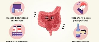
Previously, the main cause of colon diverticula was considered to be a deficiency of plant fiber, because a regular lack of vegetables and fruits in the diet leads to the development of constipation and increases pressure in the intestinal lumen [1][7][9][10]. But thanks to research, it has become known that the real causes of the formation of diverticula are disorders in the genes that encode the development of neuromuscular and connective tissue. These genetic changes disrupt the structure and contractile activity of the intestinal wall. In fact, “weak” spots are formed in which diverticula appear [1][2][12]. And fiber deficiency is one of the main risk factors for the development of diverticulosis.
Other risk factors include smoking and alcohol abuse. By avoiding these factors and consuming at least 25-30 g of vegetables and fruits per day, we reduce the risk of developing diverticular disease [3].
Diverticulosis can be diagnosed at any age:
- the proportion of patients under 40 years of age is 5%;
- 40-50 years - 5-10%;
- 50-60 years old - 14%;
- 60-80 years old - 30%;
- over 80 years old - 60-65% [1][2][7][9][19].
Statistics show that diverticula are more often found in older people. But this does not mean that the disease appears with age. It’s just that older people are more likely to undergo emergency operations for peritonitis, and are prescribed an abdominal ultrasound or colonoscopy, for example, if constipation develops. It is possible that these same people could have developed diverticula in their youth.
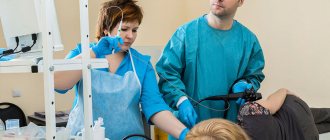
A case of ultrasound diagnosis of rectal cancer
Ultrasound machine HS40
Top seller in high class.
21.5″ high-definition monitor, advanced cardio package (Strain+, Stress Echo), expert capabilities for 3D ultrasound in obstetrics and gynecology practice (STIC, Crystal Vue, 5D Follicle), high-density sensors.
Introduction
Colorectal cancer ranks 4th in frequency among malignant neoplasms in the world. Every year, 950 thousand people in the world fall ill with this form of cancer, of which 500 thousand die. High incidence rates are observed in European and North American countries. In Russia, 46 thousand newly diagnosed patients with colorectal cancer are registered every year, and from 20 to 50% of patients are already at the stage of distant metastases. In terms of mortality, colorectal cancer ranks second after lung cancer, ahead of malignant tumors of the stomach, prostate and other localizations [1, 2]. Statistics show that rectal and anal cancer accounts for up to 10% of all cancer sites, and in different countries its frequency varies significantly - from 1.5 to 17.7 per 100 thousand population.
The survival rate of patients with colorectal cancer directly depends on the stage of the disease at the time of its discovery. Unfortunately, in 25% of patients an incurable disseminated form of the disease is detected. In 75%, colorectal cancer is curable, however, 50% of these patients die within 5 years due to tumor recurrence. All this dictates the need to develop new principles and approaches to the diagnosis of intestinal cancer.
Methods for diagnosing (including screening) for rectal cancer include digital rectal examination, stool occult blood test, anoscopy, sigmoidoscopy and colonoscopy with targeted biopsy. There is also data in the literature on the possibility of using ultrasound diagnostics through transabdominal and transrectal approaches to recognize this disease [3]. However, we did not find any descriptions of cases of visualization of rectal cancer during transvaginal ultrasound examination (TVUS).
Here is an observation
Patient S., 48 years old, was referred by a gynecologist for a transvaginal ultrasound as part of an annual clinical examination. There is a history of supravaginal amputation of the uterus without appendages due to multiple fibroids.
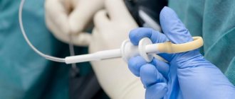
Ultrasound, performed according to the generally accepted method with a cavity sensor with a frequency of 7 MHz, visualized a cervical stump with smooth, clear contours measuring 5.2x5.3x3.7 cm. The structure of the tissue is heterogeneous, with the presence of cysts ranging in size from 0.4x0.4 to 0.7x1 cm The cervical canal is not dilated. The endocervix is not thickened. The right ovary is located typically, its size is 3.5x1.9x3.5 cm. The contours are clear and even. The structure reveals two avascular liquid formations up to 1 cm in size with thin walls and homogeneous content. The left ovary is located typically, its size is 2.8x1.6x2.4 cm. The contours are clear and even. The structure is moderately heterogeneous, follicles are not identified. No fluid was detected in the retrouterine space.
Adjacent to the posterior wall of the cervical stump is a section of the rectum up to 2.9 cm thick over a length of up to 7.4 cm. The intestinal wall is unevenly thickened to 0.5-0.6 cm, reduced echogenicity, it is not possible to differentiate the layers of the wall. Ultrasound angiography in the changed area reveals pronounced vascularization with the presence of vessels with the arterial type of spectrum (Vmax 14.4 cm/s, RI 0.74). There is no intestinal peristalsis in this segment, the internal lumen is not differentiated. To the left of the described section of the intestine in the interintestinal space, a hypoechoic formation with clear, even contours, a heterogeneous structure, measuring 2.6x1.9x2.3 cm is determined. Ultrasound angiography reveals multiple deformed vessels in it (Fig. 1, a, b, c).
Rice. 1.
Transvaginal ultrasound.
A)
B-mode. In the pelvic cavity, posterior to the uterine stump, a section of intestine measuring up to 6.8x2.9x7.4 cm with a wall thickened to 0.5-0.6 cm and reduced echogenicity is determined. There is no intestinal peristalsis in this projection, the internal lumen is not clearly differentiated. The contents of the intestine are practically not determined, the lumen is closed.
b)
Energy mapping mode.
V)
Pulse Doppler mode. Ultrasound angiography reveals pronounced vascularization in the wall with the presence of vessels with the arterial type of spectrum (Vmax 14.4 cm/s, RI 0.74).
Conclusion. Condition after supravaginal amputation of the uterus without appendages. Cysts of the right ovary. Ultrasound signs of a rectal tumor, pelvic mass (changed enlarged lymph node?).
Upon detailed questioning of the patient, it turned out that she had been suffering from constipation for more than 20 years. Over the past 6 months, she periodically experienced pain in the lower abdomen, mucus in the stool, and an increase in weakness.
To clarify the diagnosis, magnetic resonance imaging of the pelvic organs was performed. On a series of tomograms obtained in the projection of the distal parts of the sigmoid and proximal parts of the rectum, an area of an altered magnetic resonance (MR) signal is determined, measuring 5.5x4.2x5.5 cm - a suspicion of a space-occupying mass in the intestine. The external contours of the changes are unclear and uneven. The structure is heterogeneous, with the presence of solid and liquid components. The wall of the distal parts of the rectum is circularly thickened to 8 mm. The changes have a volumetric effect on the uterine stump (deviated anteriorly and to the right) and adjacent intestinal loops (unevenly dilated, filled with contents, liquid and gas). Along the course of the iliac vessels, multiple lymph nodes measuring 6-13 mm are identified. There is a change in the MR signal from the lower lumbar, sacral spine and pelvic bones - the MR signal is heterogeneously hypointense on T1-WI and mixed hypohyperdense MR signal on T2-WI.
Uterus: condition after supravaginal amputation. Cervical stump measuring 44x37 mm. The MR signal from the cervical canal is somewhat heterogeneous, the canal is widened to 5 mm, and the presence of a small polyp (3 mm) along the anterior wall cannot be excluded. In the structure of the cervix, single cysts of the nabothian glands of 3-6 mm are detected. In the projection of the right ovary there is a cyst-like formation measuring 16x17 mm, with clear, even contours. The left ovary is not clearly visualized against the background of intestinal loops. Free fluid in the pelvic cavity is not determined (Fig. 2-4).
Rice. 2.
Magnetic resonance imaging (MRI). T2-weighted image, sagittal projection. Tumor of the sigmoid colon (light long arrows). The MR signal is heterogeneously increased on T2-weighted images, the contours of the formation are lumpy. The uterine stump with a cyst (light dotted arrow) is deviated anteriorly due to the formation.
Rice. 3.
Magnetic resonance imaging (MRI). T2-weighted image, frontal projection. Tumor of the sigmoid colon (light long arrows). The MR signal from the tumor is heterogeneous, the contours of the changes are uneven and lumpy. Above the tumor, the intestinal lumen is unevenly expanded.
Rice. 4.
Magnetic resonance imaging (MRI).
A)
T2-weighted image, axial projection.
b)
T2-weighted image, axial projection.
V)
T1-weighted image, axial projection.
G)
FS, axial view.
Tumor of the sigmoid colon (light long arrows). The uterine stump pushed anteriorly and to the right (light dotted arrows) with a cyst (a). Infiltration of pararectal tissue (dark long arrows), against this background mts into the pararectal lymph node on the left (dark short arrow; b, c) and pararectal lymph nodes along the posterior contour of the intestine (light short arrow; d).
Conclusion. Suspicion of a mass formation in the distal parts of the sigmoid colon (with spread to the proximal parts of the rectum; manifestations of moderate lymphadenopathy). Suspicion of secondary changes in bone structures at the studied levels. Condition after supravaginal amputation. Cysts of the uterus of the nabothian glands. Suspicion of a polyp of the cervical canal. Cyst-like formation of the right ovary.
To clarify the diagnosis, a colonoscopy was performed: examination at 50 cm, but it was not possible to go further due to a sharp bend in the intestine. There is a lot of mucus on the walls, the folds are thickened and elastic. The mucous membrane is swollen and pink. The vascular pattern is enhanced. In an area of 15 cm, a large (5-7 cm) exophytic lobular tumor of a bluish color is visible. During biopsy, the tissue is dense and bleeding is increased.
Conclusion. Tumor of the rectosigmoid angle.
Subsequent histological examination confirmed the presence of well-differentiated dark cell adenocarcinoma with foci of necrosis and active inflammation.
The patient was hospitalized for surgical treatment, during which an anterior resection of the rectum was performed with end-to-end sigmoidal anastomosis. According to a histological examination of a 16 cm long section of the colon, a mushroom-shaped tumor with a diameter of 3.5 cm was identified in the distal part. Microscopically, a well-differentiated papillary mucus-forming adenocarcinoma with focal invasion into the muscle layer was determined. In some areas the tumor had the structure of a tubular adenoma. Cancer metastases were detected in 4 out of 9 lymph nodes. Resection was performed outside the tumor growth.
Clinical diagnosis: stage III rectal cancer T2N2M0.
Subsequently, the patient feels well and is under the supervision of an oncologist and gastroenterologist.
During the control TVUS performed 1 year after surgery (Fig. 5, a, b), no signs of recurrence of the rectal tumor, damage to the lymph nodes, or progression of changes in the cervix and appendages were detected.
Rice. 5.
Transvaginal ultrasound.
A)
B-mode.
b)
Energy mapping mode.
Pathological changes in the pelvic cavity are not clearly defined.
Conclusion
Ultrasound examination of the colon and rectum via transabdominal access is becoming increasingly widespread in clinical practice [4-7], however, it has a number of limitations in visualizing the affected part of the intestine, especially if the patient is insufficiently prepared. Transvaginal examination in rectal imaging does not require changing the patient's position, is not so dependent on flatulence, and at the same time allows one to obtain better images of the wall. The presented observation indicates the need, when conducting standard abdominal examinations of the genital organs in women, to pay attention to and describe not only the organ under study (uterus, ovaries), but also changes in the adjacent sigmoid and rectum, which makes it possible to diagnose them, as well as concomitant neoplastic pathology.
Literature
- Makaneni D. Diseases of the rectum and anal canal // General medical practice / Ed. Nobel J. Per from English. M.: Praktika 2005. pp. 986-992.
- Rivkin V.L., Bronshtein A.S., Fain S.N. Rectal cancer // Guide to coloproctology. M.: Medical practice. 2001. pp. 219-265.
- Lemeshko Z.A. Ultrasound transabdominal examination of the intestine // Clinical guide to ultrasound diagnostics / Ed. Mitkova V.V. Volume 4. M.: Vidar 1997. P. 49-81.
- Valette P.-J., Rioux M., Pilleul F., Saurin J.-S., Fouque P., Henry L. Ultrasonography of chronic inflammatory bowel diseases // Ultrasound. Categorical Course ECR. 2002. P. 205-212.
- Burkov S.G., Kokova N.I., Chistov L.V. and others. A case of ultrasound diagnosis of leiomyoma of the appendix // Sonoace International. 1999. N5. pp. 14-16.
- Burkov S.G., Burdina E.G., Gurova N.Yu. and others. A case of ultrasound diagnosis of jejunal cancer // Sonoace International. 2000. N7. pp. 11-13.
- Pimanov S.I., Vergesova E.V., Lud N.G. Screening ultrasound diagnosis of colon cancer // Sonoace International. 2002. N10. pp. 5-11.
Ultrasound machine HS40
Top seller in high class.
21.5″ high-definition monitor, advanced cardio package (Strain+, Stress Echo), expert capabilities for 3D ultrasound in obstetrics and gynecology practice (STIC, Crystal Vue, 5D Follicle), high-density sensors.