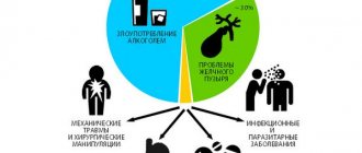
Recently, the incidence of pancreatitis has increased significantly. This is due to the spread of fast food and the abuse of low quality alcohol. The peak incidence occurs in active working age from 30 to 60 years.
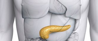
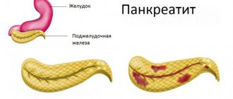
Acute pancreatitis is an inflammatory process that initially occurs in the pancreas and subsequently spreads to surrounding tissues and organs. It is difficult to diagnose early onset, therefore, despite modern medicine, the mortality rate with this diagnosis remains high.
To avoid serious consequences, we will consider in detail what acute pancreatitis is, its symptoms and treatment, and also tell you what preventive measures will help prevent its development.
Causes
Acute pancreatitis is recorded in adults 30-40 years old. The male population is more at risk than the female population. The incidence of the form is higher in people who abuse alcoholic beverages and suffer from pathologies of the biliary tract, such as:
- dyskinesia of the bile duct of the hypertensive type;
- cholecystitis in chronic or acute form;
- cholelithiasis.
Other causes of pancreatitis:
- hypertriglyceridemia;
- disruptions in the circulatory system of glandular tissue;
- cystic fibrosis;
- hemolytic-uremic syndrome;
- hyperparathyroidism;
- abdominal injuries;
- heredity;
- autoimmune diseases;
- blockage of the pancreatic or common ducts;
- damage to the canals and gland during surgery;
- uncontrolled use of drugs;
- consequences of severe acute respiratory viral infections, mumps, mycoplasmosis, pneumonia, hepatitis;
- various gastrointestinal diseases.
Acute pancreatitis can occur in two forms:
- mild – organs and systems are slightly affected. The disease responds well to treatment, recovery occurs quickly;
- severe - pronounced disorders are observed in tissues and organs, tissue necrosis, abscesses and cysts cannot be excluded.
The clinical picture of this disease in severe form may also be accompanied by:
- there is an accumulation of fluid inside the gland;
- tissue infection and necrosis;
- false cyst;
- pus accumulates in the gland or on tissues adjacent to it.
List of recommended products
The main condition for a disease such as pancreatitis of the pancreas is diet. This is the main principle of treatment. What is the essence of the diet? Use only those foods and dishes that will not irritate the mucous membrane of the pancreas. The following products can and should be used by people suffering from this type of inflammation.
- Wheat bread is stale, yesterday's bread.
- Soup made with recycled chicken or beef broth.
- Meat: chicken, veal, turkey. Method of preparation: boil, bake in the oven. The meat should not contain any seasonings.
- Fish, steamed, boiled or baked in the oven.
- Dairy products with a small percentage of fat.
- Boiled vegetables. If you want it raw, then only in grated form.
- Various types of pasta.
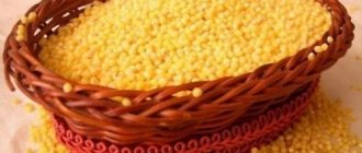
- Cereals (buckwheat, rice, oatmeal).
- Baked fruits (meaning apples and pears).
- Jelly.
- Compotes, jelly, weak tea.
- Primary meat or fish broths. That is, fatty, high-calorie.
- Millet should not be consumed from cereals.
- Fatty meats, fish, poultry.
- Among vegetables, radishes, radishes, cabbage, sorrel and spinach are taboo.
- Fresh bread or any sweet products.
- Various types of sausages, canned food.
- Alcoholic drinks.
- Ice cream.
- Strong tea, coffee.
Consumption of the above products will lead to a negative outcome, which is called “inflammation of the pancreas” (pancreatitis, simply put). In order not to play roulette with your health, remember the foods that you should not eat if you have this disease. After all, following a diet is already 60% of the positive outcome of the disease.
Development mechanism
When the pancreas functions normally, the enzymes it produces are secreted into the lumen of the duodenum and are activated under the influence of certain permissive factors. This is how the physiological process of digestion occurs - the breakdown of proteins, fats and carbohydrates into simpler components.
However, for a number of reasons described above, enzyme activation can begin within the gland itself. Lysis of its tissues occurs, followed by their death, swelling and compression of the gland tissue by intercellular fluid, spasm of the vasculature and impaired circulation in the organ. The large pancreatic duct becomes blocked. Pancreatic juice does not find its usual outlet, it stagnates and the aggression of digestive enzymes against glandular tissue increases.
The pancreas increases in size, and aseptic (non-infectious) inflammation first develops in it. There is an effusion of fluid saturated with active enzymes into the abdominal cavity, the visceral (covering the abdominal organs) and parietal peritoneum are irritated. Nerve endings are compressed and pain receptors, which the peritoneum is rich in, are irritated. First, the pain occurs directly in the projection of the pancreas itself - to the left of the navel with a return to the lower back. Then the whole stomach hurts, peritonitis develops.
Excess enzymes and necrosis products are absorbed into the vascular bed, intoxication develops, the temperature rises, the pulse quickens, and blood pressure decreases. The patient experiences toxic-pain shock. Microorganisms (Escherichia coli, clostridia, staphylococcus, Proteus, etc.) enter the area of inflammation through the lymphatic tract from the intestine. Peritonitis becomes purulent and is extremely difficult to treat; mortality at this stage reaches 70%.
In surgical practice, acute postoperative pancreatitis (APP) is one of the most dangerous and common complications after surgery on the abdominal and retroperitoneal organs [15]. Mortality when AKI occurs varies from 19.5 to 80% and depends on the severity of the process. In destructive forms of AKI, the probability of death reaches 100% [23, 28, 32].
Assessment of the incidence of AKI is controversial, since some surgeons consider a deviation from the norm in laboratory parameters (such as increased levels of urine diastase and blood amylase) to be sufficient to establish this complication, while others take into account only its severe forms with a clear clinical picture. In addition, the duration of the postoperative period in each specific case is not precisely determined [30].
That is why the incidence of AKI, according to various sources [15, 17, 32, 40, 74], varies widely from 0.08 to 100%.
Most often, AKI develops after surgical interventions on the pancreas (PG) itself and, according to various authors [11, 15, 34, 40, 67], ranges from 1.9 to 100%.
In addition, a high incidence of this complication is observed after operations on organs that have an anatomical and functional connection with the pancreas. After gastric resection for duodenal ulcer, AKI was noted in 7.9 -31.4% of patients, after surgery for gastric cancer - 1.8 - 21.8% of cases [37, 39, 62]. Duodenoplasty (DP) is complicated by AKI with an incidence of 12–35% [6, 7], and operations on the gallbladder and extrahepatic bile ducts lead to AKI in 0.2–9.6% of patients [15, 26, 40, 55]. Laparoscopic cholecystectomy is accompanied by the development of AKI in no more than 1.8% of cases [2, 55].
Surgical interventions with the application of a biliodigestive anastomosis, endoscopic retrograde cholangiopancreatography (ERCP) and associated medical procedures lead to AKI in 0.5-40.5% of patients [19, 32, 58, 71].
In the work of T. White et al. [74] indicated that in 10 out of 70 patients they observed, AKI developed when direct pancreatic injury was excluded, i.e. after orthopedic and vascular operations. According to L. Hashimoto et al. [59] and G. Sakorafas et al. [70], cardiac surgery and operations on large vessels are accompanied by the development of AKI in 0.06-27% of cases. In addition, cases of AKI have been described after excision of neck melanosarcoma, strumectomy, pneumonectomy, cystostomy, cesarean section, hemorrhoidectomy, cataract removal, radical mastectomy, tonsillectomy, and hip replacement [60, 64]. A case of the development of severe pancreatitis in a patient who underwent lithotripsy for urolithiasis was described [48].
The given figures indicate, first of all, the high prevalence of this postoperative complication, the relevance of the issue of its diagnosis, treatment and prevention.
Etiology
AKI is a multi-etiological disease. There are many mechanisms of damage to the pancreatic parenchyma that can lead to the development of this complication, alone or in combination. I.V. Maev, Yu.A. Kucheryaev [22] highlight the most significant of them:
— direct trauma to the pancreas during mobilization of the stomach, splenectomy, etc.;
— duodenal hypertension associated with the characteristics of the intervention or the course of the postoperative period;
- biliary hypertension that occurs during operations on the biliary tract;
— pancreatic ischemia due to post-traumatic systemic and regional circulatory and microcirculation disorders;
— hyperstimulation of the exocrine apparatus of the pancreas after prolonged fasting.
Many authors [9, 27, 41] consider organ trauma during surgical procedures to be the main cause of postoperative complications. As a result, tissue ischemia, stasis and hypertension of the ductal system, leakage and action of pancreatic juice on the wound surface occur [41]. A.A. Shelagurov in 1970 [45] formulated a “traumatic theory” of the occurrence of AKI, according to which, when pancreatic tissue is damaged, a cytokine is released that activates pancreatic enzymes.
However, cases have been described when surgery was performed outside the abdominal cavity, and there was no pancreatic injury, and yet pancreatitis developed in the postoperative period [3]. The etiopathogenesis of AKI in such situations is associated with impaired microcirculation, which occurs as a result of extravascular and transcapillary translocation of fluid, platelet aggregation, thrombosis of small vessels of the pancreas, leading to its ischemia. Massive blood loss with transfusion of large doses of canned blood can also be an important factor [71].
Damage to the acinar tissue of the pancreas leads to the effusion of pancreatic juice, however, enterokinase is required to activate autolysis processes, i.e. contact of juice with bile and intestinal contents, which is not always the case. Therefore, according to modern ideas about the etiology of AKI, in addition to trauma to the pancreatic parenchyma, the presence of pancreatic hypertension is considered a necessary condition for its occurrence, since under these conditions intestinal contents and bile are refluxed into the pancreatic ducts, which leads to the activation of pancreatic enzymes [18, 26, 61, 67 ]. It follows that duodenal hypertension is also an important etiological factor in the development of AKI.
Another etiological factor is considered to be an increase in pressure in the pancreatic ductal system and extrahepatic bile ducts due to post-traumatic edema of the major duodenal papilla (MDPD), which occurs due to its reflex reactions during operations on the biliary tract [18, 26, 63]. This is to a certain extent confirmed by the fact that in the majority of patients, AKI develops after operations on the extrahepatic bile ducts, especially after direct interventions on the bile duct (during its bougienage, papillosphincteroplasty), forced probing of the bile ducts with hard metal instruments, often with the formation of a “false tract” ", difficulties in removing stones fixed in the BSDPC [19, 46].
According to some authors [15, 18, 70], one of the main causes of AKI is pancreatic ischemia due to systemic or regional circulatory disorders that occurs during surgery or in the early postoperative period. E.S. Katanov [15] in the etiology of the ischemic form of AKI considers the most significant factors to be disturbances of the systemic circulation, reflex disorders of organ hemodynamics and disturbances of blood flow in the main arterial and venous vessels during their ligation, etc.
There is also an opinion that the cause of the development of AKI may be an infectious agent that enters the gland through direct intraoperative contact [30]. In clinical medicine, the results of the work of M.N. are important. Molodenkova [23], N.N. Korenevich [16], who experimentally showed the possibility of developing rapidly occurring hemorrhagic pancreatic necrosis under the influence of ovalbumin and Escherichia coli
and
Salmonella abortis
- microorganisms that are often found in bile.
According to the “allergic theory”, AKI develops as a result of sensitization of the body by exo- and endogenous factors, which leads to damage to acinar cells and the entry of antigens into the bloodstream with the subsequent formation of antibodies to them [16]. The validity of the “allergic theory” is confirmed by data [51] on an increase in the level of immune complexes circulating in the blood, a decrease in the absolute and percentage content of T-active and T-total lymphocytes.
And yet, the leading role in the genesis of AKI is played by the initial state of the pancreas, the readiness for the development of inflammatory and destructive processes in it in the early postoperative period. According to many authors [38, 46], from 7.1 to 63.8% of patients who developed this complication suffered from chronic pancreatitis.
The etiological factors listed above trigger the processes of activation of pancreatic enzymes and autocatalytic reactions, determining the further pathogenesis of the development of AKI, which is no different from that in any other form of acute pancreatitis.
Diagnostics
Several factors should be highlighted that greatly complicate the diagnosis of AKI. Firstly, the clinical manifestations of AKI develop against the background of the patient’s initially severe general condition, caused by concomitant diseases, as well as the severity of the surgical intervention itself [26]. Secondly, the clinical picture of this complication manifests itself against the background of symptoms of the postoperative period and is smoothed out by the therapy, including painkillers. Thirdly, in 25-30% of patients the severity of the condition is regarded as a manifestation of another postoperative complication [42]. Often the disease occurs under the guise of intestinal paresis, anastomositis, residual effects of peritonitis, etc. [27]
AKI very often leads to severe impairment of the motor function of the duodenum, as evidenced by dull pain in the epigastric region or left hypochondrium that is poorly controlled by conventional analgesics, accompanied by persistent nausea and repeated vomiting of gastric contents mixed with bile.
Very characteristic of pancreatitis is swelling in the epigastric region, which does not decrease with decompression and gastric lavage - Gobier's symptom. This is associated with isolated paresis of the transverse colon and is detected by plain radiography of the abdominal organs.
Percussion reveals tympanitis, especially high in the epigastrium and in the projection of the lateral canals of the abdomen. Dullness may be detected in sloping areas of the abdominal cavity, which varies depending on the position of the patient’s body (free fluid in the abdominal cavity). At the same time, exudate begins to be released through the drainages, often hemorrhagic in nature, with high amylase values.
The most common, simple and cheap biochemical method for diagnosing AKI is the determination of amylase activity in the blood and urine. However, the sensitivity and specificity of this method range from 30 to 92% and from 20 to 60%, respectively [22]. In addition, to date there is no consensus regarding the threshold values confirming the diagnosis of “acute pancreatitis”. Some authors focus on the upper limit of the norm [68], others on values 3-6 times higher [65]. In domestic medicine, values 2-4 times higher than normal are usually taken into account, i.e. amylase is above 60-120 g/h·l [36, 47].
Some authors [52], in order to increase the accuracy of AP diagnosis, propose to determine alpha-amylase activity not in blood serum, but in tears. In their opinion, this is technically much simpler and faster, since it does not require taking blood from a vein and obtaining serum. In addition, the determination of amylase in tears is more accurate, since the activity of amylase in tears is much higher than in the blood (4 times) and persists for 7 days. Amylase activity in the tears of healthy people ranges from 193.5±20.9 U/l (130-250 U/l). When amylase rises to more than 300 U/l, AP is diagnosed. However, this method has not yet found its widespread use in practice.
A more specific indicator for acute pancreatitis is an increase in blood lipase activity [22, 47]. Lipase persists in the blood longer than amylase, so using this test, acute pancreatitis can be diagnosed even several days after the onset of the disease [68]. However, hyperlipasemia, as well as increased blood amylase activity, does not have high specificity for pancreatic diseases, since it is also detected in other gastrointestinal diseases [9, 22, 47]. It is registered in 60% of patients with diseases of the hepatobiliary system, acute intestinal obstruction, renal failure, prostate cancer and other non-pancreatic diseases.
According to the results of a study by S. Raty et al. [68], a test for determining trypsinogen content in urine is sufficiently informative for early diagnosis of postoperative pancreatitis after pancreatic resections. The sensitivity and specificity of the urine trypsinogen test were estimated to be 100 and 92%, respectively. According to Russian literature [22], the sensitivity and specificity of determining the level of trypsinogen-2 in urine in the differential diagnosis of AKI and acute diseases of the abdominal organs of non-pancreatic origin reach 91-95% and 95-99%, respectively.
Based on this, the authors propose this test as a method for early diagnosis of postoperative pancreatitis.
In recent years, great importance in the diagnosis of pancreatitis, as well as in determining the prognosis and likelihood of complications, has been attached to the study of phospholipase A2 activity in the blood and urine and the determination of serum elastase-1. In pancreatitis, the sensitivity and specificity of determining phospholipase A2 activity in the blood is 100 and 90%, respectively [22]. Determination of type 1 elastase activity in the blood is considered a test for late diagnosis of pancreatitis, since its elevated level persists for 8-10 days after the attack. However, when trying to diagnose acute pancreatitis in a group of patients with various diseases of the pancreas, these indicators demonstrate low specificity, since in almost half of patients with chronic pancreatitis and pancreatic cancer their content exceeds the normal level and in some cases reaches values characteristic of patients with acute pancreatitis [ 8, 25, 52].
However, among all laboratory indicators it is difficult to single out one that could be called the “gold standard”. Many of them are highly sensitive, but they do not have sufficient specificity to reliably diagnose AKI.
Among the instrumental methods for diagnosing AKI are X-ray examination, ultrasound of the pancreas, as well as CT and MRI of the abdominal organs.
Radiological signs of AKI are: limited mobility of the domes of the diaphragm, pneumatosis of the transverse colon and left-sided pleurisy [3]. Conventional radiological signs of primary acute pancreatitis, such as pneumotization of the small intestine, regional spasm of the transverse colon, blurred contours of the left kidney, and others, are rarely found in AKI, which is associated with postoperative trauma [17].
Ultrasound examination is considered one of the most significant in the early diagnosis of AKI [33]. Among the most significant ultrasound signs of acute pancreatitis are an increase in the size of the pancreas, blurred contours, an increase in the distance between the posterior wall of the stomach and the anterior surface of the pancreas, as well as a change in its echogenicity. Dynamic monitoring using ultrasound of the state of the entire biliopancreatic system makes it possible to assess the effectiveness of conservative therapy for this complication. It is important that this method has no contraindications, and the harmlessness and painlessness of its implementation make it possible to perform multiple studies directly at the patient’s bedside.
However, according to A.L. Kostyuchenko et al. [17] the rapid development of intestinal paresis and inevitable pneumoperitoneum after laparotomy can lead to low-informative ultrasound scanning in AKI.
The use of endoscopic ultrasonography in patients with AKI allows for an objective assessment of the condition of the pancreas, to differentiate complicated forms of AKI, and also to monitor the dynamics of the pathological process in the pancreas and surrounding organs and tissues. According to M.V. Danilova et al. [9], the low invasiveness of endoscopic ultrasonography allows it to be used in the early and late period after pancreatic surgery and to identify postoperative complications. However, it is worth noting that this method is not available for most Russian clinics.
CT with bolus contrast enhancement has now established itself as the “gold standard” for assessing the severity and spread of the inflammatory process in patients with AP, as well as determining indications for surgical treatment [33]. With this method, pneumotized intestinal loops and the stomach do not interfere with the assessment of changes in the retroperitoneal tissue and the tissue of the gland itself [22].
MRI is also a promising method for diagnosing and assessing the dynamics of AKI development [74]. High-field MRI fully reflects the morphological changes in acute destructive pancreatitis [43]. The main MRI signs of acute pancreatitis are swelling, enlargement of the pancreas, necrosis of the parenchyma and surrounding fatty tissue, accumulation of free fluid in the abdominal cavity, the presence of a hemorrhagic component, infiltration of the fatty capsule of the kidney.
Computed tomography and MRI allow for accurate differential diagnosis of AKI with other postoperative conditions that determine the severity of the patient’s condition. Unfortunately, these highly informative and accurate methods are not widely used in clinical practice due to their high cost.
Prevention
Prevention of AKI is carried out both surgically and medically.
The surgical way to prevent AKI is to use gentle surgical techniques to reduce the morbidity and duration of surgery [1, 18]. In addition, it is possible to perform simultaneous surgical interventions aimed at preventing the development of AKI [44], among which biliary drainage is especially highlighted as the most effective procedure that prevents the development of post-manipulation pancreatitis [22].
Despite this, drug prevention of AKI is more common and promising, which can be divided into specific and nonspecific. Among nonspecific factors, an important role is given to preventive therapy during the preoperative preparation of the patient, including diet, the use of enzyme preparations, stabilization of water-electrolyte and protein metabolism [26]. In addition, an important component of nonspecific prevention of the development of postoperative complications, including pancreatitis, is the optimization of anesthetic management of operations [12].
Specific methods for the prevention of acute postoperative pancreatitis involve the use of drugs whose action is aimed directly at the stages of its pathogenesis. Among them are:
- protease inhibitors;
- inhibitors of exocrine secretion;
- antioxidants;
- cytostatics.
Protease inhibitors (aprotinin preparations - contrical, trasylol and gordox) are proteins that have the property of inactivating proteolytic enzymes of endogenous and exogenous origin. It has been established that the effect of these drugs is based on the inactivation of trypsin, chymotrypsin, plasmin, kallikrein; however, they have virtually no effect on their synthesis. The interaction between active pancreatic enzymes and antiproteolytic drugs occurs mainly in the blood plasma, but it is possible that this is also possible in the pancreatic tissue [18].
Great hopes were placed on a low-molecular protease inhibitor, gabexate mesylate, which, unlike drugs from the aprotinin group, has the ability to penetrate into the pancreas. Experiments by J. Wisner et al. [75] provided convincing evidence in animals for the effectiveness of its prophylactic administration. Later C. Cavallini et al. [54] demonstrated a significant decrease in the incidence of acute pancreatitis and a decrease in blood amylase levels in a group of patients who received the drug during ERCP. Other authors [50] note the ineffectiveness of gabexate mesylate administration in patients at high risk of developing pancreatitis after ERCP.
Currently, the greatest prospects in the prevention and treatment of acute postoperative pancreatitis are associated with the use of synthetic analogues of somatostatin: octreotide, stylamine, sandostatin and octreotide depot [10, 24, 26].
According to a number of authors [56, 66, 67, 76], preventive administration of somatostatin analogs is quite effective, but a positive effect was not achieved in all studies performed according to the rules of evidence-based medicine. This, in particular, was demonstrated in a group of patients at high risk of developing pancreatitis after ERCP. According to A. Andruilli et al. [50], prophylactic use of somatostatin drugs did not reduce the incidence of acute postoperative pancreatitis.
In recent years in Russia, the domestically produced drug octreotide has become widely used in the prevention of acute destructive pancreatitis, including postoperative pancreatitis [13, 14, 36].
The results of studies [14, 35, 36] showed that in terms of reducing the exocrine function of the pancreas, octreotide has a spectrum of action almost identical to the spectrum of action of natural somatostatin, and is not inferior in this criterion to another synthetic analogue of foreign production - sandostatin, which has been widely used for a long time. used in clinical practice.
The mechanism of action of octreotide is based on the suppression of adenylate cyclase and the associated decrease in cyclic adenosine monophosphate (cAMP) in the cell.
The inhibitory effect of octreotide on gastric G cells leads to inhibition of gastrin production, which leads to a decrease in the secretion of pepsin and hydrochloric acid, as well as the main stimulators of digestive enzymes and bicarbonates in the pancreas - cholecystokinin, secretin and vasointestinal peptide. A decrease in the formation of motilin leads to a decrease in the motor activity of the gastrointestinal tract [49, 65, 75].
The authors [53] suggest administering octreotide subcutaneously, the first dose of 200 mcg 1 hour before surgery, after surgery - 100 mcg 3 times a day for the next 7 days, but there are other schemes for its use. Thus, when predicting significant trauma to the pancreas and, as a consequence, the possible development of severe forms of AKI, the dosage of the drug is increased by 2 times and it is 600 mcg per day.
E.S. Sirota [29], in order to prevent postoperative pancreatitis, suggests administering octreotide subcutaneously or intravenously at 0.1 mg 3 times a day for 3 days after surgery.
However, some foreign authors [49, 57] indicate that somatostatin derivatives are not effective enough to treat AKI and reduce mortality in patients with this complication. Moreover, for preventive purposes (for example, in elective surgery on the organs of the upper abdominal cavity), inhibition of exocrine pancreatic secretion using these drugs significantly reduces the number of complications.
Recently, studies have appeared [10, 13] reporting an effective reduction in the incidence of postoperative pancreatitis when prescribed long-acting forms of synthetic somatostatin analogues (10 mg octreotide depot 7 days before surgery) in patients operated on for gastric cancer. According to Yu.S. Polushina et al. [26], the use of octreotide depot according to the above scheme reduced the incidence of pancreatitis by 4.5 times and prevented severe forms of this postoperative complication.
N.N. Krylov et al. [20] assessed the effectiveness of drug prevention of acute postoperative pancreatitis with octreotide, octreotide depot, 5-fluorouracil (5-FU) in combination with dalargin in 325 patients operated on for gastric cancer. In the 1st group of patients, octreotide was used to prevent AKI according to the following regimen: 200 mg before surgery, 100 mg 3 times a day after surgery for 6 days. In group 2, AKI was prevented with depot octreotide, which was administered at a dosage of 0.01 g 7 days before surgery. In group 3, 5-FU was used with dalargin and contrical according to the following regimen: 5-foruracil - 500 mg intraoperatively, 500 mg each for 3 days after surgery; dalargin - 5.0 ml intraoperatively and 5.0 ml for 6 days; Kontrikal was administered at a dose of 20,000 units intravenously for 6 days. According to the results of the study, when using octreotide, acute pancreatitis was observed in 13% of patients, the use of octreotide depot was the most effective and did not avoid the development of AKI in only 4.2% of patients, and in the group in which 5-FU was used to prevent AKI , contrical and dalargin, this complication developed in 20% of patients.
Of the antioxidant drugs for the prevention of AKI V.V. Shabanov [40] suggests intravenous use of thioctacid and ubiquinone-compositum. According to the author, these drugs reduce the severity of the postoperative period, as well as the absolute and relative risk of developing this complication by 11.4 and 100%, respectively, in patients with stomach and esophageal cancer.
Among the drugs that suppress the secretory activity of pancreatic cells, for a long time special attention was paid to cytostatics - pyrimidine derivatives - 5-FTU and fluorafur. These compounds are considered the most effective inhibitors of the enzyme-producing activity of pancreatic cells, since they selectively accumulate in the pancreas, inhibiting protein synthesis in it [17]. It has been proven that the synthesis of amylase and trypsin decreases 15 minutes after the start of intravenous infusion of the drug. In addition, its breakdown releases fluoroacetic acid, which can inhibit lipase [26].
A number of authors propose combination regimens for the prevention of AKI. E.S. Sirota [29] used antimetabolites (5-fluorouracil; sandostatin), antioxidants (Mexidol, vitamin E), parenteral nutrition with subsequent transfer to the enteral route of food intake, as well as antibiotic therapy to prevent postoperative pancreatitis in patients with abdominal, thoracic and thoracoabdominal injuries for the entire period of treatment. The proposed regimen for the prevention of AKI was used in 51 patients, and in none of the cases did the development of this complication occur.
Considering the significant role of impaired microcirculation in the pancreas in the pathogenesis of acute pancreatitis, V.I. Lupaltsov [21] reports on the advisability of preventing microcirculatory disorders in organ tissue during operations in the pancreaticoduodenal zone, which may be effective in preventing complications. For this, the author proposes intravenous administration of rheopolyglucin-complamine-trental mixture, which, according to his data, reduced the incidence of acute pancreatitis in the group of operated patients from 35 to 12%.
From the data presented, it follows that at present, issues of assessing the likelihood of developing AKI have not been fully developed; there are no clear recommendations on methods for preventing pancreatitis in the intraoperative period, based on the severity of certain prognostic risk factors [53]. In addition, the economic costs of preventing AKI do not always justify the hopes placed on it. Therefore, the issue of clinical and economic effectiveness of the prevention of postoperative pancreatitis remains relevant to this day. Which drug is most effective, whether preventive treatment should be carried out for all patients or those belonging only to the risk group, whether complex therapy is necessary or whether it is enough to limit ourselves to drugs with one mechanism of action - these and many other problems are still unresolved.
Symptoms of acute pancreatitis
There is no clear clinical picture of symptoms in acute pancreatitis. In this regard, an accurate diagnosis requires a number of additional studies.
Complaints of acute abdominal pain, nausea, vomiting of duodenal contents, which does not bring relief, bloating. As a rule, due to intoxication and vomiting, water-electrolyte imbalance and dehydration occur, which plays an important role in the pathogenesis of the disease. Hemorrhagic bluish spots may appear on the left side wall of the abdomen, sometimes with a yellowish tint (Gray Turner's symptom). Spots may appear at the navel (Cullen's sign).
Pancreatic pseudocysts often form after acute pancreatitis. Increasing in size and accumulating pathological fluid, the pseudocyst, due to compression of surrounding organs, can cause pain and disruption of the movement of food in the stomach and duodenum. Possible suppuration of the pseudocyst.
Sometimes edema or sclerosis in the area of the head of the pancreas leads to a clinical picture resembling compression of the bile ducts and the pancreatic duct (Wirsung duct). A similar picture is observed with tumors of the head of the pancreas, which is why this form of pancreatitis is called pseudotumorous. Impaired bile outflow in such cases can cause obstructive jaundice.
The most common cause of death in patients with acute pancreatitis in the first days of the disease is endogenous intoxication, accompanied by the development of circulatory hypovolemic shock, cerebral edema, and acute renal failure.
Complications
The following consequences are possible:
- Pancreatic necrosis.
- Pancreas cancer.
- Mechanical jaundice.
- Pancreatic coma.
- Cysts and pseudocysts of the pancreas.
- Pancreatic abscess.
- Reactive hepatitis.
- Reactive pleurisy.
With complications, the usual nature of the disease changes: the nature, location and intensity of pain changes, and it can become permanent. The development of complications in chronic pancreatitis can occur at any stage of the disease and requires immediate examination by a doctor and hospitalization in a surgical hospital, since many complications pose an immediate threat to the patient’s life.
What tests are needed to make a diagnosis?
https://www.youtube.com/watch?v=YkefLGAvbKg
In addition to the above-described methods for recognizing the disease, the doctor gives directions for the following tests:
- General blood analysis. Its results show signs of inflammation and possible anemia.
- Donating blood to determine blood sugar levels. Such an analysis is necessary to find out whether the patient has diabetes.
- General urine analysis. Shows possible kidney diseases.
- Electrocardiogram and echocardiography exclude heart disease.
After passing the above tests, the picture will become clearly visible, and the diagnosis of “pancreatitis of the pancreas” will be made or not.
Diagnostics
Diagnostics consists of the following procedures:
- collecting anamnesis, visual examination of the patient by palpation of the abdominal area, identifying the causes of severe pain;
- endoscopic ultrasonography (in addition to assessing the size and structure of the pancreas, the study examines the condition of its ducts); angiography (can confirm blood supply disorders to the inflamed pancreas);
- Ultrasound to identify the degree of enlargement of the pancreas in size, to establish the etiology of the disease in the presence of accumulation of gases or liquid in the intestinal loops; learn more about how an ultrasound scan of the pancreas is performed →
- physical examination to determine accurate visualization of a false cyst or abscess, a path of necrosis outside the pancreas closer to the intestine;
- laparoscopy (carries out a direct visual examination of the organs located in the abdominal cavity, detecting evidence of acute pancreatitis: areas of fat necrosis on the peritoneum and omentum, excess fluid in the abdomen, various hemorrhages, redness of the peritoneum, swelling of the omentum).
- CT as a more accurate diagnostic method, in contrast to ultrasound, without interference by introducing a contrast agent into the peritoneal cavity to identify complete or local visualization, the degree of enlargement of the gland in size and swelling, the presence of foci of necrosis and their localization.
Additionally, differentiated diagnostics are carried out to separate acute pancreatitis from cholecystitis, acute appendicitis, intestinal obstruction, gastrointestinal bleeding, perforation of a stomach ulcer, and abdominal ischemic syndrome.
Treatment of acute pancreatitis
In case of acute pancreatitis, treatment is possible only in a hospital setting, under the supervision of qualified specialists; this is considered a very dangerous condition. If you suspect acute pancreatitis, you should urgently call an ambulance and the person should be urgently hospitalized.
Sometimes failure to provide medical care in a timely manner can cost a person his life. The first aid that can be provided to a person with an attack of pancreatitis, the symptoms of which are obvious, is to apply cold to the stomach, take an antispasmodic - Na-shpa, Papaverine, as well as refusal to eat any food and bed rest until the ambulance arrives.
In the first 3-5 days, the patient is prescribed diet 0, which means hunger. Starting from the second day, you need to drink alkaline water (Borjomi, Essetuki No. 4) in large quantities, up to about 2 liters per day. On days 3-5, light, liquid porridges (except wheat) are allowed. On days 5-6, you can add light low-fat soups, kefir, teas, lean fish and others to your diet. Food should be warm (not hot or cold), finely ground, semi-liquid consistency.
Drug treatment of acute pancreatitis
- To improve microcirculation: intravenous administration of solutions is used (Reopoliglyukin, Hemodez and others).
- Anesthesia: due to severe pain, the introduction of only painkillers does not eliminate it, so various types of blockades are carried out (sacrospinal novocaine blockade, perinephric, epidural anesthesia with the introduction of an anesthetic through a catheter) with intravenous administration of painkillers (Tramadol, Baralgin and others).
- Elimination of signs of shock (low pressure): carried out using intravenous solutions (Polyglucin, Albumin and others).
- Correction of water and electrolyte deficiency: carried out using intravenous administration of solutions containing salt (NaCl, KCl and others).
- Prevention of purulent complications and peritonitis: carried out using broad-spectrum antibiotics (Ciprofloxacin, Imipenem, Metronidazole and others).
- Removing excess enzymes from the body: carried out using forced diuresis, after intravenous administration of solutions a diuretic (Lasix) is prescribed; plasmapheresis.
- Reduced production of enzymes by the pancreas: statins (Somatostatin), protease inhibitors (Kontrikal, Gordox). Antisecretory drugs (Kvamatel, Omeprazole) are used to neutralize gastric contents, since hydrochloric acid is a powerful stimulator of pancreatic secretion.
Approximately 10-15% of patients in whom acute pancreatitis has progressed to the stage of purulent complications require surgical treatment. It is performed under general anesthesia with intubation of the lungs; areas of necrosis (dead tissue) are removed from the pancreas.
Nutrition and medications
The use of medications and diet at the same time will help relieve the symptoms of chronic pancreatitis and eliminate the acute form of the disease. The list of required medications includes:
- painkillers and antispasmodics (No-Shpa, Baralgin, Drotaverine, Atropine, Platyfillin);
- means for reducing acidity and the action of digestive juices (Almagel, Phosphalugel);
- medications that replenish enzyme deficiency (Festal, Pancreatin, Mezim).
Salt solutions are used to restore water balance, and diuretics are used to eliminate edema. If there are signs of secondary development of cholangitis, antibiotics are also prescribed (Ampiox, Cefspan). Vitamin therapy is important.
In order to speed up the process of treatment and recovery, as well as prevent relapse of pancreatitis, it is important to follow a diet. The diet allows the consumption of the following products:
- porridges and cereal soups;
- side dishes of vegetables (stewed and fresh);
- vegetable broths and soups;
- light meat and fish products;
- dry white bread;
- light fermented milk products;
- non-acidic fruits.
It is prohibited to use:
- fried;
- smoked;
- semi-finished products;
- fast food;
- fatty meat and fish products;
- sweets, fresh baked goods and confectionery products;
- sour vegetables and fruits.
Rules for preparing and eating food:
- cook only with water, steam, bake in the oven without oil;
- Before cooking, remove rough skin (applies to vegetables), peel and excess fat (applies to fish and meat);
- eat fractionally, in portions of one hundred to two hundred grams;
- avoid prolonged fasting and overeating.
The last meal should occur no later than two hours before bedtime. Dinner should be as light as possible. It is permissible to be treated with traditional methods only with the permission of a doctor.
Rehabilitation at home
During remission, patients are advised to adhere to a work-rest regime. Smoking and drinking alcohol is prohibited. Sanatorium-resort treatment is only for stable remission and absence of symptoms. Balneological resorts with hydrocarbonate waters of low and medium mineralization are shown. These are Essentuki, Truskavets, Morshin, Zheleznovodsk, Borjomi. One must be extremely careful with physiotherapeutic procedures and carry them out only in cases of stable remission.
In acute pancreatitis, temporary disability is often prolonged. It depends not so much on the patient’s well-being, but on the complete disappearance of pathological local (palpation, sonographic, etc.) and laboratory symptoms. In some cases, subsequent temporary or permanent employment through the VKK is required. Work associated with significant physical stress, body shaking, trauma to the abdominal area, contact with poisons, and work that interferes with compliance with the dietary regime is contraindicated.
In severe, protracted forms of acute pancreatitis without surgical treatment, long-term disability occurs, leading to group III or II disability.
Prevention
The main prevention of acute pancreatitis is to follow a diet, eat small portions up to several times a day, and avoid spicy, fatty and smoked foods. It is important to undergo routine diagnostics in a timely manner, at least once a year. Do not refuse timely treatment of gastritis, cholecystitis, viral hepatitis, and congenital defects in the pancreas.
Visit a gastroenterologist at least 2 times a year and do not delay visiting a specialist if you suspect the development of acute pancreatitis. It is important to always remember that only complete and timely medical care will quickly eliminate all the unpleasant signs of acute pancreatitis and bring blood and urine test results back to normal.
If it was not possible to avoid the disease, then testing should become periodic, and observation by a gastroenterologist should be constant.
Hospitalization
If emergency doctors suggest hospitalization during an attack of illness, it is highly not recommended to refuse, since pancreatitis cannot be cured at home, and a trip to the doctor may be delayed. As soon as the patient enters the medical facility, he undergoes a medical examination consisting of the following procedures:
- measuring body pressure and temperature;
- initial examination and palpation of painful areas;
- examination of blood tests (for the level of leukocytes and enzymes);
- Ultrasound examination;
- laparoscopy (in some cases).
Other procedures and consultations with doctors in other fields may also be necessary to identify the causes and intensity of inflammatory processes. Only after a comprehensive diagnosis is therapy prescribed, and the patient is placed in a ward for long-term observation.
How long does it take to treat pancreatitis after hospitalization? In case of exacerbation, infusion treatment is usually required, after which the patient is discharged to continue therapy at home. As soon as the patient is admitted to the hospital, his blood pressure is measured and he is sent for an initial examination to the doctor. Next, it is necessary to carry out the following diagnostic measures:
- blood test to determine leukocytes and enzymes;
- laparoscopy;
- ultrasound examination.
After carrying out all the necessary procedures, the doctor evaluates the pancreas and the patient’s condition and identifies the form of pancreatitis. Based on the data obtained, a method and treatment plan are selected and medications are selected. Treatment for moderate severity is carried out in the intensive care unit; for severe pain and intoxication, resuscitation may be required.