- T
More diseases starting with the letter “T”: Tachycardia, Tendinitis, Tenosynovitis, Thyroiditis, Tyrosinemia, show more Typhoid, Toxidermia, Toxidermia, Toxicosis of pregnant women, Tracheitis, Trachoma, Trichomoniasis, Vein thrombosis, Thrombophlebitis, Thrombocytopenia, Trophic ulcer, Tuberculosis, Hearing loss, Tunnel
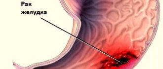
Typhlitis syndrome is an inflammation of the cecum. This problem develops extremely slowly, but often occurs with intoxication of the body and severe headaches. Typhlitis can also accompany appendicitis and other diseases of the gastrointestinal tract. As a rule, this disease occurs in older people.
Classification of typhlitis
According to morphological changes
Based on how pathologically the mucous membrane is changed, typhlitis is distinguished:
- catarrhal;
- ulcerative
Due to the occurrence
Depending on the factor that caused typhlitis, it can be:
- Infectious. Inflammation of the cecum is caused by Shigella, dysenteric amoeba, and less commonly tuberculous typhlitis occurs. It is extremely rare that the cecum is affected by sexually transmitted infections (these diseases are characterized by total damage to the colon - colitis). Mycosis of the cecum (infection with pathogenic fungi) is difficult to treat.
- Toxic. Occurs due to damage to the mucous membrane by chemicals. These can be either waste products of pathogenic and opportunistic microorganisms, or low-quality food.
- Allergic. In case of individual intolerance to foods or medications, certain components cause irritation of the mucous membrane. This is how inflammation occurs.
- Ray. Radiation therapy used to treat cancer contributes to the onset of the disease.
- Inflammation of unknown etiology. This group includes Crohn's disease, microscopic typhlitis, eosinophilic. These diseases are mainly associated with autoimmune pathological processes.
According to the form of flow
- spicy;
- chronic.
Symptoms
Main symptom
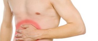
The most common complaint with typhlitis is pain . It occurs in the lower abdomen on the right, sometimes it is dull, but more often patients complain of colic in the right iliac region. Intensifies:
- 6 hours after eating;
- when standing for a long time;
- in a lying position on the left side.
Sometimes the pain radiates to the lower back.
Other symptoms
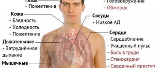
In addition to pain, typhlitis manifests itself:
- constipation or diarrhea (they often alternate);
- increased body temperature;
- rumbling in the stomach;
- flatulence;
- belching;
- feeling of fullness in the stomach;
- lack of appetite.
Features of the manifestation of various forms of typhlitis
In many ways, the clinical manifestations of typhlitis depend on its form.
| Symptom | Spicy | Chronic | Catarrhal | Ulcerative |
| Pain | Sharp, strong colic type | Dull, aching sometimes intensifies | Not intense | Cramping |
| Body temperature | Suddenly rises. Can increase up to 40 °C | Normal or subfebrile. Sometimes during periods of exacerbation it can suddenly rise to high values. | 37.1-38.3 °C | Over 38 °C. If the disease is chronic, then the temperature may be normal (up to 37.1 ° C) |
| Presence of impurities in stool | Depends on the degree of damage to the mucous membrane | Mucus, sometimes pus | Mucus, pus, blood | |
| Constipation | Typhlitis is characterized by alternating constipation with diarrhea. More often, retention of feces in the cecum is the cause of inflammation. And constipation itself occurs due to the consumption of food low in dietary fiber and a violation of the evacuation function of the large intestine. | |||
| Frequency of bowel movements | Depends on the severity of the disease:
| |||
Be sure to read: Rectal cancer: symptoms, stages, treatment and prognosis for life
Symptoms of typhlitis
The disease manifests itself as a sudden attack, which is very similar in symptoms to appendicitis. A person is worried about acute pain in the ilium on the right side. The pain can radiate to both the inner thigh and groin area. An attack develops approximately an hour after eating. The patient's temperature may rise significantly, he feels weakness and chills, which are accompanied by increased sweating and headache.
After about two hours, nausea and frequent diarrhea may occur. Vomiting with this disease is extremely rare.
In chronic typhlitis, the symptoms are even less pronounced. During the period of remission, patients show no deviations from the norm at all. An exacerbation of typhlitis is provoked by any psychological, stressful or physical activity, as well as non-compliance with diet and nutrition.
The general symptom complex of typhlitis is very similar to the manifestation of an acute attack of appendicitis, so a thorough examination of the patient is required.
Causes and factors of occurrence
Typhlitis occurs when various agents act on the cells of the mucous membrane of the cecum. Increase the risk of its development:
- intestinal infections;
- dysbacteriosis;
- poor nutrition;
- medications (cytotoxins, antibiotics);
- autoimmune pathologies;
- ileocecal valve insufficiency;
- irritation of the mucous membrane of the cecum with bile;
- violation of intestinal evacuation function;
- chronic stress;
- violation of neurohumoral regulation of intestinal function;
- frequent trauma to the mucous membrane with feces during constipation;
- defect of local immunity of the mucous membrane of the cecum.
Typhlitis is a common condition that occurs against the background of immunodeficiency (including after chemotherapy, a long course of antibacterial therapy, and blood diseases). Occurs mainly in childhood and old age. It is rarely detected in people with intact immunity.
Based on clinical manifestations, it is impossible to accurately establish a diagnosis and determine the cause of the disease.
According to statistical data, such symptoms reveal:
- 60% of cases are tumor and inflammatory diseases of the ileocecal region;
- 10% – dysfunction of the colon;
- 5% – pathologies not related to the ileocecal region (inguinal hernia, adnexitis, etc.).
It is especially difficult to determine the disease only by the nature of the pain. The same pain as with typhlitis is characteristic of appendicitis. And if inflammation of the cecum, especially catarrhal inflammation, which occurs in a mild form, can be treated conservatively, then in case of appendicitis, severe ulcerative lesions of the cecum, urgent surgery is necessary. Therefore, it is imperative to undergo diagnostics.
Diagnostics
When diagnosing, the following goals are pursued:
- Determine the cause of damage to the mucous membrane.
- Determine whether other parts of the intestine are affected.
- Carry out differential diagnosis with other diseases.
To do this, you should contact a therapist or gastroenterologist. A consultation with a surgeon is required.
Required:
- general examination;
- palpation;
- auscultation;
- blood and urine tests;
- stool analysis.
For certain indications the following is prescribed:
- Ultrasound of the abdominal organs;
- x-ray with oral administration of contrast agent;
- colonoscopy with biopsy - endoscopic examination of the colon and tissue sampling for histological examination;
- computed tomography (if intestinal neoplasms are suspected).
Women who complain of pain in the right iliac region must be referred for consultation to a gynecologist. Since most gynecological diseases (inflammation of the genitourinary system, ectopic pregnancy, etc.) occur with similar symptoms.
To clarify the cause of typhlitis, a morphological study is required. Only after a biopsy can you most accurately determine whether the disease is caused by autoimmune disorders or other factors. In the presence of ulcers, a biopsy is mandatory, since such lesions of the mucous membrane occur in the initial stages of cancer, precancerous diseases.
Be sure to read:
Main symptoms of appendicitis
Typhlitis
Clinic
Abdominal pain, first in the epigastric or periumbilical region; Often it is non-localized in nature (pain “all over the abdomen”), after a few hours the pain migrates to the right iliac region - the “movement” symptom or Kocher’s (or Kocher-Volkovich) symptom. Somewhat less often, pain appears immediately in the right iliac region. The pain is constant; their intensity is usually moderate. As the disease progresses, they somewhat intensify, although they may also subside due to the death of the nervous apparatus of the appendix during gangrenous inflammation. The pain intensifies when walking, coughing, or changing body position in bed. When the parietal peritoneum becomes increasingly irritated, the pain is localized in the right lower quadrant. This stage is called acute appendicitis. Irradiation is not observed in the typical form of acute appendicitis and is characteristic only of atypical forms. It is important to distinguish between spastic (spasmodic) and intermittent (intermittent) abdominal pain and progressive worsening pain. If a patient has nausea, vomiting or diarrhea followed by cramping and intermittent abdominal pain, there is a high likelihood of gastroenteritis. If the first manifestation is fever, appendicitis is less likely. With appendicitis, a slight increase in temperature may occur within 24 hours; a subsequent more significant increase in temperature may indicate perforated appendicitis[18];
- Lack of appetite (anorexia) [ source not specified 1626 days
]; - Nausea and vomiting occur 1-2 times and are reflexive in nature. The appearance of nausea and vomiting before the onset of pain is not typical for acute appendicitis [ source not specified 1626 days
]; - A rise in temperature to 37-38 °C (low-grade fever) (Murphy triad - anorexia, vomiting, fever) [ source not specified 1626 days
]. - Possible: loose stools, frequent urination, increased heart rate and increased blood pressure (very rare)[ unspecified source 1626 days
] - There are “atypical” manifestations of appendicitis in its atypical location, as well as in children, the elderly and during pregnancy [ source not specified 1626 days
]. - Clinical manifestations depend on the location of the appendix. If the appendix is retrocecal (located behind the cecum), the pain is muted. If the appendix is in the pelvis, atypical pain appears [ source not specified 1626 days
]. - Children may experience dysuria (urinary disorders) due to irritation by the inflamed appendix of the bladder [ source not specified 1626 days
].
Physical examination
During the examination, it is important to exclude other sources of infection. Upper respiratory tract infections can lead to mesadenitis, which can also cause abdominal pain. The patient's obesity, the retrocecally located appendix, and the small size of the intestine make the diagnosis difficult.
Clinical signs. Symptoms of peritoneal irritation
- pain in the right iliac region upon palpation. Pain with a sudden decrease in pressure during palpation in the right lower quadrant is difficult to establish in children. A simpler and more accurate method of determining the degree of peritoneal irritation is to ask the patient to walk or jump [ source not specified 1626 days
]. - increased pain at McBurney's point
[19] (the projection point of the base of the process, on the border of the outer 1/3 of the imaginary line connecting
the anterior superior iliac spine
of the right ilium with the navel); - muscle tension in the right iliac region during palpation [ source not specified 1626 days
]; - soreness of the anterior wall of the rectal ampulla due to the presence of effusion in the pouch of Douglas, or in the pouch of Douglas during rectal examination [ source not specified 1626 days
]; - Aaron's symptom - pain, a feeling of fullness in the epigastrium or precordial region when pressing at McBurney's point [20] ;
- Bartomier-Michelson's symptom (Bartomier) - pain on palpation of the cecum intensifies when the patient is positioned on the left side;
- Bassler's symptom - pain when pressing along the line from the navel to the anterior superior spine of the right ilium intensifies as it approaches the bone;
- Brown's symptom - on the anterior abdominal wall in a supine position, the place of greatest pain is noted, after which the patient is placed on the left side. After 15-20 minutes, the site of pain moves 2.5 - 5 cm more medially or the pain intensifies;
- Brando's symptom (Brindeau) - pain on the right when pressing on the left rib of the pregnant uterus;
- Brittain's symptom - upon palpation of the abdomen in the area of greatest pain in the right iliac region, muscle tension and pulling of the right testicle to the upper part of the scrotum are observed. With the cessation of palpation, the testicle descends;
- Wachenheim-Reder symptom - the appearance of pain in the right iliac region during digital examination of the rectum;
- Widmer's symptom - the temperature in the right armpit is higher than in the left;
- Voskresensky's symptom - the doctor with his left hand pulls the patient's shirt by the bottom edge (for uniform sliding). As the patient inhales, use your fingertips with moderate pressure on the abdomen to make a quick sliding movement from top to bottom towards the right iliac region. At the moment the movement ends, the patient notices a sharp increase in pain;
- Gabay's symptom - in the area of the Petit triangle on the right, apply pressure with a finger, and then quickly remove the finger. At this point, the pain increases. A sign of acute appendicitis with a retrocecal location of the appendix;
- Dolinov's symptom - increased pain in the right iliac region when the abdomen is retracted;
- Donnelly's symptom is the appearance of pain on palpation above and below McBurney's point while the patient straightens his right leg. Sign of acute appendicitis with retrocecal localization of the appendix;
- Dieulafoy's triad of symptoms - pain, muscle tension and skin hyperesthesia in the right iliac region;
- Sattler's symptom - pain in the right iliac region when a sitting patient raises his straightened right leg;
- Ivanov's symptom - the distance from the navel to the anterior superior iliac spine on the right is less than on the left, due to muscle contraction in the right iliac region;
- Ikramov's symptom - increased pain in the right iliac region when the right femoral artery is pressed;
- Klemm's symptom - accumulation of gas in the ileocecal intestine, determined by x-ray examination;
- Cope's symptom (Soret) - increased pain in the right iliac region when the hip is extended in a position on the left side (with pelvic localization of the appendix);
- Kocher's symptom (Kocher, Volkovich-Kocher) - pain initially occurs in the epigastric region directly under the xiphoid process, and after 1-3 hours moves to the right iliac region;
- Krymov's symptom - the appearance or intensification of pain in the right iliac region when examining the external opening of the right inguinal canal with a finger;
- Krymov-Dumbadze symptom - pain on palpation of the umbilical ring, indicating irritation of the peritoneum;
- Larock's symptom - a tightened position of the right or both testicles, occurring spontaneously or upon palpation of the anterior abdominal wall;
- Lennander's symptom - a difference in axillary and rectal temperatures of more than 1 °C;
- Murphy's symptom - due to the presence of effusion in the right iliac region, dullness is determined upon percussion in this area;
- Michelson's symptom - increased pain in the right half of the abdomen in pregnant women in a position on the right side, when the uterus presses on the source of inflammation;
- Obraztsov's symptom - increased pain when pressing on the cecum and simultaneous raising of the right leg straightened at the knee joint;
- Ostrovsky's symptom - the patient raises his straightened right leg up and holds it in this position. The doctor quickly unbends her and lays her horizontally. Pain appears in the right iliac region;
- Payr's symptom is hyperesthesia of the anal sphincter with tenesmus and spasms during defecation. Positive for the pelvic location of the process;
- Przewalsky's symptom - the patient has difficulty raising his right leg;
- Razdolsky's (Mendel-Razdolsky) symptom - upon percussion of the abdominal wall, pain is detected in the right iliac region;
- Rizvan's symptom - increased pain in the right iliac region with deep inspiration;
- Rovsing's symptom - the appearance or intensification of pain in the right iliac region with compression of the sigmoid colon and push-like pressure on the descending colon;
- Sumner's symptom - increased tone of the muscles of the anterior abdominal wall with mild palpation;
- Sitkovsky's symptom - the occurrence or intensification of pain in the right iliac region with the patient positioned on the left side;
- Soresi's symptom - pain in the right iliac region that occurs when coughing and simultaneous palpation in the right hypochondrium in a patient lying on his back with bent legs;
- Horn's sign - pain in the right testicle when slightly pulling at the base of the scrotum;[21]
- Chase's symptom (Chase) - pain that occurs in the right iliac region with quick and deep palpation along the transverse section of the colon when pressing the descending section with the other hand;
- Cheremskikh-Kushnirenko (Karavaeva) symptom - increased pain in the right iliac region when coughing;
- Chugaev's symptom - when palpating the anterior abdominal wall, tense bundles of the external oblique abdominal muscle are felt (“strings of appendicitis”);
- Shilovtsev's symptom - in a supine position, the place of greatest pain in the right iliac region is determined and then the patient is asked to turn on his left side. The pain shifts lower and to the left;
- Shchetkin-Blumberg symptom - reverse sensitivity, increased pain with sudden removal of the hand, compared to palpation;
- Yaure-Rozanov symptom - pain when pressing with a finger in Petit's triangle.
Laboratory signs
The diagnosis of “acute appendicitis” is clinical
(usually it is performed by a surgeon, determining
the indications
for emergency surgery).
Laparoscopy
The exact morphological form of the disease (catarrhal, phlegmonous, gangrenous) is revealed only intraoperatively, during diagnostic laparoscopy or the first stage of laparotomy (in the domestic tradition, such a diagnosis is called “Postoperative diagnosis”).
- The removed appendix is examined histologically (usually 5-7 working days are required) to confirm and detail the intraoperative diagnosis.
- Reliable laboratory changes in blood, urine, and other biological fluids that allow the diagnosis of acute appendicitis to be determined without surgical intervention currently do not exist.
In acute appendicitis, nonspecific changes in blood tests are observed, characteristic of the inflammatory reaction as such: an increase in the number of leukocytes in the blood, an increase in the erythrocyte sedimentation rate, an increase in C-reactive protein after the first 12 hours, a small number of red blood cells and leukocytes in the urine (“toxic changes in urine” ). A significant increase in the number of leukocytes may indicate perforation [ source not specified 1626 days
].
- In the foreign literature of the last 2-3 years, reports have appeared that in destructive forms of acute appendicitis an increase in total serum bilirubin of more than 18.5 µmol/l may be characteristic. A possible explanation for hyperbilirubinemia is the absorption of toxic products from the appendix into the veins of the appendix, entry through the portal system into the liver, toxic changes in hepatocytes, which lead to an increase in bilirubin levels. Currently, this information needs confirmation. In any case, these data are of an auxiliary nature and are not specific to acute appendicitis
, as they can be present in a number of acute and chronic diseases.
Instrumental examination
| This section is missing references to information sources. Information must be verifiable, otherwise it may be questioned and deleted. You can |
Ultrasonography
- dilatation of the lumen (diameter more than 6 mm), lack of peristalsis, sometimes coprolite can be located. An ultrasound examination should be the first instrumental examination if appendicitis is suspected. A fluid-filled, incompressible tubular structure with a diameter exceeding 6 mm, appendicolith, and peri-appendicular pericecal fluid are detected.
- The most common echo sign of acute appendicitis is the presence of free fluid in the right iliac fossa (that is, around the appendix) and (or) in the pelvic cavity (the most sloping part of the abdominal cavity) - symptoms of local peritonitis.
- Ultrasound examination for acute appendicitis is not always specific. To locate (detect) the appendix, the following are required: the presence of obturation of the appendix, experience and an expert-class apparatus. With gangrenous-perforated appendicitis, the contents of the appendix pour into the abdominal cavity, dilatation disappears, and the appendix may not be located. At the same time, free fluid is necessarily located in the abdominal cavity, “free gas” can be located in the abdominal cavity, and paretic loops of the small intestine.
Intestinal gases, obesity, protective fixation, and movements complicate ultrasound examination. The finding of a normal appendix on ultrasound is grounds for excluding appendicitis.
Radiography
abdominal cavity in the early stages of the disease is not informative; it is possible to identify only indirect signs of a pathological process in the abdominal cavity (the “sentinel loop” symptom). With the development of widespread peritonitis (according to Simonyan’s classification - at the paralytic and terminal stages of peritonitis), signs of paralytic intestinal obstruction appear: “Kloiber cups”, “small intestinal arches”, pneumatization of the colon disappears. In 10% - 20% of cases, radiography shows coprolite.
X-ray (irrigoscopy)
indicated for suspected chronic appendicitis. Signs of chronic appendicitis are the absence of filling the lumen of the appendix with a contrast agent, whether the appendix has a clear contour or not with contrast; it can be soldered to adjacent loops of the intestine (check displacement by palpation)
Diagnostic laparoscopy
indicated in doubtful cases, can proceed to therapeutic laparoscopy if technically possible, when there are conditions for laparoscopic appendectomy; The patient's written consent is required for removal of the appendix.
CT scan
informative in the presence of a spiral tomograph, when obstruction of the appendix, expansion of its lumen, wall thickening (> 1 mm) and signs of free fluid (inflammatory effusion) in the abdominal cavity are detected.
Radionuclide research
with leukocytes labeled with 99Tc.
Observation
If the medical history, physical and laboratory examination do not confirm or exclude appendicitis, it is recommended to adhere to a diet and repeat a complete blood count with a repeat clinical examination with palpation the next morning. In most cases, in the absence of appendicitis, improvement occurs and the patient can be sent home. Children with appendicitis experience increased pain, so intravenous antibiotics are used until an appendectomy is performed[22].
Appendicitis during pregnancy
| This section is missing references to information sources. Information must be verifiable, otherwise it may be questioned and deleted. You can |
Acute appendicitis is the most common cause of emergency surgery in pregnant women. Frequency of acute appendicitis in pregnant women: 1 case in 700-2000 pregnant women.
The anatomical and physiological characteristics of the female body make timely diagnosis of appendicitis difficult. This leads to a higher frequency of development of complicated forms, which can lead to termination of pregnancy and fetal death.
The correct surgical tactic is early appendectomy in pregnant women. It avoids complications and saves both the life of the mother and the child.
Features of a woman’s body during pregnancy that influence diagnosis and surgical tactics:
- erased clinical picture of “acute abdomen” due to hormonal, metabolic, physiological changes;
- progressive weakening of the muscles of the anterior abdominal wall due to their stretching by the growing uterus;
- displacement of internal organs by the growing uterus: the appendix and cecum are displaced cranially, the abdominal wall rises and moves away from the appendix.
Pregnant women with acute appendicitis experience acute abdominal pain, which acquires a constant aching character and moves to the location of the appendix (right lateral abdomen, right hypochondrium). The presence of a positive Taranenko symptom is noted - increased abdominal pain when turning from the left side to the right.
Treatment
Treatment of the disease depends on:
- degree of damage;
- causes of pathology;
- flow forms.
In mild and moderate cases, treatment is carried out conservatively, and radical methods are resorted to if conservative therapy does not help. To avoid surgery, the patient must:
- stick to a strict diet;
- take the medications prescribed by the doctor;
- use folk remedies as a complementary method, having previously discussed the possibility of such treatment with a doctor.
If typhlitis occurs with complications, surgical intervention is necessary.
Diet
Patients are recommended to use:
- Meat of dietary grades. It is a source of protein and iron.
- Dairy products. Necessary to compensate for the lack of protein and calcium. It is better to use low-fat, non-acidic cottage cheese, bio-kefir or bio-yogurt (to restore microflora in case of dysbacteriosis), and hard varieties of cheese. Milk in its pure form is acceptable if the patient tolerates it normally. Usually, with intestinal diseases, fresh milk causes bloating and abdominal pain.
- Raw vegetables and fruits can only be thoroughly ground as a source of vitamins. Better in the form of homogenized purees.
- Boiled porridge. For constipation, it is better to eat buckwheat, oatmeal, and barley. If the patient has diarrhea, then rice and semolina are recommended.
During illness or during exacerbation of a chronic process, it is recommended to exclude from the diet:
- foods rich in dietary fiber (although they help eliminate constipation);
- smoked meats;
- spicy dishes;
- canned food;
- carbonated drinks;
- kvass;
- alcohol (even low-alcohol drinks).
Meals should be fractional (5-6 times). During the period of exacerbation of the disease, treatment table No. 3 is recommended (if the disease is accompanied by constipation); in case of diarrhea, diet No. 4 is prescribed.
Nutritional therapy does not replace medications. But nutrition also plays an important role in the treatment of inflammation of the cecum.
Medicines
Medicines are prescribed depending on the cause and severity of symptoms:
- Antibiotics. They are necessary for proven bacterial etiology of typhlitis. If the disease is caused by fungi, antifungal drugs are prescribed.
- Antipyretic and anti-inflammatory drugs are recommended at temperatures above 38.3 °C.
- Antispasmodics are prescribed if the disease is caused by a spasm of smooth muscles. In this case, antispasmodics even relieve pain.
In addition, they prescribe:
- absorbents;
- astringents;
- anti-inflammatory;
- motor modulators.
Self-prescribing medications is not recommended. Only a doctor can choose the safest and most effective drugs, depending on the cause and severity of the disease.
Surgery
Indications for surgery:
- lack of effect from conservative therapy;
- development of complications (general peritonitis, intestinal perforation, bleeding);
- detection of neoplasms in the cecum.
The volume of the operation depends on the extent of the process. Resection of the affected area and restoration of intestinal integrity is performed. An examination of the abdominal organs is indicated.
Traditional methods
Traditional methods can be used as an additional component of complex treatment of typhlitis.
- For constipation, cleansing enemas with a decoction of chamomile and calendula are recommended (see here).
- A decoction of chamomile, yarrow, St. John's wort, and rose hips will help relieve inflammation and restore the water-salt balance disturbed by diarrhea and vomiting (see more about the use of herbs for the treatment of gastrointestinal diseases).
- Thermal procedures (warming up with heating pads, warm compresses) are indicated if there is no purulent process.
- Tea made from motherwort, lemon balm, valerian, and peppermint will help with typhlitis caused by prolonged stress.
It is extremely dangerous to treat typhlitis using exclusively folk methods, especially without preliminary diagnosis. The disease will lead to serious consequences.
Symptom of cecal irritation
If excessive mobility of the cecum
is easily recognized by palpation, then, using the same techniques, one can easily ascertain the loss of normal mobility, i.e., pathological fixation of the cecum. This fixation always indicates an inflammatory process around the intestine, which is most often caused by appendicitis, and in women it is often perisalpingitis.
But of course, in general
, any local peritonitis in this area, no matter what its origin, may result in limitation or loss of intestinal motility.
Regarding soreness
of the cecum during palpation, then remembering that under normal conditions palpation of the intestine is not accompanied by painful sensations, any pain during palpation should be considered a pathological phenomenon. It gives an indication of diseases of the intestine itself or its appendage or the peritoneum covering them, or the suffering of neighboring organs, and in women, mainly, the inflammatory process in the right appendages. Of course, the ability to determine whether the intestine is painful on palpation from a disease itself or from an inflammatory process in the neighborhood is of great diagnostic importance. This can only be achieved through experience. If the doctor is experienced and familiar with the normal palpation sensations of the cecum, he will soon determine whether the wall of the cecum is normal and whether the intestine itself is painful, or whether palpation is painful because it disturbs the neighboring inflamed organ.
Usually an inflammatory process
in the intestine itself, as, for example, in acute influenza, stercoral, dysenteric, typhoid typhlitis or intestinal tuberculosis, it causes compaction of its walls, their tension, increased resistance in the intestine itself; the main thing is that by palpating near the intestine, and not the intestine itself, we do not notice pain.
On the contrary, severe pain
outside the intestine and a decrease in sensitivity upon palpation of the intestine itself, in the absence of any changes in the properties of the intestine itself, rather indicates a disease of neighboring organs. Needless to say, with careful palpation we sometimes find a diseased organ nearby - for example, an appendage, inflamed glands, affected by an ulcerative process ileum in typhus, tuberculosis, etc., and in a woman, adding a gynecological examination to the general one, we are convinced of the presence diseases of the right tube and ovary (Salpyngoophoritis dextra).
Finally, we will have to consider
those acoustic phenomena that are heard in the intestine itself when it is palpated. Normally, when the intestine is pressed against the back wall of the abdominal cavity during palpation and the fingers palpating roll through the intestine, a rumbling sound is heard, caused by the mixing of the natural mushy contents of the intestine with the gases in it and squeezing them into the ascending colon.
But if the contents happen
If it is thicker and contains less gases, then there is no rumbling at all. The absence of rumbling upon palpation of the coeci indicates thickening of the contents, which occurs in rare cases of habitual constipation. In other cases, individual thickened lumps of feces (scybala) can be felt in the cecum, similar to what is observed in S. Romanum. This phenomenon, it must be said, is generally observed rarely and only in the case when the entire colon is filled with fecal dense masses, when, therefore, upon palpation we find them both in S. Romanum and colon transversum.
Source
Possible consequences and prognosis for life
Untreated typhlitis for a long time is dangerous because the inflammatory process will affect the small intestine, other parts of the colon, and the peritoneum. In addition, other complications may arise:
- intestinal bleeding;
- abscess;
- peritonitis;
- perforation of the ulcer;
- sepsis.
Be sure to read:
Rectal fissure: causes, symptoms and treatment of pathology Typhlit leads to other equally serious consequences. So, with ulcerative hemorrhagic typhlitis, anemia will develop due to frequent bleeding. Inflammatory processes contribute to tissue necrosis and gangrenous changes. Due to the inflammatory process, absorption processes are disrupted, resulting in:
- Iron-deficiency anemia;
- hypo-, avitaminosis;
- hypocalcemia.
If typhlitis is accompanied by diarrhea, vomiting disrupts the water-salt balance.
Typhlitis, especially if it occurs in a mild form, can be treated with conservative therapy if treated in a timely manner. If you follow your doctor's recommendations, a quick recovery is possible. But if complications arise, the prognosis for life is somewhat worse. There is a high probability of death with peritonitis, massive intestinal bleeding, and perforation.
Prevention
- Prevention of constipation. One of the main causes of inflammation of the mucous membrane of the cecum is its frequent trauma associated with fecal retention. Therefore, it is imperative to prevent the development of constipation.
- Observe basic hygiene rules. Infections that cause intestinal inflammation are transmitted mainly through the fecal-oral route.
- Eat properly. Eating foods containing fiber reduces the risk of developing constipation.
- Treat other diseases in a timely manner.
- Do not abuse laxatives, antibiotics and other medications, especially if they were prescribed by friends and not by your doctor.
To prevent the development of complications, you should seek medical help at the first symptoms.
In continuation of the topic, be sure to read:
- Rectal fissure: causes, symptoms and treatment of pathology
- Rectal cancer: symptoms, stages, treatment and prognosis for life
- Causes of bloating and increased gas formation, treatment methods
- More about hemorrhoids: causes, symptoms and treatment methods
- Details about bowel cancer: stages, symptoms, treatment and prognosis
- Intestinal colitis: symptoms and treatment in adults
- Irritable bowel syndrome: symptoms and treatments
- Sigmoiditis (inflammation of the sigmoid colon): symptoms and treatment methods
- Duodenitis (inflammation of the duodenum): symptoms and treatment methods
- Diseases of the cecum: symptoms, treatment and prevention
Diagnostic methods
If the symptoms described above are detected, the patient should urgently seek professional medical help. At an appointment with a specialist, the patient will receive directions for the following clinical activities:
- Consultation with a gastroenterologist.
- Stool collection.
- Carrying out irrigoscopy and parietography.
One of the most pronounced complaints with this disease, clearly visible when scanning with an X-ray machine, is changes or shortening of the intestine. In certain situations, barium dots appear on the inside of the mucosa, indicating the presence of ulcers and erosions. More accurate examinations involve identifying the disease and distinguishing it from diverticulitis of the large intestine, oncology, appendicitis and other unfavorable processes.