brief information
The content of the article:
1.1 Definition
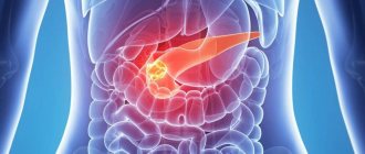
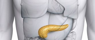
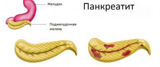
Acute pancreatitis (AP) is an initially aseptic inflammation of the pancreas, which may damage surrounding tissues and distant organs.
1.2 Etiology and pathogenesis
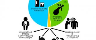
Etiological forms of pancreatitis:
- Acute alcohol-nutritional – 55%
- Acute biliary – 35%
- Acute traumatic 2 – 4%
- Other etiological forms – 6 – 8%
Factors of aggression
Primary:
- trypsin, chymotrypsin cause proteolysis of proteins;
- phospholipase A2 destroys cell membranes;
- lipase leads to lipolytic necrosis in the gland, fiber and mesentery of the intestine;
- elastase destroys the vascular wall and connective tissue structures, which leads to necrosis.
Secondary:
- activation of the kallikrein–kinin system by enzymes disrupts microcirculation.
Tertiary:
- macrophages, mononuclear cells, neutrophils produce cytokines that suppress immunity.
Quaternary:
- cytokines, enzymes, metabolites increase the permeability of the intestinal wall, which facilitates the entry of toxins into the bloodstream and lymph and damage target organs.
Factors of aggression and organ dysfunction create a syndrome of “mutual burden.”
Phases of acute pancreatitis:
- Edematous (interstitial) pancreatitis 80-85% is mild with the rare development of local complications or systemic disorders, has no phases.
- Necrotizing pancreatitis (pancreatic necrosis) in 15-20%, moderate or severe, phase course with 2 peaks of mortality - early (1 week each of phases IA and IB) and late (weeks and months).
Phase I A – up to 3 days, the formation of foci of necrosis in the parenchyma and the development of endotoxemia with multiple organ failure.
Phase I B – the body’s reaction to foci of necrosis with resorptive fever and the formation of a peripancreatic infiltrate.
Phase II – aseptic and septic sequestration.
1.3 Epidemiology
- Prevalence 32-389 per 1 million population.
- Mortality rate is 6-12 per 1 million population.
- Overall mortality 2.5%-3.5%; postoperative 20%-25%
- Since 2009, in the structure of “acute abdomen” AP has moved from 1st place to 2nd place with a share of 25%-35%, giving way to acute appendicitis.
1.4. Coding according to ICD-10
Acute pancreatitis (K85):
- K85.0 – Idiopathic acute pancreatitis;
- K85.1 – Biliary acute pancreatitis: cholelithiasis pancreatitis;
- K85.2 – Alcoholic acute pancreatitis;
- K85.3 – Drug-induced acute pancreatitis;
- K85.8 – Other types of acute pancreatitis;
- K85.9 – Acute pancreatitis, unspecified.
1.5 Classification
Mild AP – pancreatic necrosis does not form (edematous pancreatitis) and organ failure does not develop. Moderate AP - with one of the local manifestations (infiltrate, pseudocyst, abscess), or/and the development of transient - no more than 48 hours of organ failure. Severe AP - with either not limited infected pancreatic necrosis (purulent-necrotic parapancreatitis) and/or the development of persistent organ failure.
Diagnostics
Clinical manifestations depend on the morphological form, phase of the disease, the severity of the systemic inflammatory response syndrome and organ failure.
Each phase of the disease corresponds to a clinical and morphological form, so diagnosis is carried out depending on the phase of the disease.
Scale of criteria for primary express assessment of the severity of AP (St. Petersburg Research Institute of SP, 2006):
- peritoneal syndrome;
- oliguria (less than 250 ml in 12 hours);
- skin symptoms (facial hyperemia, marbling, cyanosis);
- SBP less than 100 mmHg;
- encephalopathy;
- Hb more than 160 g/l;
- leukocytes more than 14 x109/l;
- blood glucose more than 10 mmol/l;
- urea more than 12 mmol/l;
- metabolic disorders according to ECG;
- cherry or brown-black enzymatic exudate during laparoscopy/laparocentesis;
- during laparoscopy, widespread enzymatic parapancreatitis extending beyond the boundaries of the omental bursa and spreading along the flanks;
- common steatonecrosis during laparoscopy;
- lack of effect from basic therapy.
- Scale rating:
5 signs – with 95% probability a severe form of AP. 2-4 signs – moderate AP, hospitalization in the ICU. 0 – 1 sign – mild form of AP, hospitalization in the surgical department.
The SOFA scale is used to assess organ and multiple organ dysfunctions.
If it is impossible to determine the severity of AP using scales, clinical and laboratory criteria for AP are:
- signs of systemic inflammatory response syndrome (SIRS);
- hypocalcemia <1.2 mmol/l,
- hemoconcentration: Hb> 160 g/l or Ht> 40 units, glucose> 10 mmol/l;
- CRP >120 mg/l;
- shock
- renal failure (oligo-anuria, creatinine >177 µmol/l);
- liver failure (hyperfermentemia);
- cerebral insufficiency (delirium, stupor, coma);
- gastrointestinal bleeding (more than 500 ml/day);
- coagulopathy
Urgent EPST with lithoextraction for impaction of a stone in the major duodenal papilla (MDP):
- intense pain syndrome that is not relieved by narcotic analgesics;
- rapidly progressing jaundice;
- absence of bile in the duodenum during FGDS;
- signs of biliary hypertension on ultrasound.
Indications for CT/MSCT (MRI):
- uncertainty of diagnosis and differential diagnosis;
- the need to confirm severity using clinical prognostic features;
- lack of effect from conservative treatment.
Time frame for MSCT (MRI):
- for the diagnosis of pancreatic necrosis on days 4–14 of the disease;
- as the disease progresses;
- in the absence of treatment effect;
- to clarify the localization of foci of suppuration before drainage interventions.
The Balthazar CT pancreatitis severity index is not a mandatory study, but is used to predict the severity of the disease.
Acute pancreatitis
- Severe acute pancreatitis. The incidence is 5%, mortality is 50-60%. The morphological substrate of severe AP is widespread pancreatic necrosis (large focal and total-subtotal), which corresponds to severe endotoxicosis.
Mild acute pancreatitis. The incidence is 95%, mortality is 2-3%. Pancreatic necrosis in this form of acute pancreatitis either does not form (swelling of the pancreas) or is limited in nature and does not spread widely (focal pancreatic necrosis - up to 1.0 cm). Mild AP is accompanied by endotoxemia, the severity of which does not reach a severe degree.
Phase II is reactive (2nd week of the disease), characterized by the body’s reaction to the formed foci of necrosis (both in the pancreas and in the parapancreatic tissue). The clinical form of this phase is peripancreatic infiltrate.
Phase III - melting and sequestration (starts from the 3rd week of the disease, can last several months). Sequesters in the pancreas and retroperitoneal tissue begin to form from the 14th day from the onset of the disease. There are two possible options for the course of this phase:
- aseptic melting and sequestration - sterile pancreatic necrosis; characterized by the formation of postnecrotic cysts and fistulas;
- septic melting and sequestration - infected pancreatic necrosis and necrosis of parapancreatic tissue with further development of purulent complications. The clinical form of this phase of the disease is purulent-necrotic parapancreatitis and its own complications (purulent-necrotic leaks, abscesses of the retroperitoneal space and abdominal cavity, purulent omentobursitis, purulent peritonitis, arrosive and gastrointestinal bleeding, digestive fistulas, sepsis, etc.) .
Patients diagnosed with acute pancreatitis should, if possible, be sent to multidisciplinary hospitals.
Protocols for the diagnosis and treatment of acute pancreatitis in the enzymatic phase (the first five days of the disease)
I Primary protocol for diagnosis and tactics for acute pancreatitis
As a rule, it is carried out in the emergency department or emergency department.
1) The basis for establishing a diagnosis of acute pancreatitis (after excluding other surgical pathologies) is a combination of at least two of the following identified signs: a) a typical clinical picture (intense girdle pain that is not controlled by antispasmodics, uncontrollable vomiting, bloating; consumption of alcohol, spicy food or the presence of history of cholelithiasis, etc.); b) Ultrasound (increase in size, decreased echogenicity, blurred contours of the pancreas; presence of free fluid in the abdominal cavity); c) laboratory parameters (hyperamylasemia, hyperamylasuria); d) high amylase activity of the enzymatic exudate (2-3 times higher than the activity of blood amylase) obtained during laparocentesis; e) laparoscopic signs of acute pancreatitis (see protocol IV). Methods a), b), c) are mandatory when diagnosing AP, and d) and e) (laparoscopy and laparocentesis) are performed according to indications (see protocol IV).
2) In parallel with the diagnosis of acute pancreatitis, it is necessary to determine the severity of the disease (severe or non-severe). The most important thing is the early detection of severe pancreatitis, the results of treatment of which are largely determined by the timing of its onset. Signs characteristic of severe AP are the following: a) clinical: - peritoneal syndrome; - unstable hemodynamics - tachycardia (>120 per 1 min) or bradycardia (<60 per 1 min); decrease in systolic blood pressure below 100 mmHg; - oliguria (less than 250 ml in the last 12 hours); — encephalopathy (lethargy or agitation, delirium); — presence of “skin” symptoms (facial hyperemia, marbling, etc.); b) general blood test: - hemoglobin above 150 g/l; — leukocytosis above 14x109/l; c) biochemical blood test: - glucose above 10 mmol/l; — urea above 12 mmol/l; d) ECG - myocardial ischemia or severe metabolic disorders.
3) The presence of at least two signs listed in paragraph 2 allows one to diagnose severe AP, which is subject to referral to the intensive care unit. For the remaining patients (non-severe pancreatitis), hospitalization in the surgical department is indicated.
4) Intense pain syndrome, not relieved by narcotic analgesics, rapidly progressing jaundice, absence of bile in the duodenum during FGDS, signs of biliary hypertension according to ultrasound data indicate the presence of an impacted stone in the major duodenal papilla (MDP). In this case, the patient needs urgent restoration of the passage of bile and pancreatic juice, the optimal method of which is EPST. In case of an impacted BDS stone and in acute pancreatitis, EPST is performed without ERCP.
5) The optimal type of treatment for AP in the enzymatic phase is intensive conservative therapy .
II Protocol for the treatment of non-severe acute pancreatitis
1) For the treatment of non-severe pancreatitis, a basic treatment complex is sufficient: - hunger; — probing and aspiration of gastric contents; — local hypothermia (cold on the stomach); - analgesics; - antispasmodics; — infusion therapy in a volume of 40 ml per 1 kg of patient’s body weight with forced diuresis for 24-48 hours. It is advisable to enhance basic therapy with antisecretory and antienzyme therapy (see protocol III).
2) If there is no effect from the basic therapy (p. 1) within 6 hours and there is at least one of the signs of severe pancreatitis (protocol I, p. 2), severe pancreatitis should be diagnosed and the patient should be transferred to the intensive care unit and treated , corresponding to severe acute pancreatitis (protocol III).
III Protocol for intensive care of severe pancreatitis
The main type of treatment is intensive conservative therapy. The above basic treatment complex for severe AP is not effective enough and should be supplemented with a specialized treatment complex (see below). The effectiveness of the latter is maximum when treatment is started early (the first 12 hours from the onset of the disease). Surgical intervention in the form of laparotomy is indicated only in the event of the development of surgical complications that cannot be eliminated by endoscopic methods (destructive cholecystitis, gastrointestinal bleeding, acute intestinal obstruction, etc.).
Specialized treatment. 1) Antisecretory therapy (the optimal period is the first three days of the disease): - the drug of choice is sandostatin (octreotide) 100 mcgx3r subcutaneously; - reserve drugs - quamatel (40mgx2r i.v.), 5-fluorouracil (5% 5ml i.v.). 2) Rheologically active therapy (heparin, rheopolyglucin, refortan, etc.). 3) Compensation for plasma loss (correction of water-electrolyte, protein losses, etc.: in total, at least 40 ml of appropriate infusion agents per 1 kg of body weight; the ratio of colloid and crystalloid solutions is 1:4). 4) Histoprotection: - antienzyme therapy (contrical - no less than 50 thousand units, gordox - no less than 500 thousand units intravenously; the optimal period is the first 5 days of the disease); - antioxidant and antihypoxic therapy. 5) Detoxification: - in case of severe AP, extracorporeal detoxification methods are indicated, of which the most effective is serial therapeutic plasmapheresis (after replenishment of blood volume and in the absence of endotoxin shock) followed by plasma exchange (1-3 sessions every 24-48 hours, average volume of plasma exfusion is about 1 liter ); each session of extracorporeal detoxification (in addition to direct plasmapheresis) should be accompanied by rehydration and correction of water-salt metabolism in the mode of forced diuresis; — the detoxification process in severe AP can also be achieved by evacuation of toxic exudates (peritoneal, and especially retroperitoneal) during laparoscopic (or using laparocentesis) drainage of the abdominal cavity and during laparoscopic decompression of the retroperitoneal tissue (see standard IV). 6) Broad-spectrum antibiotic therapy (cephalosporins of III-IV generations or fluoroquinolones of II-III generations in combination with metronidazole).
IV Laparoscopic surgery protocol
Laparoscopy is indicated for: - patients with peritoneal syndrome, including those with ultrasound signs of free fluid in the abdominal cavity; — if it is necessary to differentiate the diagnosis from other diseases of the abdominal organs.
The objectives of laparoscopic surgery can be diagnostic, prognostic and therapeutic. If laparoscopy is not possible, laparocentesis is indicated, which partially solves the problems.
The objectives of laparoscopic surgery are: a) confirmation of the diagnosis of acute pancreatitis (and, accordingly, exclusion of other diseases of the abdominal cavity, primarily acute surgical pathology - mesenteric thrombosis, etc.); Signs of AP include: - the presence of edema of the root of the mesentery of the transverse colon; - presence of effusion with high amylase activity (2-3 times higher than blood amylase activity); — presence of steatonecrosis; b) identification of signs of severe pancreatitis: - hemorrhagic nature of the enzymatic effusion (pink, raspberry, cherry, brown); - widespread foci of steatonecrosis; - extensive hemorrhagic permeation of the retroperitoneal tissue, extending beyond the pancreas;
Verification of serous (“vitreous”) edema in the first hours of the disease (especially against the background of the patient’s severe general condition) does not exclude the presence of severe pancreatitis, since laparoscopy in the early stages may not reveal signs of severe pancreatitis, i.e. the disease may further progress.
c) therapeutic objectives: - removal of peritoneal exudate and drainage of the abdominal cavity; — laparoscopic decompression of retroperitoneal tissue (indicated in cases of hemorrhagic penetration into the retroperitoneal tissue along the ascending and descending colon in areas of maximum damage); — cholecystostomy is indicated in the presence of progressive biliary hypertension with hyperbilirubinemia more than 100 µmol/l and no earlier than 24 hours from the start of intensive therapy; - when acute pancreatitis is combined with destructive cholecystitis, in addition to the listed measures, cholecystectomy with drainage of the common bile duct is indicated;
Laparoscopy is contraindicated in case of: - unstable hemodynamics (endotoxin shock); - after multiple operations on the abdominal cavity (pronounced scarring of the anterior abdominal wall and giant ventral hernias).
Protocols for the diagnosis and treatment of acute pancreatitis in the reactive phase
I Protocol for diagnosis and monitoring of peripancreatic infiltrate
The reactive (intermediate) phase occupies the second week of the disease and is characterized by the onset of a period of aseptic inflammatory reaction to foci of necrosis in the pancreas and parapancreatic tissue, which is clinically expressed by peripancreatic infiltrate (local component) and resorptive fever (systemic component of inflammation). Peripancreatic infiltrate (PI) and resorptive fever are natural signs of the reactive phase of destructive (severe or moderate) pancreatitis, while in edematous (mild) pancreatitis these signs are not detected.
1. In addition to clinical signs (peripancreatic infiltrate and fever), the reactive phase of ADP is characterized by: 1.1 laboratory indicators of systemic inflammatory response syndrome (SIRS): leukocytosis with a shift to the left, lymphopenia, increased ESR, increased concentration of fibrinogen, C-reactive protein, etc.; 1.2 Ultrasound signs of PI (continued increase in the size of the pancreas, blurred contours and the appearance of fluid in the peripancreatic tissue). 2. Monitoring of peripancreatic infiltrate consists of a dynamic study of clinical and laboratory parameters and repeated ultrasound data (at least 2 studies in the second week of the disease). 3. At the end of the second week of the disease, computed tomography of the pancreatic area is advisable, since by this time the vast majority of patients experience one of three possible outcomes of the reactive phase: 3.1 Resorption, in which there is a reduction in local and general manifestations of the acute inflammatory reaction. 3.2 Aseptic sequestration of pancreatic necrosis resulting in a pancreatic cyst: preservation of the size of the PI with normalization of health and subsidence of the systemic inflammatory response syndrome (SIRS) against the background of persistent hyperamylasemia. 3.3 Septic sequestration (development of purulent complications).
II Protocol for the treatment of peripancreatic infiltrate
In the vast majority of patients, treatment of acute pancreatitis in the reactive phase is conservative. Laparotomy in the second week of ADP is performed only for surgical complications (destructive cholecystitis, gastrointestinal bleeding, acute intestinal obstruction, etc.) that cannot be eliminated by endoscopic methods.
Composition of the treatment complex:
1. Continuation of basic infusion-transfusion therapy aimed at replenishing water-electrolyte, energy and protein losses as indicated. 2. Medical nutrition (table No. 5 for moderate AP) or enteral nutritional support (severe AP). 3. Systemic antibiotic therapy (cephalosporins of III-IV generations or fluoroquinolones of II-III generations in combination with metronidazole, reserve drugs - carbapenems). 4. Immunomodulation (two subcutaneous or intravenous injections of roncoleukin at 250,000 units (for body weight less than 70 kg) - 500,000 units (for body weight more than 70 kg) with an interval of 2-3 days);
Protocols for the diagnosis and treatment of acute pancreatitis in the phase of purulent complications
I Protocol for the diagnosis of purulent complications of acute pancreatitis
The clinical form of acute destructive pancreatitis in the phase of septic melting and sequestration (the third week from the onset of the disease or more) is infected pancreatic necrosis (IP) and purulent-necrotic parapancreatitis (NPP) of varying degrees of prevalence.
Criteria for IP and GNPP: 1. Clinical and laboratory manifestations of a purulent focus: 1.1 Progression of clinical and laboratory indicators of acute inflammation in the third week of ADP. 1.2 Acute inflammatory markers (increased fibrinogen by 2 times or more, high C-reactive protein, precalcitonin, etc.). 2. CT, ultrasound (increase in the process of observation of liquid formations, identification of devitalized tissues and/or the presence of gas bubbles). 3. Positive results of bacterioscopy and bacterial culture of the aspirate obtained by fine-needle puncture. The decision about the presence of GNP in patients is made based on the laboratory and clinical minimum (clause 1.1). The remaining signs are additional.
II Protocol for the treatment of purulent complications of acute pancreatitis
1. In case of purulent complications of ADP, surgical intervention is indicated, the purpose of which is to sanitize the affected retroperitoneal tissue. The object of surgical intervention is purulent-necrotic parapancreatitis and/or infected pancreatic necrosis. The intervention includes opening, sanitation and drainage of the affected retroperitoneal tissue. The main method of sanitation of purulent-necrotic foci is necrosequestrectomy, which can be either single-stage or multi-stage, and is achieved by both traditional and minimally invasive methods. 2. In the postoperative period, complex therapy is indicated: 2.1 Enteral nutritional support (through a probe inserted into the small intestine through the ligament of Treitz). 2.2 Systemic antibiotic therapy according to indications (the choice of antibacterial drug depends on the sensitivity of the isolated microorganisms) in combination with the prevention of dysbacteriosis and other complications. 2.3 Immunocorrection, the options of which are determined individually depending on clinical and laboratory parameters:
- with severe sepsis and, especially with the threat of septic shock,
- immunoglobulin replacement therapy for intravenous administration in combination with the use of hormones;
- for persistent and severe SIRS - anti-cytokine therapy (protease inhibitors, efferent procedures);
- with a low absolute number of peripheral blood lymphocytes (calculated by the formula: absolute number of leukocytes x percentage of lymphocytes in the leukocyte formula / 100%)
- cytokine therapy with roncoleukin at a dose of 250,000 - 1,000,000 units until the indicator is restored (an average of 2-5 injections).
Protocol for diagnosis and monitoring of peripancreatic infiltrate in phase IB.
Peripancreatic infiltrate (PI) and resorptive fever are signs of severe or moderate pancreatitis.
Define:
- indicators of systemic inflammatory response syndrome: leukocytosis with a shift to the left, lymphopenia, increased ESR, increased fibrinogen, CRP, etc.;
- Ultrasound signs of PI (continued enlargement of the gland, blurred contours and the appearance of fluid in the tissue).
To monitor PI it is recommended:
- dynamic study of clinical and laboratory parameters;
- at least 2 ultrasound scans in the 2nd week of the disease;
- at the end of the 2nd week, CT scan of the pancreas.
Possible outcomes by the end of phase IB:
- resorption for up to 4 weeks;
- aseptic sequestration of pancreatic necrosis with the formation of a pseudocyst: stabilization of the size of the PI with normalization of health and subsidence of SIRS against the background of hyperamylasemia;
- septic sequestration (purulent complications).
annotation
Acute pancreatitis, being an exotic disease at the beginning of the 20th century, currently occupies one of the first places in the list of “acute abdomen”. However, there is still ongoing debate about treatment tactics caused by the lack of a unified classification and diagnostic and tactical algorithm. Existing foreign standards (European, British, etc.) are poorly adapted to the conditions of domestic healthcare. These protocols for the diagnosis and treatment of acute pancreatitis were developed in the pancreatology clinic of the St. Petersburg Research Institute of Emergency Medicine (headed by A.D. Tolstoy, MD) and are based on the experience of treating more than 10,000 patients with verified destructive pancreatitis, as well as the results of our own experimental research. The central point of the protocols is the refusal of early surgical interventions in the vast majority of patients with acute pancreatitis, the alternative to which is early “terminating” intensive care. Thus, achieving an aseptic course of the disease is the main way to improve the results of treatment of patients with destructive pancreatitis.
These protocols were approved by the Association of Surgeons of the North-West of the Russian Federation on March 12, 2004 and presented to the medical community of St. Petersburg on March 17, 2004 at the Round Table meeting in St. Petersburg Research Institute of Emergency Medicine named after. I.I. Dzhanelidze. A five-year (1999-2003) testing of the tactics set out in the protocols made it possible to reduce the mortality rate from destructive pancreatitis from 25% to 13.6%, the incidence of purulent complications from 30% to 14%, and the incidence of sepsis from 9.5% up to 4.8%.
The authors of the recommendations invite surgical teams to take part in testing the proposed protocol. You can discuss the presented material on the specialized forum of the School of Pancreatology project.
© St. Petersburg Research Institute of Emergency Medicine named after. I.I. Dzhanelidze, 2004
Acute pancreatitis (AP) is characterized by the development of pancreatic edema (edematous pancreatitis) or primary aseptic pancreatic necrosis (destructive pancreatitis) followed by an inflammatory reaction.
Acute destructive pancreatitis has a phase course, and each phase corresponds to a specific clinical form.
I is enzymatic , the first five days of the disease, during this period the formation of pancreatic necrosis of varying extent, the development of endotoxemia (the average duration of hyperenzymemia is 5 days), and in some patients, multiple organ failure and endotoxin shock. The maximum period for the formation of pancreatic necrosis is three days, after this period it does not progress further. However, with severe pancreatitis, the period of formation of pancreatic necrosis is much shorter (24-36 hours). It is advisable to distinguish two clinical forms: severe and non-severe AP.
• Severe acute pancreatitis. The incidence is 5%, mortality is 50-60%. The morphological substrate of severe AP is widespread pancreatic necrosis (large focal and total-subtotal), which corresponds to severe endotoxicosis.
• Mild acute pancreatitis. The incidence is 95%, mortality is 2-3%. Pancreatic necrosis in this form of acute pancreatitis either does not form (swelling of the pancreas) or is limited in nature and does not spread widely (focal pancreatic necrosis - up to 1.0 cm). Mild AP is accompanied by endotoxemia, the severity of which does not reach a severe degree.
II is reactive (2nd week of the disease), characterized by the body’s reaction to the formed foci of necrosis (both in the pancreas and in the parapancreatic tissue). The clinical form of this phase is peripancreatic infiltrate.
III – melting and sequestration (starts from the 3rd week of the disease, can last several months). Sequesters in the pancreas and retroperitoneal tissue begin to form from the 14th day from the onset of the disease. There are two possible options for the course of this phase:
• aseptic melting and sequestration – sterile pancreatic necrosis; characterized by the formation of postnecrotic cysts and fistulas;
• septic melting and sequestration – infected pancreatic necrosis and necrosis of parapancreatic tissue with further development of purulent complications. The clinical form of this phase of the disease is purulent-necrotic parapancreatitis and its own complications (purulent-necrotic leaks, abscesses of the retroperitoneal space and abdominal cavity, purulent omentobursitis, purulent peritonitis, arrosive and gastrointestinal bleeding, digestive fistulas, sepsis, etc.) .
Patients diagnosed with acute pancreatitis should, if possible, be sent to multidisciplinary hospitals.
Protocol for the diagnosis of purulent complications in phase II of the disease in the phase of septic sequestration.
Infection of the site of destruction occurs at the end of the 2nd – beginning of the 3rd week.
With late admission, inadequate treatment or too early surgery, infection may bypass the period of aseptic destruction (“crossover phases”).
Infected pancreatic necrosis:
- limited – pancreatic abscess (PA);
- not delimited – purulent-necrotic parapancreatitis (NPP).
Verification of purulent complications:
- progression of clinical and laboratory indicators of acute inflammation in the 3rd week;
- on MSCT, MRI, ultrasound, growth of devitalized tissue and/or gas bubbles;
- positive bacterioscopy and bacterial culture of aspirated samples during fine-needle puncture.
Examination by a surgeon during the first hour of hospitalization.
3. Treatment.
Each phase of the disease corresponds to a specific clinical and morphological form of AP; it is most advisable to consider treatment tactics for AP in the corresponding phases of the disease.
3.1 Conservative treatment.
Treatment of acute pancreatitis in phase IA of the disease Intensive conservative therapy is recommended.
Laparotomy is indicated only in the event of the development of surgical complications that cannot be eliminated by minimally invasive technologies.
I. Treatment of mild AP.
Hospitalization in the surgical department.
Basic treatment complex:
- hunger;
- probing and aspiration of gastric contents;
- local hypothermia (cold on the stomach);
- analgesics;
- antispasmodics;
- infusion therapy up to 40 ml/1 kg of body weight with 24-48-hour forced diuresis.
Basic therapy is enhanced with inhibitors of pancreatic secretion.
If there is no effect from the therapy within 6 hours and at least one more sign of the express assessment scale, moderate (severe) pancreatitis is stated.
In case of moderate (severe) pancreatitis, treatment is carried out in the intensive care unit according to protocols III, IV.
II. Intensive therapy of moderate acute pancreatitis.
Hospitalization in the ICU; in the absence of organ failure and progression within 24 hours, transfer to the surgical department is possible.
The main type of treatment is conservative therapy.
The basic treatment complex is supplemented by a specialized treatment complex.
Specialized treatment:
- pancreatic secretion inhibitors, optimally the first three days;
- active rheological therapy;
- infusion therapy of at least 40 ml/1 kg of body weight with forced diuresis in case of organ dysfunction (in the absence of contraindications);
- antioxidant and antihypoxic therapy;
- evacuation of toxic exudates according to indications;
- for enzymatic peritonitis - sanitary laparoscopy, percutaneous drainage under ultrasound guidance or laparocentesis is allowed.
Prophylactic antibiotics are not recommended.
III. Intensive therapy of severe acute pancreatitis.
Hospitalization in the ICU.
The main type of treatment is intensive therapy.
The basic treatment complex is supplemented by a specialized treatment complex.
Recommended:
- extracorporeal detoxification methods: plasmapheresis; hemofiltration;
- nasogastric tube for decompression;
- if possible, nasogastrointestinal intubation for early enteral support;
- correction of hypovolemic disorders;
- epidural block;
- disaggregant antithrombotic therapy.
Antibiotics are not recommended for prophylactic purposes in the first three days of the disease.
Phase IB treatment, i.e. peripancreatic infiltrate.
In most patients, treatment is conservative.
Laparotomy in the 2nd week of AP only in case of surgical complications that cannot be eliminated by minimally invasive technologies: destructive cholecystitis, gastrointestinal bleeding, acute intestinal obstruction, etc.
Composition of the treatment complex:
- continuation of basic infusion-transfusion therapy;
- therapeutic nutrition table No. 5 (moderate AP);
- nutritional support: oral, enteral or parenteral (severe AP);
- systemic antibiotic prophylaxis (cephalosporins of III-IV generations or fluoroquinolones of II-III generations in combination with metronidazole, reserve drugs - carbapenems);
- immunotherapy (correction of cellular and humoral immunity).
Late (II) phase (sequestration)
Treatment of AP in the phase of aseptic sequestration, i.e. treatment of pancreatic pseudocyst.
It is not recommended to operate on small pseudocysts – less than 5 cm; dynamic observation by a surgeon.
Large pseudocysts - more than 5 cm - are operated on routinely in the absence of complications.
The operation of choice for an immature pseudocyst (less than 6 months) is external drainage.
A mature pseudocyst (more than 6 months) is subject to surgical treatment as planned.
Complications of pancreatic pseudocyst:
- infection;
- bleeding into the cyst cavity;
- perforation with a breakthrough into the abdominal cavity with the development of peritonitis;
- compression of neighboring organs with the development of obstructive jaundice, gastric stenosis, intestinal obstruction, etc.
pancreonecrosis.ru
Strength of recommendation "C"
2) If there is no effect from the basic therapy (item 1) within 6 hours and the presence of at least one more sign of the express assessment scale (protocol I.2), moderate-to-severe (severe) pancreatitis should be identified and the patient should be transferred to the intensive care unit and intensive care and carry out treatment appropriate for moderate-to-severe (severe) acute pancreatitis (protocols III, IV).
Strength of recommendation "C"
III
Protocol for intensive therapy of moderate acute pancreatitis
The main type of treatment is conservative therapy. The above basic treatment complex for moderate-severe AP must be supplemented with a specialized treatment complex (see below). The effectiveness of the latter is maximum when treatment is started early (the first 24 hours from the onset of the disease). Upon admission, patients with moderate AP must be hospitalized in the intensive care unit (ICU). To eliminate diagnostic errors, it is advisable for the duty service to observe patients with moderate AP during the day in the ICU. In the absence of signs of organ failure and progression of the disease during the day, patients with moderate AP can be transferred to the surgical department. Subject to the appearance of signs of organ dysfunction or failure in patients with moderate AP who are in the surgical department, which indicates the progression of the disease - hypotension (BP <100 mm Hg), respiratory failure (RR> 30 per minute), delirium and others (see protocol I.2, I.3) - the latter must be transferred to the ICU.
Strength of recommendation "C"
Specialized treatment
1) Inhibition of pancreatic secretion (the optimal period is the first three days of the disease).
Strength of recommendation " D "
2) Active rheological therapy.
Strength of recommendation " D "
3) Infusion therapy with a total of at least 40 ml of appropriate infusion agents per 1 kg of body weight with forced diuresis in the presence of organ dysfunction (in the absence of contraindications).
Strength of recommendation "C"
4) Antioxidant and antihypoxic therapy.
Strength of recommendation " D "
5) Evacuation of toxic exudates according to indications (see standard V). For enzymatic peritonitis - sanitation laparoscopy.
Percutaneous drainage of the abdominal cavity under ultrasound guidance or laparocentesis is acceptable.
Strength of recommendation " D "
6) The use of antibiotics for prophylactic purposes is not mandatory.
Strength of recommendation "C"
IV
Protocol for intensive therapy of severe acute pancreatitis
The main type of treatment is intensive therapy. The above basic treatment complex for severe AP is not effective enough and should be supplemented with a specialized treatment complex (see below). The effectiveness of the latter is maximum when treatment is started early (the first 12 hours from the onset of the disease). Upon admission, patients with severe AP should be hospitalized in the intensive care unit. The treatment and diagnostic complex for patients with severe AP must be carried out in an ICU; after relieving the symptoms of organ failure and stabilizing the condition (relieving delirium, hemodynamic disorders, respiratory activity, etc.), it is possible to transfer the patients to the surgical department.
Strength of recommendation "C"
Specialized treatment
to protocols
II and III :
1. Extracorporeal detoxification methods - according to indications:
a) plasmapheresis;
b) hemofiltration
Strength of recommendation " D "
2. Nasogastric intubation for decompression and, if possible, nasogastrointestinal intubation for early enteral support.
Strength of recommendation " D "
3. Correction of hypovolemic disorders.
Strength of recommendation "C"
4. It is advisable to perform an epidural block.
Strength of recommendation " D "
5. The use of antibiotics for prophylactic purposes in the first three days of the disease is not mandatory.
Strength of recommendation "C"
6. It is advisable to prescribe disaggregant antithrombotic therapy.
strength of recommendation " D "
V
Laparoscopic surgery protocol
Laparoscopy is indicated:
— patients with peritoneal syndrome, including those with ultrasound signs of free fluid in the abdominal cavity;
Strength of recommendation "C"
— if necessary, differential diagnosis with other diseases of the abdominal organs.
Strength of recommendation " C "
The objectives of laparoscopic surgery can be diagnostic, prognostic and therapeutic. It is possible to perform percutaneous drainage of the abdominal cavity under ultrasound guidance or laparocentesis.
Objectives of laparoscopic surgery:
a) confirmation of the diagnosis of acute pancreatitis (and, accordingly, exclusion of other diseases of the abdominal cavity, primarily acute surgical pathology - mesenteric thrombosis, etc.); Signs of OP include:
— the presence of edema of the root of the mesentery of the transverse colon;
- presence of effusion with high amylase activity (2-3 times higher than blood amylase activity);
— presence of steatonecrosis;
Strength of recommendation " D "
b) identifying signs of severe pancreatitis:
- hemorrhagic nature of the enzymatic effusion (pink, raspberry, cherry, brown);
- widespread foci of steatonecrosis;
- extensive hemorrhagic permeation of the retroperitoneal tissue, extending beyond the pancreas;
Strength of recommendation " D "
Verification of serous (“vitreous”) edema in the first hours of the disease (especially against the background of the patient’s severe general condition) does not exclude the presence of severe pancreatitis, since laparoscopy in the early stages may not reveal signs of severe pancreatitis, i.e. the disease may further progress.
c) therapeutic tasks:
— removal of peritoneal exudate and drainage of the abdominal cavity.
Strength of recommendation "C"
VI
Computed tomography
Indications for early MSCT (MRI) are:
1. Uncertainty of diagnosis and differential diagnosis with other diseases.
2. The need to confirm the severity based on the identified clinical prognostic signs of severe AP.
3. Lack of effect from conservative treatment.
Strength of recommendation "C"
The optimal timing for performing MSCT (MRI) for diagnosing pancreatic necrosis (and assessing the entire volume of pathological changes in the chest, abdominal cavity and retroperitoneal tissue) is days 4–14 of the disease.
Strength of recommendation "C"
It is advisable to perform MSCT (MRI) on the eve of invasive intervention.
Strength of recommendation "C"
Subsequent MSCT (MRI) must be performed when the disease progresses, in the absence of treatment effect and to localize foci of suppuration before performing drainage interventions.
Strength of recommendation "C"
The use of the Balthazar CT severity index of pancreatitis in clinical practice is not a mandatory diagnostic test. It is advisable to use it to predict the severity of the disease.
Protocols for the diagnosis and treatment of acute pancreatitis
I In the phase of the disease
I
Protocol for the diagnosis and monitoring of peripancreatic infiltrate
The second week of the disease is characterized by the onset of a period of aseptic inflammatory reaction to foci of necrosis in the pancreas and surrounding tissue, which is clinically expressed by the appearance of infiltrate in the epigastric region (local component) and resorptive fever (systemic component of inflammation). Peripancreatic infiltrate (PI) and resorptive fever are natural signs of severe or moderate pancreatitis, while in mild pancreatitis these signs are not detected.
Strength of recommendation "C"
1. In addition to clinical signs (peripancreatic infiltrate and fever), the second week of the early phase of AP is characterized by:
1.1 Laboratory indicators of systemic inflammatory response syndrome (SIRS): leukocytosis with a shift to the left, lymphopenia, increased ESR, increased concentration of fibrinogen, C-reactive protein, etc.;
Strength of recommendation "C"
1.2 Ultrasound signs of PI (continued increase in the size of the pancreas, blurred contours and the appearance of fluid in the peripancreatic tissue).
Strength of recommendation " D "
2. Monitoring of peripancreatic infiltrate consists of a dynamic study of clinical and laboratory parameters and repeated ultrasound data (at least 2 studies in the second week of the disease).
Strength of recommendation " D "
3. At the end of the second week of the disease, a computed tomography scan of the pancreas is advisable, since by this time the vast majority of patients experience one of three possible outcomes of phase IB:
3.1 Resorption, in which there is a reduction in local and general manifestations of the acute inflammatory reaction.
3.2 Aseptic sequestration of pancreatic necrosis with a possible subsequent outcome in a pancreatic pseudocyst: preservation of the size of the PI with normalization of health and subsidence of the systemic inflammatory response syndrome (SIRS) against the background of persistent hyperamylasemia.
3.3 Septic sequestration (development of purulent complications).
Strength of recommendation "C"
II
Protocol for the treatment of peripancreatic infiltrate
In the vast majority of patients, treatment of peripancreatic infiltrate is conservative. Laparotomy in the second week of AP is performed only for surgical complications (destructive cholecystitis, gastrointestinal bleeding, acute intestinal obstruction, etc.) that cannot be eliminated with minimally invasive technologies.
Composition of the treatment complex:
1. Continuation of basic infusion-transfusion therapy aimed at replenishing water-electrolyte, energy and protein losses as indicated.
Strength of recommendation "C"
2. Medical nutrition: table No. 5 for moderate-severe AP; nutritional support (oral, enteral or parenteral) for severe AP.
Strength of recommendation "C"
3. Systemic antibiotic prophylaxis (cephalosporins of III-IV generations or fluoroquinolones of II-III generations in combination with metronidazole, reserve drugs - carbapenems).
Strength of recommendation "C"
4. Immunotherapy (correction of cellular and humoral immunity is desirable).
Strength of recommendation " D "
Late phase (sequestration)
Protocols for the diagnosis and treatment of acute pancreatitis in the phase of aseptic sequestration
I
Protocol for diagnosis and monitoring of pancreatic pseudocyst
The clinical form of acute pancreatitis in the aseptic sequestration phase is a postnecrotic pancreatic pseudocyst, the formation period of which ranges from 4 weeks and on average up to 6 months.
Criteria for verification of pancreatic cyst:
1. Subsidence of the systemic inflammatory response syndrome against the background of persistent hyperamylasemia.
Strength of recommendation " D "
2. By the 5th week of the disease, the size of the fluid accumulation in the parapancreatic tissue increases and the appearance of its wall according to ultrasound and CT data.
Strength of recommendation "C"
3. If there are no complications (see below), the patient can be discharged for outpatient treatment. The size of the cyst must be monitored by ultrasound (once every 2-4 weeks).
Strength of recommendation " D "
If during aseptic sequestration the pancreatic ductal system is not opened, then cyst formation does not occur. In this case, as a rule, there is resorption of the peripancreatic infiltrate (reduction of fluid accumulation in the pancreas) within a period of up to 4 weeks. During this period, patients should be under dynamic medical supervision (permissible on an outpatient basis).
Strength of recommendation " D "
II
Protocol for the treatment of pancreatic pseudocysts
Small pancreatic pseudocysts (less than 5 cm) are not advisable to operate; they are subject to dynamic observation by a surgeon. Large pancreatic pseudocysts (more than 5 cm) are subject to surgical treatment as planned in the absence of complications (see below). The operation of choice for an immature (unformed) pseudocyst (less than 6 months) is external drainage. A mature (formed) pseudocyst (more than 6 months) is subject to surgical treatment as planned.
Strength of recommendation "C"
Complications of pancreatic pseudocyst:
1. Infection.
2. Bleeding into the cyst cavity.
3. Perforation of the cyst with a breakthrough into the free abdominal cavity with the development of peritonitis.
4. Compression of neighboring organs with the development of obstructive jaundice, gastric stenosis, intestinal obstruction, etc.
Protocols for the diagnosis and treatment of acute pancreatitis in the phase of septic sequestration
I
Protocol for the diagnosis of purulent complications of acute pancreatitis
Infection of the focus of pancreatogenic destruction occurs, on average, at the end of the 2nd - beginning of the 3rd week from the onset of the disease. However, with late admission of the patient, inadequate treatment, or after too early and hasty surgery, infection of areas of pancreatic necrosis and purulent-destructive complications may develop earlier, bypassing the period of aseptic destruction (“cross-phase”). The clinical form of acute pancreatitis in the phase of septic sequestration (the third week from the onset of the disease or more) is infected pancreatic necrosis: limited - pancreatic abscess (PA) or not limited - purulent-necrotic parapancreatitis (NPP) of varying degrees of prevalence. An important point is the timely diagnosis of infection and verification of clinical and morphological forms of pancreatogenic infection.
PA and GNPP criteria:
1. Clinical and laboratory manifestations of a purulent focus:
1.1 Progression of clinical and laboratory parameters of acute inflammation in the third week of AP.
Strength of recommendation "C"
1.2 Markers of acute inflammation (increased fibrinogen by 2 times or more, high “C”-reactive protein, procalcitonin, etc.).
Strength of recommendation "C"
1.3 CT, ultrasound (increase in the process of observation of liquid formations, identification of devitalized tissues and/or the presence of gas bubbles).
Strength of recommendation "C"
1.4 Positive results of bacterioscopy and bacterial culture of the aspirate obtained by fine-needle puncture.
Strength of recommendation "C"
In the event that the methods of clauses 1.3 and 1.4 fail to identify signs of infection, the decision about the presence of purulent complications in patients and indications for surgical treatment is made on the basis of the laboratory and clinical minimum (clause 1.1).
Strength of recommendation " D "
II
Protocol for the treatment of purulent complications of acute pancreatitis
1. For purulent complications of AP, surgical intervention is indicated, the purpose of which is to sanitize the affected retroperitoneal tissue. The intervention includes opening, sanitation and drainage of the affected retroperitoneal tissue. The main method of sanitation of purulent-necrotic foci is necrosequestrectomy, which can be either single-stage or multi-stage, and is achieved using both minimally invasive and traditional methods.
Strength of recommendation "C"
1. When deciding on primary drainage of a pancreatic abscess or purulent-necrotic parapancreatitis, preference should be given to minimally invasive interventions (ultrasound-guided drainage, retroperitoneoscopy, minilaparotomy using the “Mini-Assistant” kit, etc.). If minimally invasive drainage is ineffective, the operation of choice is sanitary laparotomy with necrosequestrectomy. Drainage is preferably performed via extraperitoneal approaches. The optimal timing for performing the first sanitary laparotomy with necrosequestrectomy is 4-5 weeks of illness. If complications develop that cannot be treated with minimally invasive interventions, it is necessary to perform open surgery, including through a mini-access.
Strength of recommendation "C"
2. After surgery, most patients develop an external pancreatic fistula, which, after stopping the inflammatory process, is treated conservatively and closes on its own in an average of 2-4 months. A persistent pancreatic fistula that does not close after more than 6 months is usually associated with large pancreatic ducts. The patient is subject to surgical treatment as planned.
Strength of recommendation "C"
3. In the postoperative period, complex therapy is indicated:
· Parenteral or enteral nutritional support (through a tube inserted into the small intestine through the ligament of Treitz) if oral nutrition is not possible.
Strength of recommendation "C"
· Systemic antibiotic therapy according to indications (the choice of antibacterial drug depends on the sensitivity of the isolated microorganisms) in combination with the prevention of dysbacteriosis and other complications.
Strength of recommendation " B "
· Immunocorrection, the options of which are determined individually depending on clinical and laboratory parameters.
Strength of recommendation " D "
METHODOLOGY
Rating scheme for assessing the strength of recommendations (Scheme 1)
Prevention and follow-up
- At the outpatient stage, D II and D III groups.
- Scheduled examinations by a therapist and gastroenterologist 1-4 times a year, depending on the frequency of exacerbations.
- If indicated, consult a surgeon.
- If blood sugar increases, consult an endocrinologist.
- Clinical blood and urine analysis, blood amylase and urine diastase, blood and urine sugar.
- Ultrasound of the liver, gallbladder, pancreas,
- Cholecystography according to indications.
- Preventive courses of drug treatment 2-4 times a year, according to indications - physiotherapy, exercise therapy.
- Diet correction, rational employment, selection for sanatorium treatment.
- If there are signs of disability - MSEC.
- Indications for sanatorium-resort treatment are recurrent chronic pancreatitis with rare relapses without a tendency to frequent exacerbations.
- Treatment at balneological resorts: Zheleznovodsk, Essentuki, Morshin, Truskavets, Izhevsk Mineral Waters, etc.
- Climatotherapy: air baths, swimming in open water (stable remission).
- Medical nutrition – table No. 5.
- Non-carbonated, low- and medium-mineralized mineral waters, with bicarbonate and sulfate ions, alkaline waters.
Procedures:
- carbon dioxide-hydrogen sulfide baths;
- carbon dioxide-radon sulfide;
- pearl (every other day);
- heat therapy;
- mud and ozokerite applications;
- electrophoresis of dirt;
- sinusoidal-modeled currents;
- SMV therapy;
- DMV therapy;
- diadynamic currents.
Therapeutic physical education according to a gentle tonic regimen with morning hygienic exercises, sedentary games, dosed walking.
Number of people who read this article: 4,648