Volgograd
CHRONIC ULCER.
Penetrates into the stomach wall to varying depths. Destroys all layers, including the muscle layer, and sometimes reaches the serous layer.
- The bottom of the ulcer is smooth, sometimes rough.
- The edges are wavy raised, dense.
- The serous membrane in the area of the ulcer is thickened.
- Localized mainly on the lesser curvature. The lower third of the body and the angle of the stomach.
- Sizes from 1 to 4 cm.
- And it was noted that the more proximal the ulcer is located, the larger its size.
Microscopic picture:
- during an exacerbation: a zone of fibrinoid necrosis appears in the area of the bottom and edges of the ulcer. The necrosis zone is delimited by granulation tissue with a large number of thin-walled vessels. Deeper, following the granulation tissue, there is coarse fibrous scar tissue.
- During the period of remission, the opposite microscopic picture is observed: granulation tissue grows into the necrosis zone, which matures into coarse fibrous scar tissue, and epithelization of the ulcer often occurs. In the vessels located in the area of the ulcer, wall sclerosis and obliteration of the lumen develop.
Thus, gastric ulcer leads to increased cicatricial changes in the stomach and aggravates the disruption of the trophism of its tissues.
The newly formed scar tissue is easily destroyed during the next exacerbation.
ENDOSCOPIC SIGNS OF CHRONIC ULCERS.
1. The shape is often oval or round, less often linear, slit-like.
2. The edges are smooth, clear, evenly delimited from the surrounding mucosa.
3. Lack of infiltration of the surrounding mucous membrane - edema!
4. Identical coloration of the edges and the mucous membrane surrounding the ulcer, often hemorrhagic / submucosal spots.
5. The bottom is smooth, often covered with a yellow or gray coating.
6. The bottom and edges of the ulcer are sharply delimited from each other along the circumference.
7. Bleeding more often than the bottom of the ulcer.
8. The convergence of the folds of the mucous membrane towards the ulcer is visible evenly along the entire circumference and reaches its edges.
9. Deformation of the wall in the area of the ulcer occurs quite often, but is more limited in nature; retraction of the wall in the area of the ulcer is often observed - in the form of a “tent”.
10. During targeted biopsy, rigidity of the edges of the ulcer is rarely observed. There is no fragmentation when taking a biopsy.
Ways to heal an ulcer, 4 options:
1. Healing by epithelization from the periphery to the center, and the ulcer retains a round or oval shape.
2. Healing through the stage of a linear ulcer perpendicular to the lesser curvature.
3. Healing by dividing into 2 “mirror” or “kissing” ulcers.
4. Healing through the stage of a linear ulcer parallel to the lesser curvature /for deep ones/. Ulcers that heal according to type 2-3 are more likely to recur.
A callous ulcer is an ulcer that is not prone to healing, with dense edges and a bottom. Prone to degenerating into cancer. In the direction from the pylorus to the cardiac region, the tendency to malignancy increases 5 times, i.e. The higher the ulcer is located in the stomach, the more likely it is to be malignant.
A senile ulcer is single, flat, with mild signs of inflammation around it. It occurs against the background of atrophic gastritis, similar to ulcerated cancer. Heals in 1-2 months. without wall deformation. Not prone to relapse.
A slit-like or trench-like ulcer occurs in patients over 60 years of age with preserved gastric secretion. Localized along the lesser curvature of the stomach. It can reach 10cm in length.
Features of ulcerations of the stomach and duodenum depending on the type of chronic gastritis
| Morphological changes in the gastric mucosa | Interpretation |
| Normal mucous membrane | Peptic ulcer disease is impossible. If an ulcer is present, it is most likely caused by NSAID use. |
| Chronic antral or pangastritis, no atrophy in the fundus (+ bulbitis) | Peptic ulcer disease is possible or even present; the risk of its occurrence is high |
| Chronic pangastritis with atrophy in the fundus | Peptic ulcer of the 12th intestine is impossible. Stomach ulcers are possible, although the likelihood is low |
| Chronic pangastritis or fundal gastritis + severe atrophy in the fundus | Peptic ulcer disease is impossible; if there is an ulcer, then most likely it is malignant |
In connection with the above, and after the discovery of H. pylori, a concept arose that is finding more and more supporters of “ gastritis-associated peptic ulcer disease ” as its most common variant. G. Borsch (1987) in this “gastritis-ulcer” tandem puts gastritis in first place and formulates it as follows:
- “ulcers of the stomach and duodenum are not just a violation of the integrity of the epithelium, but an episodic and recurrent complication superimposed on more pronounced and diffuse lesions of the mucous membrane, in the form of type B gastritis or gastroduodenitis.”
An indispensable condition for the chronicization of ulcers is the development of scar tissue in the bottom and edges, which disrupts the trophism of the newly formed mucous membrane and contributes to the recurrence of ulcers.
When healing, a chronic ulcer goes through 4 stages.
The identification of these stages is based on a comparison of endoscopic and histological pictures:
1. Initial healing stage.
Endoscopically and when studied using a stereoscopic microscope, it is characterized by creeping of the epithelium in the direction from the edges to the center and the presence of spindle-shaped protrusions on the surface. Histologically, growth of the prismatic epithelium is noted.
2. Stage of proliferative healing (membranous regeneration).
Endoscopic and stereomicroscopic examination reveals low fusiform protrusions; histological examination reveals regenerating epithelium covering these protrusions in one layer.
3. Palisade scar stage.
The ulcer crater is not visible; in its place are palisade-like strands, converging towards the center of the ulcer. There are many capillaries in the scar tissue, and immature pseudopyloric glands appear.
4. Cobblestone scar stage.
It is named because of the characteristic appearance that the newly formed mucous membrane has when viewed through an endoscope or stereoscopic microscope. Small pits are also visible. Histologically, many pseudopyloric glands are detected.
Healing of ulcers is considered complete only when a “cobblestone” scar is formed.
Quality of ulcer healing.
Peptic ulcer disease is characterized not only by the presence of a long-term non-healing (chronic) ulcer, but also, no less important, by its tendency to recur. In this case, relapses usually occur at the site of a healed ulcer.
The optimal outcome of any reparative regeneration is restitution , i.e. complete restoration of the structure of lost tissue.
In the stomach, restitution occurs during healing of erosions and arterial damage after biopsy or endoscopic polypectomy.
Unlike erosions, ulcers destroy not only the mucous membrane, but also the underlying layers. At the same time, the type and completeness of regeneration changes fundamentally.
All ulcers heal by secondary intention with the help of granulation tissue. The structure of the mucous membrane is not completely restored.
This picture of the mucous membrane at the site of a healed ulcer is designated by the term “ substitution. ” Usually, substitution refers to a healing outcome in which the area of necrosis is replaced by connective tissue, which subsequently undergoes scarring.
Thus, the concept of quality of healing determines not just the completeness of regeneration, but also the prognosis.
In recent years, it has been established that the leading role in the restoration of the extracellular matrix after damage belongs to the transforming growth factor (TGF).
Increased production of TGF causes excess scarring. The importance of TGF for the quality of ulcer healing has recently been established. Local injection of TGF into the area of ulcers accelerated their healing, but was accompanied by the development of severe and severe sclerosis at the site of the healed ulcer.
At the same time, injection of TGF-neutralizing antibodies also accelerated healing, but the severity of sclerosis was significantly less, and the quality of healing was therefore higher.
It is possible that this way of improving the quality of ulcer healing could find clinical application. Here, however, it is important to find the moment when it is advisable to introduce this growth factor into the bottom of the ulcer. In the experiments, TGF was administered prophylactically, immediately after applying 100% acetic acid to the serosa.
It is important to decide whether TGF should be administered in the active phase or in the cleansing phase of the ulcer base.
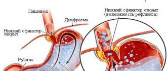
Zollinger-Ellison syndrome
Zollinger-Ellison syndrome is characterized by hypergastrinemia, hypersecretion of HCL, the presence of persistently recurrent duodenal or jejunal ulcers, diarrhea, and impaired digestion and absorption.
In the USA, the incidence varies between 0.1-3 cases per 1 million inhabitants.
There are 2 types of Zollinger-Ellison syndrome.
In type 1, there is pronounced hyperplasia of G-cells in the antrum of the stomach.
Type 2 is a hormonally active tumor (gastrinoma).
In 80% of patients, gastrinomas are located in the so-called “ gastrinomic triangle ”, limited by the pancreas, duodenum and the place where the cystic duct enters the common bile duct.
Gastrinomas of the stomach, liver, ovaries, parathyroid glands and even lymph nodes have also been described.
There are no gastrin-producing cells in the normal pancreas, and therefore the source of gastrinoma development remains unclear.
In almost 1/3 of patients, gastrinoma metastasizes to the lymph nodes, and in 10-20% to the liver.
Hypergastrinemia , an essential component of Zollinger-Ellison syndrome, causes parietal cell hyperplasia. This is due to the well-known trophic effect of gastrin.
Macroscopically:
- Ulcers in most patients are single, in 25% they are multiple.
- Their sizes usually do not exceed 2 cm,
- they are often complicated by bleeding and perforation,
- as a rule, recur after gastric resections.
Only total gastrectomy or removal of gastrinoma can prevent relapses.
- Almost a third of patients with Zollinger-Ellison syndrome experience diarrhea.
If it is possible to completely eliminate all sources of excess gastrin production, Zollinger-Ellison syndrome undergoes reverse development. In this case, not only the ulcers heal, but the normal structure of the fundic glands is restored.
Giant stomach ulcers.
Stomach ulcers whose diameter exceeds 3 cm are commonly called giant ulcers.
Features of giant stomach ulcers:
- are considered to be the most severe forms of peptic ulcer disease.
- They usually respond poorly to drug therapy,
- often (40-50%) complicated by bleeding,
- and penetrate 40-70% into neighboring organs.
- Often with giant gastric ulcers, perforations and gastrointestinal fistulas occur.
- The incidence of giant gastric ulcers according to endoscopic examination is 8.6%.
Endoscopically:
In all giant gastric ulcers, two zones can be distinguished - central and peripheral.
The presence of central and peripheral zones gives giant gastric ulcers a peculiar trapezoidal shape with a wide base facing the lumen of the stomach. The narrow part is directed towards its outer surface.
The value of biopsies in the differential diagnosis of gastric ulcers.
The main task facing a morphologist when studying biopsies of patients with gastric ulcers is differential diagnosis between a chronic ulcer and cancer . As is well known, in many patients this problem can be solved only after a biopsy.
Clinical and endoscopic differential diagnosis is also complicated by the fact that an ulcerated gastric cancer tumor can, like an ordinary benign ulcer, undergo healing, although such healing is rarely complete, but it is observed in 70% of patients with early cancer. At the site of ulceration, normal granulation tissue and mucous membrane may form. The surrounding tumor again grows into it, which soon undergoes re-ulceration.
Due to the fact that stomach cancer grows relatively slowly, such cycles can be repeated several times.
There is evidence that cancer takes almost 10 years to develop from microscopic to early stage.
And from early to severe with clinical manifestations - 16-27 years.
And that early type 1 cancer doubles its size in 6.5 years, and early cancers of other types in 2-3 years.
If gastric juice destroys the tumor, and the resulting defect, in the process of reparative regeneration, is replaced by a “non-cancerous” mucous membrane, then the assumption arises about the possibility of “self-healing” of superficial cancer.
Apparently, this can explain those rare sectional observations when the pathologist finds metastases in the liver or lymph nodes, and in the stomach - a benign ulcer or postulcerous scar.
The possibility of healing of ulcerated stomach tumors also requires a new attitude to the indications for surgery.
Until recently, it was generally accepted that surgical treatment of patients after 4-6 weeks was necessary. unsuccessful conservative therapy.
It was meant that if the ulcer does not heal within this time frame, then it is either cancerous or may become malignant. However, it is now quite well known that completely benign ulcers may not heal for months, but a cancer ulcer may “heal” within the usual time frame.
Therefore, the main method in differential diagnosis and in determining treatment tactics is gastroscopy with multiple biopsies.
Biopsies must certainly be multiple, both from the edges and from the bottom of the ulcer . It is well known that carcinomatous changes can be observed only in certain areas of the bottom and edges of the ulceration, which may not be included in the excised material. Very expressive data supporting this well-known, but unfortunately not always fulfilled requirement is illustrated by the materials of A. Misumi et al. (1978).
They found that the accuracy of the histological diagnosis of cancer was 100% when taking at least 6 biopsies. If the biopsy was performed only from, the number of positive findings decreased to 48.5%, from the outer edge to 19.6% and from the area around the “lesion” to 1.6%;
The doctor should know that excision of 1-2 pieces for ulcers is unacceptable . If there are no tumor elements in them, the medical documents will indicate that “histological examination did not reveal any signs of malignant growth.” It is well known how such a recording removes the much-needed oncological alertness of the clinician and how this can delay the establishment of a true diagnosis for a long time.
Therefore, in practical work one should proceed from a position that can be formulated as follows:
“A single biopsy from an ulcer can be not only useless, but also harmful to the patient.”
It should be borne in mind that even an experienced endoscopist is not always able to excise pieces from the edges and bottom of the ulceration for various reasons. In these cases, the pathologist should not limit itself to simply describing what was brought to the laboratory. In the “Specialist’s Conclusion” it should be noted that the delivered material does not contain tissue from the bottom and (or) edges of the ulceration.
Such a record tells the clinician that the biopsy was uninformative, and the task that the clinician set when ordering the biopsy was not completed.
This conclusion serves as an indication for a repeat biopsy.
And briefly about
COMPLICATIONS OF Peptic Ulcer
With peptic ulcer disease, the following complications are distinguished:
- bleeding,
- penetration,
- malignancy,
- perforation,
- ulcerative cicatricial contractions.
- perivisceritis (perigastritis, periduodenitis).
Bleeding occurs due to arrosion of the walls of blood vessels, usually during an exacerbation of peptic ulcer disease. It manifests itself with clear clinical symptoms: vomiting of blood, “coffee grounds”, tarry stools, hemodynamic disturbances.
Classification of the degree of bleeding activity according to Forrest is essentially the criteria for endoscopic prognosis of recurrent bleeding:
Forrest I. Continued bleeding:
Ia) massive jet arterial bleeding from a large vessel;
Ib) moderate, when the spilled blood from a venous or small arterial vessel quickly floods the source after it is washed off and flows down the wall of the organ in a wide stream; jet arterial bleeding from a small vessel, the jet nature of which periodically stops;
Ic) weak (capillary), slight leakage of blood from a source that can be covered by a clot.
Forrest II. Current bleeding:
IIa) the presence at the source of bleeding of a thrombosed vessel, covered with a loose clot, with a large amount of altered blood with clots or contents like “coffee grounds”;
IIb) a visible vessel with a brown or gray blood clot, the vessel may protrude above the bottom level, a moderate amount of “coffee grounds” type contents;
IIc) the presence of small pinpoint thrombosed brown capillaries that do not protrude above the bottom level, traces of contents like “coffee grounds” on the walls of the organ.
Forrest III. No signs of bleeding visible at the time of examination.
Perforation occurs during an exacerbation, when the ulcer increases in size and destroys all the walls of the stomach. There are observations when perforation occurs when receiving a closed abdominal injury, as well as after endoscopic manipulations. Perforation of the ulcer leads to peritonitis and the need for urgent surgery.
Endoscopically, in the center of the ulcer, a “black hole” or serosis of adjacent organs, the omentum, is determined. The lumen of the organ is poorly expanded due to the discharge of air through the perforation into the abdominal cavity.
It makes sense to perform urgent endoscopy in patients in whom the presence of perforation is beyond doubt, to determine the location of the ulcer and the degree of cicatricial stenosis, since the expected volume and method of surgical treatment depend on this.
Ulcer penetration is the growth of ulcerative infiltrate beyond the stomach wall into neighboring organs:
- small seal,
- head and body of the pancreas,
- hepatoduodenal ligament,
- liver,
- transverse colon,
- gallbladder.
Cicatricial pyloric stenosis - against the background of ulcerative dyspepsia, signs of gastrostasis are determined, depending on the degree of compensation, electrolyte disturbances. Sometimes the scar tightens the stomach in the middle part and divides it like an “hourglass”.
Malignization of chronic ulcers occurs in 15-25% of cases.
The danger of a perforated ulcer and how to treat it.
The incidence of perforated ulcers in the Republic of Belarus is almost twice as high as in Europe.
What is the reason for this? Mainly due to poor motivation of the patients themselves for treatment. About 60% of patients diagnosed with peptic ulcer and perforated ulcer do not follow doctors’ recommendations for treatment both at the “therapeutic stage” and after the perforation episode.
Patients often come to the operating table and find out that they are suffering from a peptic ulcer only at the moment of perforation of the ulcer. Perforated ulcers often develop in young and middle-aged people, with males predominating among patients. Recently, perforated ulcers have increasingly begun to develop in women (the peak incidence occurs in the age group of 50-60 years).
Why exactly these categories of people turned out to be so vulnerable? Young men are generally less likely to see a doctor. Consequently, they do not receive adequate drug therapy for peptic ulcer disease, which would help avoid complications such as bleeding, penetration, and perforation of the ulcer. Aggressive advertising of over-the-counter medications “against heartburn and stomach discomfort” plays a negative role in this. As a rule, these are inexpensive, but far from the most effective drugs needed for the treatment of peptic ulcers. As a result, a significant proportion of young men with peptic ulcer disease do not see a doctor until the ulcer perforates. Regarding the reasons for the increase in the number of women suffering from perforated ulcers, there is a connection with the transition of a woman from one hormonal status to another.
Once perforation of the gastric ulcer is confirmed, urgent surgical treatment is required, otherwise the prognosis is poor.
Causes of perforated ulcers: non-compliance with the prescribed diet, constant stress, mental and nervous strain, physical activity incommensurate with capabilities, malfunctions of the immune system.
With a perforated ulcer, severe weakness and intense pain in the abdominal area, chills, nausea occur almost instantly, vomiting often occurs, and dry mouth appears. The condition worsens, which is expressed by paleness of the skin. Against this background, the heart rate increases and then decreases. Subsequently, a drop in pressure is observed, which often provokes loss of consciousness. The initial period of ulcer perforation can last from 3 to 6 hours. During the latent period, which can last up to 12 hours, the patient’s condition stabilizes somewhat. Blood pressure and heartbeat are normalized, and the skin tone improves. But at the same time, dry mouth and shallow breathing remain. Painful sensations subside and become less intense. The danger at this point is that, as their condition improves, patients may refuse to undergo surgery. This increases the risk of serious complications, including death.
It is important to remember that if perforation of a stomach ulcer is confirmed, you cannot hesitate, you should immediately agree to the operation.
If surgical intervention is not performed, then purulent peritonitis develops. Unbearable pain returns, constant nausea and severe vomiting occur, abdominal bloating and fever occur. In this case, the likelihood of a positive prognosis is significantly reduced.
Since signs of perforation of an ulcer are always obvious, if they appear, you should urgently call an ambulance . Emergency care for a perforated ulcer at home is to ensure complete rest. It is advisable to persuade the person to go to bed and it is better not to do anything until the ambulance specialists arrive. To minimize pain, you can try to take the most comfortable position. As a rule, the pain is slightly reduced if you lie on your side and press your legs to your stomach. It is advisable to try to convince the person not to change position too often. It is strictly forbidden to consume any food or water. This is due to the fact that food and liquid immediately enters the abdominal cavity. Before the ambulance arrives, you should regularly monitor your pulse and blood pressure, and also observe the person’s appearance. A drop in blood pressure, increased heart rate, and pale skin are symptoms of an approaching painful shock.
Diagnostics. A general blood test, radiography of the abdominal organs, ultrasound of the abdominal cavity, and an endoscopic examination are urgently performed to determine the exact location of the defect and assess the degree of damage.
Treatment . It is impossible to cure a perforated stomach ulcer using any conservative methods. Surgical treatment is the only way to eliminate a perforated gastric ulcer . There are three main approaches: suturing the ulcer, excision of the ulcer, and gastric resection. In most patients, the perforation is closed by suturing the defect. Excision of a perforated gastric ulcer is performed only in every tenth patient. Indications for gastric resection arise in the case of a complicated course of a peptic ulcer (callous and penetrating ulcers, multiple ulcers, stenosis of the gastric outlet), suspicion of a malignant process, repeated perforation of a gastric ulcer, or a large size of the perforation. In approximately 10% of patients, minimally invasive – laparoscopic surgical techniques are used. The use of laparoscopic operations can significantly reduce the incidence of postoperative complications.
Surgery is only the initial stage of treating a perforated ulcer. A positive prognosis after surgical treatment depends entirely on proper rehabilitation.
Diet. Its goal is to reduce the load on the digestive organ and restore its motility. Only by following a strict diet for at least six months can a relapse be prevented. On days 2-3 after surgery, you are allowed to drink small amounts of mineral water, weak tea or sweet fruit jelly. On the 4th-5th day, ground boiled cereal soups, as well as rosehip decoction, are introduced into the diet. Fish soufflé and steamed cutlets made from dietary meats are allowed in small quantities. On day 10, a small amount of mashed potatoes is included in the diet. From now on, boiled pumpkin and soft carrots should be gradually introduced into the menu. Salt should be avoided.
Dairy products can be introduced into the diet after 2 weeks, but it is necessary to monitor the body’s reaction. If you feel well, you can regularly take low-fat cottage cheese and natural yogurt. Bread should first appear in the diet no earlier than in a month.
During the postoperative period, it is recommended to eat up to 6-7 times a day, but in very small portions. Dishes in the diet should be puree or semi-liquid. Food should be steamed or boiled.
The prognosis depends on various factors. The main factors are age, the presence of concomitant pathologies, the patient’s condition, and the duration of the disease.
Of course, a perforated ulcer is a very serious complication that quite often takes you by surprise. But you should never despair, because modern treatment methods allow you to fully recover after a complex operation. To do this, you need to show willpower, follow all the doctor’s recommendations and a strict diet throughout a long period of rehabilitation.
Surgeon 1 x/o
Nikolaeva O.S.
Stomach ulcer: how to treat it
Why suddenly?
Peptic ulcer disease
is not an accidental “acquisition”. According to research, in 75% of cases, a defect in the gastric mucosa is formed as a result of Helicobacter pylori infection. This spiral-shaped bacterium thrives in the acidic environment of the stomach and provokes inflammatory processes. Almost half of humanity is infected with it, but for the vast majority the infection does not cause any trouble. Only 20% of them will experience all the insidiousness of Helicobacter on their body.
Another common cause of peptic ulcers is the use of nonsteroidal anti-inflammatory drugs (NSAIDs). They disrupt the permeability of the barrier of the gastric mucosa, which becomes defenseless against the damaging effects of hydrochloric acid. According to statistics, almost 30% of people forced to take aspirin, ibuprofen, nimesulide, diclofenac and other NSAIDs for a long time become victims of stomach ulcers.
Smoking, stress or depression, anemia and some other less significant factors play a certain role in the development of the disease.
Eradication from the word “eradication”
It is much easier to ask the nearest pharmacy for a medicine for stomach pain, but it is unlikely to help you if you have an ulcer. Therefore, the first thing you need to do is go to the doctor. Moreover, the treatment regimen for peptic ulcer disease directly depends on its underlying causes. Immediately after the diagnosis is made or during gastroscopy, which depicts the picture of gastric ulceration, a test for Helicobacter pylori is performed. If the results confirm infection - and most likely they do - the patient is indicated for eradication therapy. Otherwise, treatment for ulcers consists only of blocking the production of hydrochloric acid.
The name “eradication” itself provides a comprehensive description of the method: eradication is translated from English as “eradication”. The first, simplest eradication therapy scheme was proposed by the discoverers of Helicobacter pylori, Australians John Warren and Barry Marshall in the 80s of the last century. It included only two drugs: bismuth salt and metronidazole. A little later, triple and then quadruple therapy for peptic ulcer disease appeared. They became the “gold standard” in her treatment.
Medicines for the treatment of stomach ulcers: what and why?
The eradication therapy regimen contains drugs of two or three pharmacological groups.
- Proton pump inhibitors (PPIs).
They quickly and very effectively suppress the secretion of hydrochloric acid. Within 12 hours after taking the medicine, the pH in the stomach is guaranteed not to fall below 4. This is quite enough to create favorable conditions that allow the mucous membrane to begin restoration work. Proton pump inhibitors include omeprazole (Omez), lansoprozole (Epicure), pantoprozole (Controloc), rabeprazole (Pariet), and esomeprazole (Nexium). Despite the noticeable difference in price between some representatives of the group, they are all considered to be equally effective. - Antibiotics.
Antibacterial drugs are the basis of eradication therapy. The clever bacterium, which can survive in highly acidic conditions, is treated with only a few drugs. Among them are amoxicillin (Flemoxin Solutab), tetracycline and clatrithromycin (Klacid). Due to the widespread use of eradication therapy, doctors in recent years have begun to notice an increase in strains of Helicobacter pylori infection resistant to clarithromycin. If the infection is classified as particularly “malignant” and does not respond to standard therapy, levofloxacin may be added to the treatment regimen. - Antimicrobial drugs.
Medicines with antimicrobial properties, unlike antibiotics, are active against not only bacteria, but also some other microorganisms, in particular protozoa. Two drugs are used as antimicrobial agents: metronidazole and ornidazole. They destroy the helical structure of the bacteria's DNA, and it dies. - Bismuth preparations.
Bismuth has a bactericidal effect by disrupting the integrity of the cell wall of Helicobacter pylori. The traditional bismuth preparation, to which there has been no alternative for several decades, is bismuth tripotassium dicitrate (De-Nol).
Strength is in combination
Despite the effectiveness of each drug in the fight against Helicobacter, a positive result cannot be achieved alone. To overcome a microbe, you need to deliver an accurate, targeted strike from several sides at once. In modern gastroenterology, several eradication therapy regimens are used.
- First-line therapy (i.e., given first)
includes a proton pump inhibitor, clarithromycin, and amoxicillin and lasts 7–10 days. The effectiveness of this combination does not exceed 75%. - Second-line therapy
is prescribed if the first course is unsuccessful. In this case, clarithromycin is changed to tetracycline. Quadruple therapy consists of a PPI, metronidazole or amoxicillin, tetracycline and a bismuth drug, which should be taken for 2 weeks. After such medicinal loads, Helicobacter dies in 93% of patients.
Those few patients whose germs survive this time will have to undergo “rescue therapy,” most often containing levofloxacin. When Helicobacter bacteria die, the ulcerative lesion in the gastric mucosa quickly heals, often leaving no trace.
A common mistake that often leads to failure in the treatment of peptic ulcers is the replacement of one drug from the regimen with another, even from the same group. The only exception is proton pump inhibitors.
Life after an ulcer
A few weeks after the end of the course of treatment, a repeat gastroscopy and analysis for Helicobacter pylori infection are performed. If a study that exhausts the soul, and at the same time the gastrointestinal tract, confirms healing, you can almost forget about your peptic ulcer. Why almost?
Because people who have overcome a stomach ulcer need to take non-steroidal anti-inflammatory drugs with caution throughout their lives. Ideally, they are used only “under the guise” of proton pump inhibitors - they additionally protect the stomach. Fortunately, these are all restrictions that can remind us of a disease that just thirty years ago was considered intractable.
Marina Pozdeeva
Photo thinkstockphotos.com
Products by topic: [product](omeprazole) ([product](Omez)), [product](lansoprozole) ([product](Epicure)), [product](pantoprozole) ([product](Controloc)), [ product](rabeprazole) ([product](Pariet)), [product](esomeprazole) ([product](Nexium)), [product](amoxicillin) ([product](Flemoxin Solutab)), [product](tetracycline and clatrithromycin) ([product](Klacid)), [product](bismuth tripotassium dicitrate) ([product](De-Nol)), [product](metronidazole), [product](ornidazole), [product](levofloxacin )
Modern approaches to the treatment of gastric and duodenal ulcers
Controlling stomach acid production is the cornerstone of peptic ulcer treatment. The classic formula of the early 20th century “no acid - no ulcer” has not lost its relevance; the most effective groups of drugs, according to their mechanism of action, are aimed at combating acidity. Antacid drugs Antacid drugs have been known since ancient times. This is a group of drugs that reduce the acidity of gastric contents through chemical interaction with acid in the stomach cavity. Currently, preference is given to non-absorbable antacids, which are relatively insoluble salts of weak bases. Non-absorbable antacids usually contain a mixture of aluminum hydroxide and magnesium hydroxide (Almagel, Maalox) or are aluminum phosphate (Phosphalugel). Unlike absorbable antacids (soda), they have much fewer side effects. They interact with hydrochloric acid, forming non-absorbable or poorly absorbed salts, thereby increasing the pH inside the stomach. At a pH greater than 4, pepsin activity decreases and it can be adsorbed by some antacids. Acid production in duodenal ulcers ranges between 60 and 600 mEq/day, in two thirds of patients - between 150 and 400 mEq/day. The total daily dose of antacids should be in the range of 200-400 mEq for neutralizing ability, for gastric ulcers - 60-300 mEq. Deciphering the mechanism of parietal cells and the regulation of acid secretion has made it possible to create new classes of drugs. The secretion of hydrochloric acid is under the stimulatory control of three classes of parietal cell receptors: acetylcholine (M), histamine (H2), gastrin (G) receptors. The path of pharmacological action on muscarinic receptors turned out to be historically the earliest. Non-selective M-anticholinergic blockers (atropine) and selective M1-antagonists (pirenzepine) have lost their importance in the treatment of peptic ulcers with the progress of drugs of other classes that act at the molecular level, interfere with intimate intracellular processes and provide a more powerful antisecretory effect. Histamine H2 receptor blockers Clinical studies have shown that there is a direct relationship between ulcer healing and the ability of drugs to suppress acidity. Ulcer healing is determined not only by the duration of administration of antisecretory agents, but also by their ability to “hold” intragastric pH above 3 for a given time. The meta-analysis made it possible to establish that a duodenal ulcer will heal within 4 weeks in 100% (!) of cases if the intragastric pH is maintained above 3 for 18-20 hours during the day. Despite the fact that patients with gastric ulcer have moderate levels of gastric secretion, antisecretory therapy is mandatory for them as well. Gastric ulcers are characterized by slower healing than duodenal ulcers. Therefore, the duration of prescription of antisecretory drugs should be longer (up to 8 weeks). It is assumed that we can expect scarring of gastric ulcers in 100% of cases if intragastric pH is maintained above 3 for 18 hours a day for about 8 weeks. Such control of acid secretion was achieved thanks to blockers of histamine H2 receptors in parietal cells. These drugs significantly influenced the course of peptic ulcer disease: the duration of ulcer scarring was reduced, the frequency of ulcer healing increased, and the number of complications of the disease decreased. Ranitidine for exacerbation of peptic ulcer is prescribed at a dose of 300 mg per day (once in the evening or 2 times a day, 150 mg), for duodenal ulcers, usually for 4 weeks, for gastric ulcers for 6-8 weeks. To prevent early relapses of the disease, it is advisable to continue taking a maintenance dose of ranitidine 150 mg/day. Famotidine (Quamatel) is used in a lower daily dose than ranitidine (40 and 300 mg, respectively). The antisecretory activity of the drug is more than 12 hours with a single dose. Famotidine is prescribed at a dose of 40 mg for the same duration as ranitidine. To prevent relapses of gastric ulcer – 20 mg/day. Histamine H2 receptor blockers are of particular importance in the treatment of bleeding from the upper gastrointestinal tract. Their effect is due to inhibition of hydrochloric acid production and an indirect decrease in fibrinolysis. In case of massive bleeding, drugs with parenteral forms of administration (Kvamatel) have an advantage. The effectiveness of histamine H2 receptor antagonists is due primarily to their inhibitory effect on acid secretion. The antisecretory effect of cimetidine lasts up to 5 hours after taking the drug, ranitidine - up to 10 hours, famotidine, nizatidine and roxatidine - 12 hours. Proton pump inhibitors A new step in the creation of antisecretory drugs has become inhibitors of H+,K+-ATPase of parietal cells - the enzyme that actually ensures the transfer of hydrogen ions from the parietal cell into the lumen of the stomach. These benzimidazole derivatives form strong covalent bonds with the sulfhydryl groups of the proton pump and permanently disable it. Acid secretion is restored only when new H+,K+-ATPase molecules are synthesized. The most powerful drug inhibition of gastric secretion today is provided by this group of drugs. This group includes drugs: omeprazole (Gastrozole), pantoprazole, lansoprazole and rabeprazole. Benzimidazole derivatives maintain pH values in a range favorable for the healing of gastric or duodenal ulcers for a long period of time in 1 day. After a single dose of a standard dose of a proton pump inhibitor, the pH above 4 is maintained for 7-12 hours. The consequence of such an active reduction in acid production is the amazing clinical effectiveness of these drugs. Data from numerous clinical trials regarding omeprazole therapy are shown in Table 2. Anti-Helicobacter therapy In parallel with the development of the latest generation of antisecretory drugs, scientific data and clinical experience were accumulated, which indicated the decisive importance of the Helicobacter pylori organism in the pathogenesis of peptic ulcer disease. Treatment that kills H. pylori is effective not only in healing the ulcer, but also in preventing recurrence of the disease. Thus, the strategy for treating peptic ulcer disease by eradicating H. pylori infection has an undeniable advantage over all groups of antiulcer drugs: this strategy provides long-term remission of the disease, and possibly a complete cure. Anti-Helicobacter therapy has been well studied in accordance with the standards of evidence-based medicine. A large number of controlled clinical trials provide grounds for confident use of certain eradication regimens. The clinical material is extensive and makes it possible to conduct meta-analysis. I will cite the results of just one of the meta-analyses conducted under the auspices of the US Food and Drug Administration: RJ Hopkins et al. (1996) concluded that in case of duodenal ulcer after successful eradication of H. pylori, relapses during long-term observation occur in 6% of cases (compared to 67% in the group of patients with persistent bacteria), and in case of gastric ulcer - in 4 % of cases versus 59%. Modern approaches to the diagnosis and treatment of H. pylori infection, meeting the requirements of evidence-based medicine, are reflected in the final document of the conference, which took place in Maastricht on September 21-22, 2000. For the second time, the European Helicobacter pylori Study Group organized an authoritative meeting to adopt modern guidelines on the problem of H. pylori. The first Maastricht Agreement (1996) played a significant role in regulating the diagnosis and treatment of H. pylori in the European Union. Over the past 4 years, significant progress has been made in this area of knowledge, which has forced the updating of previous recommendations. The Second Maastricht Agreement puts gastric ulcer and duodenal ulcer in first place among the indications for anti-helicobacter therapy, regardless of the phase of the disease (exacerbation or remission), including their complicated forms. It is especially noted that eradication therapy for peptic ulcer disease is a necessary therapeutic measure, and the validity of its use in this disease is based on obvious scientific facts. Indeed, eradication of H. pylori infection radically changes the course of the disease, preventing its relapse. Anti-Helicobacter therapy is accompanied by successful healing of the ulcer. Moreover, the ulcer-healing effect is due not only to the active antiulcer components of eradication regimens (for example, proton pump inhibitors or ranitidine bismuth citrate), but also to the actual elimination of H. pylori infection, which is accompanied by the normalization of the processes of proliferation and apoptosis in the gastroduodenal mucosa. The Second Maastricht Agreement emphasizes that in uncomplicated duodenal ulcers there is no need to continue antisecretory therapy after a course of eradication therapy. A number of clinical studies have shown that after a successful eradication course, ulcer healing actually does not require further medication. It is also recommended to diagnose H. pylori infection in patients with peptic ulcer disease who are receiving maintenance or course therapy with antisecretory drugs, with the prescription of antibacterial treatment. Carrying out eradication in these patients provides a significant economic effect due to the cessation of long-term use of antisecretory drugs. The outcome document of the 2000 Maastricht conference suggested for the first time that treatment for H. pylori infection should be planned without excluding the possibility of failure. Therefore, it is proposed to consider it as a single block, providing not only first-line eradication therapy, but also in case of persistence of H. pylori - second line at the same time (Table 3). It is important to note that the number of possible anti-helicobacter therapy regimens has been reduced. For triple therapy, only two pairs of antibiotics are offered; for quadruple therapy, only tetracycline and metronidazole are provided as antibacterial agents. First-line therapy: Proton pump inhibitor (or ranitidine bismuth citrate) at a standard dose 2 times a day + clarithromycin 500 mg 2 times a day + amoxicillin 1000 mg 2 times a day or metronidazole 500 mg 2 times a day. Triple therapy is prescribed for at least 7 days. The combination of clarithromycin with amoxicillin is preferable to clarithromycin with metronadzole, as it can help achieve a better result when prescribing second-line treatment - quadruple therapy. If treatment is unsuccessful, second-line therapy is prescribed: Proton pump inhibitor at a standard dose 2 times a day + bismuth subsalicylate/subcitrate 120 mg 4 times a day + metronidazole 500 mg 3 times a day + tetracycline 500 mg 4 times a day. Quadruple therapy is prescribed for at least 7 days. If bismuth preparations cannot be used, triple treatment regimens based on proton pump inhibitors are proposed as a second course of treatment. If the second course of treatment fails, further tactics are determined on a case-by-case basis. The treatment regimen of proton pump blocker + amoxicillin + nitroimidazole derivative (metronidazole) was excluded from the recommendations of the Second Maastricht Agreement. This combination is common in Russia, where metronidazole, due to its low cost and “traditional” use as a “reparant” for peptic ulcer disease, is an almost unchanged anti-Helicobacter agent. Unfortunately, in the presence of a H. pylori strain resistant to nitroimidazole derivatives, the effectiveness of this treatment regimen is significantly reduced, which has been proven not only in European studies, but also in Russia. Based on the results of a randomized controlled multicenter study, the purpose of which was to evaluate and compare the effectiveness of two triple therapy regimens: 1) metronidazole, amoxicillin and 2) omeprazole and azithromycin, amoxicillin and omeprazole in the eradication of H. pylori infection during exacerbation of duodenal ulcer. Eradication of infection in the group receiving metronidazole 1000 mg, amoxicillin 2000 mg and omeprazole 40 mg per day for 7 days was achieved in 30% of cases (confidence interval for the 95% probability was 17%-43%). We can only join the opinion of our European colleagues who excluded this scheme from the recommendations. Unfortunately, eradication therapy for H. pylori infection is not 100% effective. Not all provisions of the Second Maastricht Agreement can be unequivocally agreed with and without thoughtful analysis they can be transferred to our country. Bismuth-based eradication therapy regimens are currently not very widely used in Europe. However, the frequency of use of bismuth preparations in H. pylori eradication regimens varies across countries and continents. In particular, in the United States, triple therapy regimens containing bismuth are used to treat about 10% of patients. In China, regimens with a bismuth preparation and two antibiotics are in first place in terms of frequency of prescription. In his editorial in the European Journal of Gastroenterology and Hepatology, Wink de Boer (1999) rightly noted that “bismuth-based triple therapy is perhaps the most widely used in the world, as it is the only anti-Helicobacter therapy that is effective and economically accessible in developing countries.” countries of the world in which the majority of the world's population is concentrated.” Bismuth is also recommended for widespread use in the treatment of H. pylori infection in children. In Russia, the most widely used bismuth preparation is colloidal bismuth subcitrate (De-nol); Research is being conducted to determine the effectiveness and safety of eradication regimens using it. In 2000, the results of a study conducted by the Russian group studying H. pylori were published. In this study, eradication therapy included colloidal bismuth subcitrate (240 mg 2 times a day) + clarithromycin (250 mg 2 times a day) + amoxicillin (1000 mg 2 times a day). The duration of therapy was 1 week, eradication of H. pylori was achieved in 93% of patients. A list of other possible regimens, based on data from various clinical trials, is given in Table 4. Anti-Helicobacter therapy needs to be improved, and these recommendations are essential for its optimization. Antibiotics specifically directed against H. pylori, probiotics and vaccines may be included in the arsenal of anti-Helicobacter therapy in the future, but at present these drugs and treatment approaches are under development, and practical recommendations do not exist. Some new antibacterial drugs are of great interest, which have every chance of soon taking their rightful place in generally accepted eradication therapy regimens. A good example to illustrate the possibilities of optimizing a triple therapy regimen is azithromycin, a new drug from the macrolide group. Macrolide antibiotics, presented in triple eradication regimens mainly with clarithromycin, are perhaps the most effective. Therefore, azithromycin has been tried for a number of years as one of the possible components of therapy, but in early studies a relatively low dose of the drug was used. Increasing the course dose to 3 g led to an increase in the effectiveness of the standard seven-day triple regimen based on a proton pump inhibitor to the required level of more than 80%. In this case, the undoubted advantage is that as part of a weekly course, the full dose of azithromycin is taken over three days, once a day. This is convenient for the patient and reduces the percentage of side effects. In addition, in Russia the cost of azithromycin is lower than that of other modern macrolides. Ributin, a derivative of rifamycin S, has demonstrated very high activity against H. pylori in vitro. In combination with amoxicillin and pantoprazole, ributin led to 80% eradication in patients treated at least twice (!) with a standard triple regimen. Despite the fact that the reputation of nitroimidazoles is “tarnished” due to the high percentage of H. pylori strains resistant to them, research into this group of drugs continues. In in vitro experiments, the new nitroimidazole, nitazoxanide, proved to be highly effective against H. pylori, and the development of secondary resistance was not observed. In vivo studies should show how this drug can compete with metronidazole. As an alternative to multicomponent schemes, several approaches were theoretically offered for a long time, for example, the drug blockade of Ureaza - an enzyme, without which the existence of a bacteria is impossible, or the blockade of adhesion of microorganism to the surface of the epithelial cells of the stomach. The drug inhibiting urease has already been created, its activity in laboratory studies is indicated, including in relation to the enhancement of the effect of antibiotics used in anthelicobacter therapy. H.Pylori adhesions, such as rebepide or ekabeth, have been examined in combination with traditional anthelicobacter drugs. They statistically reliably increased the percentage of eradication compared to the same scheme without mucoprotective support. The use of double therapy (proton pump inhibitor + amoxicillin) was refused due to low efficiency, and the connection of a rebepide or ecabeth significantly increases the percentage of infection eradication. When the strains with the phenomenon of polyuretinity, stable both to the metronidazole and to clarithromycin, the combination of ecabet or rebuben with double therapy can become the treatment of the choice of choice. Opportunities that can discover a successful human vaccination against H.Pylori infection is difficult to evaluate due to their scale. Successes in the field of creating a vaccine allow us to hope that vaccination will be available in the coming years. The tested vaccines in animal experiments protect them from H.Pylori infection and related species of the Helicobacter genus, and in some cases lead to the elimination of microorganism. It was established that for successful immunization several H.Pylori antigens are needed. Due to the complete decoding of the microorganism genome, the selection of these antigens is greatly simplified. In addition, a number of studies are aimed at improving the adyuvant system, which is essential for improving the tolerance of the vaccine.
Aluminum hydroxide + magnesium hydroxide – Almagel (trade name) (Balkanpharma)
Omeprazole–Gastrozole (trade name) (ICN Pharmaceuticals)
Colloidal bismuth subcitrate – De-nol (trade name) (Yamanouchi Europe)
Famotidine–Kvamatel (trade name) (Gedeon Richter)