Chronic cholecystitis, what is it? Symptoms and treatment
In the practice of gastroenterologists, patient complaints regarding inflammation of the gallbladder (or cholecystitis) occupy an important place. The disease is differentiated into two large groups, determined by the presence (absence) of stones - calculous and non-calculous forms. Each type is characterized by a chronic course with periodic exacerbations.
Chronic acalculous cholecystitis occurs approximately 2.5 times less frequently than the calculous form, accompanied by the deposition of stones in the bladder. This disease affects 0.6%-0.7% of the population, mainly middle-aged and older. Let's look at what acalculous cholecystitis is, the symptoms and treatment of this disease.
What is the danger of pathology?
A low-grade inflammatory process affects the gallbladder. Pathology during periods of remission does not particularly annoy the patient; the person often does not realize that the digestive organs are in serious danger.
Despite rare attacks, damage to the gallbladder is quite serious:
- the outflow of bile is disrupted, the biochemical composition of the fluid changes;
- cells cope poorly with the load, food digestion occurs more slowly than expected;
- a sluggish inflammatory process causes degeneration of the walls of the gallbladder and inhibits immune mechanisms;
- improper operation of an element of the digestive system worsens the general condition of the patient.
In the absence of competent therapy or untimely seeking medical help, damage to the inflamed walls of the gallbladder is so severe that the problem organ has to be removed.
Causes and risk factors
Factors that predispose to the appearance of a chronic form of cholecystitis include the following:
- bile stagnation;
- prolapse of internal organs;
- pregnancy;
- disruption of the blood supply to the organ;
- entry into the bile ducts of pancreatic juice;
- being overweight;
- excessive fatigue;
- the presence of intestinal infections in the body;
- chronic form of pancreatitis;
- insufficiently active lifestyle;
- excessive consumption of alcoholic beverages;
- eating disorders;
- foci of infection in the body;
- eating a lot of spicy and fatty foods;
- hypoacid gastritis;
- hypothermia;
- Stressful situations, endocrine disorders, autonomic disorders can lead to problems with the tone of the gallbladder.
The causative agents of cholecystitis, as a rule, are pathogenic microorganisms - staphylococci, streptococci, helminths, fungi. They can enter the gallbladder from the intestines, as well as through the blood or lymph.
ETIOLOGY OF CHRONIC AALCTIC CHOLECYSTITIS
Causes of CBC:
- Biliary system disorders;
- Penetrating infection (Escherichia coli, enterococci, staphylococci, etc.);
- Endocrine disorders;
- Inflammatory diseases of a bacterial or viral nature;
- Diseases caused by parasites (giardiasis, opisthorchiasis);
- Pathologies of the gallbladder, various injuries;
- Impaired blood supply to the gallbladder;
- Unbalanced diet;
- Nervous overstrain.
Classification
The disease is characterized by a chronic course and a tendency to alternate exacerbations and remissions. Taking into account their number throughout the year, experts determine the nature of the disease: mild, moderate or severe.
There are 2 main types of chronic cholecystitis:
- non-calculous (stone-free) – (inflammation of the walls of the gallbladder without the formation of stones);
- calculous (with the formation of hard concretions - stones).
Depending on the course of the disease, there are 3 forms of the disease - sluggish, recurrent and purulent.
Symptoms
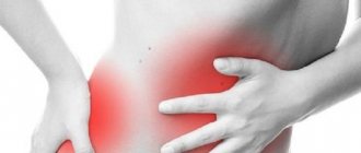
The main symptom of chronic cholecystitis is a dull pain in the right hypochondrium, which can last for several weeks, it can radiate to the right shoulder, and the right lumbar region, and be aching. Increased pain occurs after consuming fatty, spicy foods, carbonated drinks or alcohol, hypothermia or stress; in women, an exacerbation may be associated with PMS (premenstrual syndrome).
The main symptoms of chronic cholecystitis:
- Bitterness in the mouth, bitter belching;
- Heaviness in the right hypochondrium;
- Low-grade fever;
- Possible yellowing of the skin;
- Indigestion, vomiting, nausea, lack of appetite;
- Dull pain on the right under the ribs, radiating to the back, shoulder blade;
- Very rarely, atypical symptoms of the disease occur, such as heart pain, difficulty swallowing, bloating, constipation.
Chronic cholecystitis does not occur suddenly, it develops over a long period of time, and after exacerbations, against the background of treatment and diet, periods of remission occur; the more carefully you follow the diet and supportive therapy, the longer the period of absence of symptoms.
Diagnostics
In a conversation with the patient and when studying the medical history, the doctor draws attention to the reasons that could lead to the development of chronic cholecystitis - pancreatitis and other pathologies. When palpating the right side under the ribs, painful sensations occur.
Instrumental and hardware methods for diagnosing chronic cholecystitis:
- Ultrasound;
- cholegraphy;
- scintigraphy;
- duodenal intubation;
- arteriography;
- cholecystography.
Laboratory tests reveal:
- In the bile, if there are no stones, there is a low level of bile acids and an increase in the content of lithocholic acid, cholesterol crystals, an increase in bilirubin, protein and free amino acids. Bacteria that cause inflammation are also found in the bile.
- In the blood - increased erythrocyte sedimentation rate, high activity of liver enzymes - alkaline phosphatase, GGTP, ALT and AST/
DIAGNOSTICS OF CHRONIC AALCTIC CHOLECYSTITIS
This disease is difficult to diagnose due to the lack of specific signs. The clinical picture is often similar to symptoms of other pathological conditions. Only on the basis of a carefully collected anamnesis can a preliminary diagnosis be made.
Main types of research:
- Clinical and biochemical blood test: determines elevated levels of transaminases, alkaline phosphatase, γ-glutamyl transpeptidase;
- General urine analysis, coprogram;
- Duodenal intubation with bacteriological and biochemical examination of bile: allows you to determine the degree of inflammation;
- Ultrasound diagnostics: show deformation of the gallbladder, changes in its size, thickening or atrophy of the walls, unevenness of the internal contour, the presence of inhomogeneous contents with inclusions of heterogeneous bile;
- Cholecystography: evaluates the motor and concentration function of the gallbladder, its shape and position.
Treatment of chronic cholecystitis
Treatment tactics for chronic cholecystitis vary depending on the phase of the process. Outside of exacerbations, the main treatment and preventive measure is diet.
During the period of exacerbation, the treatment of chronic cholecystitis is similar to the treatment of the acute process:
- Antibacterial drugs for the sanitation of inflammation;
- Enzyme products - Panzinorm, Mezim, Creon - to normalize digestion;
- NSAIDs and antispasmodics to eliminate pain and relieve inflammation;
- Drugs that enhance the outflow of bile (choleretics) – Liobil, Allohol, Holosas, corn silk;
- Droppers with sodium chloride, glucose to detoxify the body.
In the presence of stones, litholysis (pharmacological or instrumental destruction of stones) is recommended. Medicinal dissolution of gallstones is carried out using drugs deoxycholic and ursodeoxycholic acids, instrumentally - extracorporeal shock wave methods, laser or electrohydraulic effects.
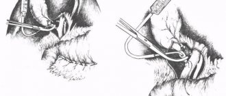
In the presence of multiple stones, persistent recurrent course with intense biliary colic, large size of stones, inflammatory degeneration of the gallbladder and ducts, surgical cholecystectomy (cavitary or endoscopic) is indicated.
Description example
Example of description: The gallbladder is enlarged in size ... cm, the contours are somewhat uneven, unclear, the wall is unevenly thickened to ... cm, the contents are heterogeneous with the phenomenon of sedimentation ..., in the projection of the neck of the gallbladder an inclusion of an ovoid shape (calculus) is visualized, with smooth, sometimes unclear contours , homogeneous structure /hypointense in all modes/, dimensions...see. The common bile duct is not dilated ... cm (the norm is up to 0.8 cm). In the area of the bladder bed, perifocally, around the descending part of the duodenum, as well as locally in the abdominal tissue on the right, an area of inflammatory infiltration without clear contours, with an approximate length of ... cm, without signs of abscess formation, was identified. Similar changes to a lesser degree of severity (reactive) are visualized in the area of perinephric fat on the right, along the lateral contour of the right kidney. Conclusion: MR picture of acute cholecystitis with inflammatory infiltration of paravesical and partially perinephric (right) tissue; GSD (gall bladder stone).
The gallbladder is pear-shaped, significantly reduced in size, the contours are uneven, the wall is thickened to ... cm, the contents are heterogeneous due to the presence of multiple small stones, bile in the form of small inclusions of high density. The common bile duct is not dilated... see Conclusion: MR signs of cholelithiasis, chronic calculous cholecystitis, blocked gallbladder.
Diet for chronic cholecystitis
In case of illness, it is necessary to strictly adhere to table No. 5 even during the period of remission for prevention. Basic principles of diet for chronic cholecystitis:
You cannot eat during the first three days of an exacerbation. It is recommended to drink rosehip decoction, still mineral water, sweet weak tea with lemon. Gradually, puree soups, porridges, bran, jelly, lean meat, steamed or boiled, fish, and cottage cheese are introduced into the menu.
Then you need to follow these recommendations:
- You need to eat small portions at least 4-5 times a day.
- Preference should be given to vegetable fats.
- Drink more kefir and milk.
- Be sure to eat a lot of vegetables and fruits.
- What can you eat if you have chronic cholecystitis? Boiled, baked, steamed, but not fried dishes are suitable.
- For the acalculous form of chronic disease, you can eat 1 egg per day. In case of calculosis, this product should be completely excluded.
The use of:
- alcohol;
- fatty foods;
- radish;
- garlic;
- Luke;
- turnips;
- spices, especially hot ones;
- canned food;
- legumes;
- fried foods;
- smoked meats;
- mushrooms;
- strong coffee, tea;
- butter dough.
Neglect of nutritional principles can cause serious consequences of chronic cholecystitis, leading to relapse of the disease and progression of inflammatory-destructive changes in the walls of the gallbladder.
Pathogenesis
Pathogenesis: The main pathogenetic link of cholecystitis is considered to be stasis of gallbladder bile. Due to dyskinesia of the biliary tract, obstruction of the bile duct, the barrier function of the epithelium of the bladder mucosa and the resistance of its wall to the effects of pathogenic flora are reduced. Stagnant bile becomes a favorable environment for the proliferation of microbes, which form toxins and promote the migration of histamine-like substances to the site of inflammation. With catarrhal cholecystitis, swelling and thickening of the organ wall occurs in the mucous layer due to its infiltration by macrophages and leukocytes.
The progression of the pathological process leads to the spread of inflammation to the submucosal and muscular layers. The contractility of the organ decreases to the point of paresis, and its drainage function worsens even more. An admixture of pus, fibrin, and mucus appears in infected bile. The transition of the inflammatory process to adjacent tissues contributes to the formation of a perivesical abscess, and the formation of purulent exudate leads to the development of phlegmonous cholecystitis. Due to circulatory disorders, foci of hemorrhage occur in the wall of the organ, areas of ischemia and then necrosis appear. These changes are characteristic of gangrenous cholecystitis.
Complications of chronic cholecystitis
Timely treatment of chronic cholecystitis allows you to maintain quality of life and avoid such serious complications as:
- internal biliary fistulas;
- acute form of pancreatitis;
- hepatitis;
- cholangitis;
- peritonitis - extensive inflammation of the peritoneum, which can occur as a result of perforation of the gallbladder and bile ducts;
- purulent abscesses in the abdominal cavity, including those localized on the liver.
Rehabilitation for chronic cholecystitis after treatment requires timely administration of medications, a gentle daily routine and strict adherence to a dietary diet. If you follow all the specialist’s recommendations, you don’t have to worry about possible complications or subsequent relapses of the disease.
Radiation diagnostics
Cholecystocholangiography: allows you to obtain information about the functioning of the gallbladder, assess the functional state of the liver, the sequence of filling of large intrahepatic and main extrahepatic bile ducts, determine their width, the state of the contours, the degree of homogeneity of the contrast pattern and, finally, the nature of the x-ray anatomical state of the end section of the common bile duct. Cholecystocholangiography usually gives a clearer, more intense, and by the end of filling the gallbladder (90 - 120 minutes after administration of the contrast agent) a homogeneous shadow of the latter, which makes it possible to more clearly identify the features of the contours of the gallbladder and the presence of stones in it. The latter are determined in the form of separate foci of clearing, often in a “suspended”, “floating” state, which is due to the lower specific gravity of the stones in relation to bile saturated with iodide preparations. The main advantage of this method is the indisputable establishment of the fact of blockage of the gallbladder, i.e. the absence of its contrast image when contrasting the main branches of the bile ducts.
Gallbladder scintigraphy (HIDA scan): allows you to objectively assess the bile synthetic and bile excretory functions of the liver. Scintigraphy is based on recording the passage of short-lived radionuclides through the biliary tract. Normally, the gallbladder, common bile duct and intestine are visualized within 1 hour. Complete obstruction of the cystic duct is characterized by the absence of visualization of the gallbladder on scinticagrams, which, with an appropriate clinical picture (and preserved bile outflow into the intestine), is a radiodiagnostic sign of acute cholecystitis. Delayed visualization of the gallbladder between 1 and 4 hours is a reliable sign of chronic cholecystitis.
CT signs of acute cholecystitis:
1. Cholelithiasis (visualization of stones directly depends on their chemical composition, which includes three components: bile pigments, cholesterol and calcium; the amount of calcium plays a decisive role in the sensitivity of CT);
2. Stretching of the gallbladder up to 5 cm in diameter or more, a symptom of stretching the bottom of the gallbladder - is positive when the stretched bottom of the gallbladder protrudes the anterior abdominal wall;
3. Circular or total thickening of the gallbladder wall;
4. Swelling or accumulation of effusion around the bladder, which is usually slightly enlarged in size. Subserosal edema may manifest as a thin, approximately 1 mm, hypodense outer edge around the denser inner layer of the bladder walls. Intramural edema is extremely rarely detected on CT. In this case, the inner mucous membrane and the outer serous are separated by a hypodense layer, which is called the “sandwich” symptom;
5. Poor visualization of the walls of the gallbladder and the phenomenon of pericholecystitis, according to most scientists, are the most important and reliable CT signs of acute inflammation. Perivesicular changes in the densitometric parameters of the liver parenchyma to the liquid level indicate the presence of inflammation and edema;
6. The wall intensively accumulates contrast;
7. The only pathognomonic sign of acute cholecystitis is gas in the cavity of the gallbladder, which indicates the emphysematous nature of the inflammatory process.
CT signs of complicated acute cholecystitis: The occurrence of gangrenous cholecystitis can be assumed with local accumulation of fluid near the gallbladder, thickening of the walls and perivesicular edema.
Phlegmonous perivesicular abscess formation is accompanied by compaction of the contents with densitometric indicators exceeding the density of bile. In this case, stones outside the bladder indicate its perforation. These stones can lead to wall erosion and fistula formation of surrounding hollow organs, such as the duodenum (most common), colon, and common bile duct. A direct sign of erosion of the biliary tract is aerocholia, detected by CT. The phenomena of partial small intestinal obstruction associated with the penetration of stones into the jejunum are described.
Empyema of the gallbladder is detected on CT scans extremely rarely, since pus has the same absorption coefficient as bile. At the same time, hemorrhagic cholecystitis is easily diagnosed due to a significant increase in the density of the contents, especially against the background of perivesicular edema and fluid.
Mirvis et al proposed diagnostic CT criteria for acute cholecystitis:
Main criteria: 1. Gallstones; 2. Thickening of the gallbladder wall; 3. Subserous edema; 4. Accumulation of fluid in the gall bladder bed.
Secondary criteria: 1. Stretching of the gallbladder; 2. Sludge syndrome.
CT signs of chronic cholecystitis : semiotic signs of chronic cholecystitis include: uneven thickening of the walls of the gallbladder, increased densitometric indicators, the presence of stones in it. The bubble is often contracted around the stones rather than expanded. Its deformation and kinks are often observed.
“Porcelain” gallbladder is an atypical manifestation of chronic cholecystitis associated with parietal calcification of the mucous membrane or smooth muscles of the organ.
MRI signs of acute cholecystitis are similar to CT signs: 1. Cholelithiasis;
2. Stretching of the gallbladder more than 5 cm in diameter; 3. Thickening of the gallbladder wall with its swelling, a double wall effect is possible; 4. Swelling or accumulation of effusion around the bladder; 5. Sludge syndrome; 6. The wall intensively accumulates contrast.
MRI signs of chronic cholecystitis: 1. Cholelithiasis; 2. Thickening of the wall of the gallbladder, slight swelling is possible; 3. Sludge syndrome.
MRCP: in acute cholecystitis, against the background of thickening of the gallbladder wall, MRCP additionally makes it possible to identify its layering due to intramural edema, a minimal amount of free fluid in the bladder bed, as well as minor filling defects in the lumen of the organ and in the cystic duct. In chronic cholecystitis, MRCP allows us to detect a defect in the filling of the gallbladder in the absence of changes in its wall. Also, with a long course of chronic calculous cholecystitis, with frequent relapses, deformation of the gallbladder is often detected, heterogeneous bile and a significant reduction in the size of the organ (the so-called “wrinkled” gallbladder) are visualized in the lumen of the bladder.
Prevention of exacerbations
To prevent the occurrence of the disease or avoid its exacerbation, general hygiene rules should be followed. Nutrition plays an important role. You need to eat food 3-4 times a day at approximately the same time. Dinner should be light and you should not overeat. Excessive consumption of fatty foods in combination with alcohol should be especially avoided. It is important that the body receives a sufficient amount of fluid (at least 1.5-2 liters per day).
In order to prevent chronic cholecystitis, it is necessary to allocate time for physical activity. This could be exercise, walking, swimming, cycling. In the presence of chronic foci of infection (inflammation of the appendages in women, chronic enteritis, colitis, tonsillitis), they should be treated in a timely manner, the same applies to helminthiasis.
If you follow the above measures, you can prevent not only inflammation of the gallbladder, but also many other diseases.
