Classification
There are congenital and acquired diverticula of the esophagus. Congenital ones can be found quite rarely. Acquired ones can be found more often, and they, in turn, are divided into several types:
- pharyngoesophageal or Zenker's - pusillon diverticula, which arise from the posterior part of the pharynx;
- bifurcation - traction diverticula that arise from the right wall of the esophagus at the level of the trachea. They act as a result of the inflammatory process in the pleura or mediastinum;
- epiphrenic or supradiaphragmatic are pulsion diverticula that arise from the right wall of the esophagus in the area of its lower part;
- multiple diverticula, which are usually functional, that is, they arise during the contraction of the esophageal wall and disappear after its relaxation.
Esophageal diverticula most often appear in the thoracic region.
Symptoms of the disease
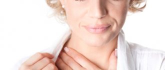
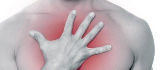
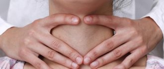
Diverticula of the upper esophagus are characterized by disturbances in swallowing food (dysphagia), regurgitation of undigested food, soreness or sensation of a foreign body in the throat, dry cough, increased salivation, nausea, and change in voice. Small diverticula of the middle part of the esophagus, as a rule, do not manifest themselves in any way; large ones cause dysphagia, regurgitation of undigested food, chest pain, nausea, and night cough. With diverticula of the lower part of the esophagus, these symptoms are supplemented by reflex shortness of breath, palpitations, bronchospasm, and heart pain caused by irritation of the vagus nerve.
Symptoms
Diverticula that are small in size usually resolve without symptoms. As they enlarge, food accumulates in the esophagus and it becomes compressed. In the presence of a disease such as esophageal diverticulum, symptoms may be: belching, dysphagia, regurgitation, bad breath. On the left side of the neck, protrusions of the esophagus may be noticed, which rumble when pressed on them. Diverticula can cause structures in the mediastinum that are adjacent to the esophagus - the heart, lungs, and large vessels. Swelling of the veins in the neck, strong heartbeat, shortness of breath, and voice changes are also evident.
Treatment of esophageal diverticula
Diverticula that do not bother the patient do not require treatment. However, it is necessary to adhere to a gentle diet that does not irritate the mucous membrane of the esophagus, and also to take certain measures to improve the emptying of the diverticulum (drinking after meals, finding an individual position that facilitates the passage of food through the esophagus, etc.). If large diverticula are present or there is a risk of complications, surgery is performed to excise the mass and suture the esophagus. In some cases, it is necessary to perform plasty of the esophageal wall with a flap of the diaphragm or pleura. After surgical treatment, patients should be observed for a long time by a gastroenterologist.
Esophageal diverticulum: symptoms and treatment
Pathologies of the digestive system can be caused by various reasons. In addition to the stomach and intestines, the esophagus also undergoes changes. The phenomenon of esophageal diverticulum is rare, so its symptoms are not immediately clear and treatment rarely begins on time. As a result, it is impossible to get rid of the disease in any other way, except for surgery.
What is it - esophageal diverticulum?
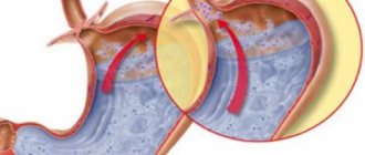
An esophageal diverticulum is a change in the walls of the organ that results in a protrusion. That is, the wall is deformed and a pouch forms on it in the mediastinum. The process may involve one layer of esophageal tissue or all three.
The protrusion can be located anywhere in the tubular organ, but is more often observed in the thoracic region. The most common carriers of the disease are men who have crossed the 50-year mark and suffer from other diseases of the digestive system:
- cholecystitis;
- gastritis;
- peptic ulcer;
- cholelithiasis and others.
Zenker's diverticulum affects the pharyngoesophageal part of the gastrointestinal tract, but belongs to the esophageal group because it has similar symptoms and development mechanisms.
With a traction diverticulum, constant stretching of the walls of the esophagus occurs, which causes a weakening of the muscle layer. The mechanical factor is adhesions with neighboring organs and lymph nodes. With pulsion, there is an external influence, for example, the pressure of a bolus of food.
Why does protrusion of the esophageal wall occur and why is it dangerous?
The causes of esophageal diverticulum are very diverse and are associated both with the anatomical characteristics of a person and with the action of external factors.
Thus, the congenital type of pathology occurs due to the weakness of the muscle tissue that forms the walls of the organ.
The incorrect lifestyle of the mother during the period of bearing the baby contributes its share, which does not allow his developing body to form correctly.
But more common is an acquired diverticulum, which forms as a consequence of:
When exposed to any of these factors, a change occurs in the walls of the esophagus: spastic contractions of the walls and an increase in internal pressure contribute to the formation of protrusions.
Among the factors that provoke the development of pathology are:
- alcoholism and smoking;
- unfavorable working conditions (inhalation of dust, toxic substances);
- imbalance in metabolic processes that develops against the background of illness or malnutrition;
- regular consumption of hot and spicy foods;
- eating large amounts of food at night.
If left untreated, the disease progresses, contributing to the development of complications:
- abscesses – localized purulent processes;
- wall erosion;
- stenosis (narrowing of the lumen);
- perforation (rupture) of the walls of the organ;
- phlegmon - diffuse purulent process;
- the appearance of aspiration pneumonia, which occurs as a result of the contents of the esophagus entering the respiratory tract;
- malignant neoplasms.
As complications develop, a person’s general well-being decreases, quality food intake becomes impossible, and the patient begins to lose weight (a symptom that signals the beginning of exhaustion of the body). In addition, the pathology affects nearby organs, causing structural and functional changes in them.
Classification
In addition to dividing into congenital and acquired, esophageal diverticula are distinguished by location:
- pharyngoesophageal;
- suprabronchial;
- supradiaphragmatic;
- abdominal.
Depending on the participation of the mucous membrane, the following types are distinguished:
- true, affecting all three layers of the walls of the esophagus;
- false, formed only due to protrusion of the mucous membrane.
All these varieties belong to the same disease, which has an ICD code - Q 36.9, respectively, their treatment and symptoms are similar, but have their own characteristics.
Esophageal diverticulum: symptoms and signs
Let us now consider the symptoms of esophageal diverticulum. If the protrusion is less than 2 centimeters in size, it usually does not appear in any way. Larger sizes reveal themselves with the following signs:
- belching with an unpleasant odor;
- difficulty passing food through the organ;
- sensations of soreness and scratching in the throat;
- production of large amounts of saliva;
- nausea and vomiting after eating;
- constipation;
- heartburn;
- a sharp increase in temperature is a symptom of infection;
- severe cough, not accompanied by sputum production;
- in a horizontal position, food can move in the opposite direction;
- feeling of a lump in the throat and others.
Source: https://stomach-diet.ru/divertikul-pischevoda-simptomyi-i-lechenie/
Sources
- Samanta J., Mandavdhare HS., Kumar N., Kumar-M P., Jafra A., Chauhan R., Gupta P., Kumar KH., Singh H., Dutta U., Kochhar R. Per Oral Endoscopic Myotomy for the Management of Large Esophageal Diverticula (D-POEM): Safe and Effective Modality for Complete Septotomy. // Dysphagia - 2021 - Vol - NNULL - p.; PMID:33533970
- Miutescu B.P., Khan S., Mony S., Khashab M.A. Role of Peroral Endoscopic Myotomy (POEM) in the Management of Esophageal Diverticula. // Clin Endosc - 2021 - Vol53 - N6 - p.646-651; PMID:33238358
- Gupta V., Darling G. Commentary: Measure Twice, Cut Once: Understanding the Anatomy and Physiology of Esophageal Epiphrenic Diverticula Guides Optimal Surgical Management. // Semin Thorac Cardiovasc Surg - 2021 - Vol33 - N1 - p.249-250; PMID:33171244
- Kamal F., Khan MA., Lee-Smith W., Sharma S., Marella HK., Iqbal U., McDonough S., Aslam A., Ismail MK., Tombazzi C., Adler DG. Peroral Endoscopic Myotomy Is a Safe and Feasible Option in the Management of Esophageal Diverticula: Systematic Review and Meta-Analysis. // Dig Dis Sci - 2021 - Vol - NNULL - p.; PMID:33123940
- Demeter M., Ďuriček M., Vorčák M., Hyrdel R., Kunda R., Bánovčin P. S-POEM in treatment of achalasia and esophageal epiphrenic diverticula - single center experience. // Scand J Gastroenterol - 2021 - Vol55 - N4 - p.509-514; PMID:32251609
- Basile P., Gonzalez JM., Le Mouel JP., Irarrazaval R., Caillo L., Barthet M. Per-oral endoscopic myotomy with septotomy for the treatment of distal esophageal diverticula (D-POEM). // Surg Endosc - 2021 - Vol34 - N5 - p.2321-2325; PMID:32144556
- Jacobs C., Draganov PV., Yang D. Two birds, one scope: peroral endoscopic myotomy as a treatment for achalasia and esophageal diverticula. // Endoscopy - 2021 - Vol52 - N2 - p.153; PMID:31991470
- Maydeo A., Patil GK., Dalal A. Operative technical tricks and 12-month outcomes of diverticular peroral endoscopic myotomy (D-POEM) in patients with symptomatic esophageal diverticula. // Endoscopy - 2021 - Vol51 - N12 - p.1136-1140; PMID:31614371
- Liu XY., Li QL., Jiao TW., Sun MJ., Zhou PH. Peroral endoscopic myotomy regains anatomical structure and improves emptying for achalasia with multiple esophageal diverticula. // Endoscopy - 2019 - Vol51 - N12 - p.E392-E393; PMID:31344726
- Sivanes S., Chang J. Gastrointestinal: A rare cause of upper gastrointestinal bleeding: Epiphrenic esophageal diverticula. // J Gastroenterol Hepatol - 2021 - Vol35 - N10 - p.1665; PMID:31273838
Diverticula of the middle third of the esophagus
Saccular protrusions of the esophageal wall of such localization most often do not manifest themselves in any way. Sometimes moderate dysphagia, chest pain, and regurgitation of food are observed, which most authors interpret as signs of diverticulitis or peridiverticulitis. Without disputing this point of view, it should be noted that in some cases, with bifurcation diverticula of the esophagus, a very peculiar clinical picture occurs. We observed a patient in whom a saccular protrusion of the middle third of the esophagus measuring up to 7 cm in length was manifested exclusively by paroxysms of intense dysphagia caused by complete compressive obstruction of the esophagus. During the period between attacks, the patient felt practically healthy and could even eat food quite freely. At first, such symptoms were regarded as the result of intermittent esophagospasm, the diagnosis of which was rejected only during repeated long-term recordings of motor activity of the esophagus.
Course and complications of bifurcation diverticula. In the vast majority of cases, the disease is benign. Complications are extremely rare. The most dangerous of them is perforation of the diverticulum with the subsequent formation of fistulous communications with neighboring organs and tissues, most often with the trachea, bronchi and pleural cavity. The etiological factors of such a complication can be diverticulitis, microperforations and trauma to the diverticular sac, on the one hand, as well as a chronic inflammatory process in the lungs and mediastinal organs, on the other. The formation of fistulous communications between the esophagus and the above-mentioned organs is accelerated under the influence of esophageal dyskinesia, hiatal hernia, and reflux esophagitis. It should also be added that suppuration, bleeding from the diverticulum cavity, and stenosis of the esophagus are possible.
Malignancy of saccular protrusions of the esophagus is observed in 1.08-2.3% of cases of all diverticula of this organ. Of this number, cancer of traction pockets, which include bifurcation pockets, accounts for 7.8%. The source of the malignant tumor is the so-called foci of disembryogenesis around the diverticular crypts: poorly differentiated epithelium, papillomatosis, heterotopic tissue, in particular cartilaginous tissue. Typically, diverticula become malignant in people over 45 years of age. In 6.6% of cases, esophageal diverticula are associated with malignant neoplasms of the stomach that occur against the background of anacid conditions. The presence of the latter is explained by the fact that in some patients with traction diverticula, the adhesive process involves branches of the vagus nerve.
Treatment of diverticulum at ON CLINIC
The doctor prescribes a diet that reduces the impact on the affected area. Teaches how to empty a diverticulum. The diverticulum is washed with antiseptic solutions. If there is a risk of complications, surgical intervention is possible.
ON CLINIC is a modern medical center where diagnosis and treatment of esophageal diverticulum takes place at the highest level available to modern world medicine. Basic information that patients with gastrointestinal diseases should know:
What are esophageal diverticula and why are they dangerous?
This is a protrusion of the wall of the esophagus with the formation of a cavity in the form of a bag or tube. Over time, diverticula can cause very serious complications.
What to do if signs of the disease appear?
If you experience any signs indicating the presence of esophageal diverticulum, you should immediately visit a doctor. ON CLINIC has a specialized department where consultations are carried out by specialized specialists of the highest category every day.
How is the disease diagnosed?
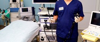
At the appointment, the gastroenterologist collects anamnesis and then prescribes the necessary diagnostics. The specialist may suggest undergoing an X-ray contrast study using barium, plain radiography, computed tomography of the chest, or endoscopic examination. The examination is carried out using modern, high-quality imported equipment (France), Olympus (Japan), Siemens (Germany), Pentax (Japan).
How to get tested?
The clinic has its own laboratory, equipped with the most modern equipment. You can even undergo a comprehensive examination in one day.
How is esophageal diverticulum treated?
The treatment program is selected individually, based on the examination results. Doctors at ON CLINIC use proprietary treatment methods, including specialized diets. Diagnosis and treatment take place without pain, in comfortable conditions, allowing you to achieve the highest results.
How affordable is treatment for esophageal diverticulum at ON CLINIC?
Treatment for diverticula at ON CLINIC is available to patients of any income level. There are programs of discounts and benefits, and it is possible to receive treatment on credit.
Diagnosis of esophageal diverticulum
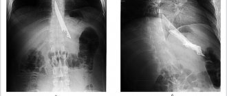
X-ray of the esophagus with contrast is the main method for diagnosing esophageal diverticula. The patient is given a barium suspension, which is opaque to X-rays, to drink, and a series of photographs are taken. This allows you to see the contours of the esophagus and, in fact, the diverticulum.
If a diverticulum is suspected, esophagoscopy is used with extreme caution so as not to cause perforation (rupture) of the diverticulum. It allows you to assess the condition of the mucous membrane in the area of protrusion, the presence of erosions, ulcers and other pathological changes.
Esophageal manometry - measuring pressure in the esophagus - allows you to study the motility of the organ. In situations where the clinical manifestations of diverticulum are similar to the symptoms of cardiovascular diseases, ECG and Holter monitoring (24-hour ECG monitoring) are prescribed.
Classification of esophageal diverticula
For reasons of appearance:
- pulsion - arise due to increased intraesophageal pressure, which provokes protrusion of a section of the wall;
- traction - appear against the background of prolonged inflammation, in place of which scar tissue is formed, tightening the wall of the esophagus around the diverticulum.
By location:
- Pharyngeal-esophageal (pharyngoesophageal, Zenker's): formed on the posterior wall of the pharynx in the area of its connection with the esophagus. Occur against the background of increased pressure inside the esophagus;
- Bifurcation (parabronchial): located in the area of bifurcation (bifurcation) of the trachea into two main bronchi. Usually result from chronic inflammation in this area of the chest (bronchitis, pleurisy, pericarditis);
- Supradiaphragmatic (epiphrenal) - usually a consequence of increased intraesophageal pressure.