Indications for examination
It is recommended to perform an abdominal ultrasound if the following complaints are present:
- abdominal pain;
- bloating;
- bitterness in the mouth;
- jaundice;
- increased gas formation;
- tendency to constipation;
- feeling of heaviness and fullness in the stomach;
- bad breath;
- palpable organ changes;
- injuries;
- vomit;
- regurgitation.
Other indications for ultrasound include long-term use of medications, alcohol abuse, and suspicion of the presence of tumor diseases. An abdominal ultrasound is also performed to monitor the effectiveness of the treatment.
Causes
Gastric cancer is a malignant tumor with uncontrolled growth that develops from the cells of the mucous membrane of the organ. Separately, lymphoma is distinguished - a tumor that is formed from lymphatic tissue (there is a lot of it between the layers of the wall). However, during the initial diagnosis before biopsy, they cannot be separated, since symptoms and signs often overlap.
Among the causes of oncological pathology it is necessary to highlight:
- infection with Helicobacter pylori infection;
- chronic inflammatory and erosive processes (gastritis and peptic ulcer);
- long-term presence of Crohn's disease;
- genetic predisposition;
- decreased reactivity of the immune system;
- smoking and alcohol abuse;
- irregular meals;
- exposure to ionizing radiation and environmental pollution.
What organs are examined on an abdominal ultrasound?
During the examination, doctors evaluate the size, contours, location and structure of:
- gallbladder;
- liver;
- spleen;
- pancreas.
In addition, based on the results of an ultrasound of the abdominal organs, the presence or absence of retroperitoneal lymph nodes, fluid and neoplasms is determined.
General practitioners, gastroenterologists, pediatricians, oncologists, and family doctors can refer you for examinations. You can also get detailed information from specialists on how to prepare for the examination.
Classification
A classification of stomach cancer is often used depending on the degree of progression and spread of the cancer process:
- Stage 0 – the process affects only the mucous membrane;
- Stage 1 – growth of pathologically altered cells into the submucosal layer is observed, distant foci cannot be detected;
- Stage 2 - the process spreads to the muscular and serous layers of the wall, several foci were also found in the nearest lymph nodes;
- Stage 3 – the tumor actively metastasizes through the regional lymphatic system (more than 3 confirmed foci), but without involving other organs;
- Stage 4 - when performing an ultrasound, CT or MRI, it was possible to find foci of the process in other organs (liver, spine, brain, spleen, kidneys or others).
Stages of development of stomach cancer
What can be revealed on an abdominal ultrasound
Using ultrasound diagnostics, a doctor can detect the following conditions:
- gallstones;
- hepatitis;
- liver dystrophy;
- cirrhosis;
- neoplasms;
- lymphadenopathy;
- congenital or acquired structural organ disorders;
- post-traumatic disorders.
In addition, the procedure allows you to study blood flow in the area under study. Circulatory disorders are diagnosed in a similar way.
Factors distorting the results
In order to obtain the most accurate ultrasound results, it is necessary to eliminate possible causes leading to distortion of the picture. Among them are:
- drinking alcohol, strong coffee, energy drinks, which contribute to muscle spasms;
- taking antispasmodics;
- presence of gases in the intestines;
- recent endoscopic examination;
- residual contrast agent in the body.
Excess body weight makes the examination difficult. This is due to the poor permeability of ultrasonic waves through the fat layer. Therefore, it is important to properly prepare for the examination.
Preparing for the examination
Preparation for ultrasound of the abdominal organs and kidneys begins 3-4 days before the study. Recommendations on how to prepare for the examination include the following:
- Following a diet aimed at reducing gas formation.
- Ultrasound of the abdominal organs is performed on an empty stomach. Therefore, the last meal should be 4-5 hours before the examination. In case of a removed gallbladder, food and water intake is allowed.
- In case of previous radiography with contrast injection, ultrasound should be performed after 3 days.
- Inform your doctor in advance about taking medications and agree on their discontinuation if necessary.
When preparing for an ultrasound of the abdominal organs, important importance is attached to the diet.
| Authorized Products | Prohibited Products |
| Cereal porridge | Milk and any dairy products |
| Chicken breast | Cabbage, radish, radish |
| Beef | Alcohol |
| Low-fat varieties of hard natural cheese | Legumes |
| River fish | Carbonated drinks |
| Boiled eggs | Pasta and flour products |
| Soups | Fresh apples, pears, grapes |
| Turkey meat | Coffee and caffeinated drinks |
| Stewed or steamed vegetables | Sweet desserts |
| Boiled potatoes | Chewing gum |
Adult patients are allowed to take certain medications a few days before the examination. The prescription must be carried out by the attending physician.
- If there is increased gas formation or bloating, medications may be taken.
- To normalize digestion, enzyme preparations are allowed.
- On the eve of the procedure, it is allowed to take an adsorbent.
Additional Methods
A number of other diagnostic methods are used to diagnose stomach cancer. Their comparative characteristics are given in the table:
| Ultrasound | FGDS with biopsy and cytological examination | CT with contrast | MRI | |
| Characteristic | Non-invasive ultrasound examination | Endoscopic examination with tissue sampling | X-ray examination with intravenous contrast | Using the phenomenon of magnetic resonance |
| Symptoms of stomach cancer | Changes in echogenicity, wall thickness, contour evenness | Violation of the integrity of the mucous membrane, change in its color, proliferation of tissue of various shapes, deformation | “Defect” of contrast accumulation, wall deformation, change in its thickness and shape | Thickening of the wall, changes in the shape of the stomach, changed lymph nodes |
| Information content of primary tumor diagnosis | ++ | ++++ | ++++ | +++ |
| Information content of metastasis detection | ++ | – | +++ | ++++ |
Special preparation cases
In some cases, preparation recommendations may change. This applies to people with certain health problems, special physiological conditions and children.
- Pregnant
Metabolic processes in pregnant women may occur more intensely than in other people. Therefore, the last meal before the examination can be 1.5-2 hours. However, it is not recommended to exceed the standard serving size.
The serving size and timing of ultrasound may be revised for patients with multiple pregnancies.
- Patients suffering from diabetes mellitus
Patients with diabetes follow a special diet. Long breaks between meals can worsen the condition. Therefore, the preparation stage for this category of people has some peculiarities: immediately before the study it is permissible to eat a light breakfast.
- Children
For infants, the last feeding is allowed 3 hours before the procedure. Children under 15 years of age are allowed to eat 4 hours before the test. In cases where the child persistently asks to eat, you can give him a few sips of water. However, it is worth noting that to obtain the most accurate results, ultrasound should be performed on an empty stomach.
- Gallbladder function test
Involves performing an ultrasound with a test breakfast. Initially, the examination is carried out on an empty stomach, then 30 minutes after eating. The following products are allowed as a trial breakfast: yogurt, sour cream, 2 yolks, boiled egg.
Clinical picture
The symptoms of the disease also differ greatly depending on the stage of the process. But the problem with this oncological pathology is that the clinical picture, even in stage 3, may not differ from gastritis or peptic ulcer. In patients with first detected stomach cancer, the following complaints are most common:
- periodic pain of varying intensity in the upper abdomen (on an “empty” stomach, or after eating);
- feeling of abdominal discomfort or fullness after a small snack
- nausea;
- a sharp change in diet, the appearance of aversion to certain types of dishes;
- decreased appetite and decreased body weight (even with an unchanged diet);
- black feces;
- belching with an unpleasant odor.
Important If you have the symptoms listed above, you should immediately seek qualified medical help.
How is an abdominal ultrasound performed?
The essence of the method is to direct ultrasonic vibrations to various tissues, which are characterized by varying degrees of reflectivity and absorption. A special sensor detects reflected rays. Thanks to this, it is possible to obtain a visual image of the area under study on the monitor screen.
During the examination, the patient sits on a comfortable couch. A special gel is applied to the skin of the abdomen. Scanning is performed using a sensor. The doctor sees an image of the internal structures in real time. The examination procedure is absolutely painless and comfortable. The average duration of the study is up to 20 minutes. In some cases, the time may be increased at the discretion of the diagnostician. If necessary, the specialist may ask the patient to change his body position or hold his breath for a few seconds.
Decoding the results
The results of the examination are delivered 10-15 minutes after its completion. If any changes are detected, the diagnostician will give detailed comments in an accessible form and recommend further observation by a specialized specialist. The conclusion reflects the following parameters:
- Size, shape, structure of the abdominal organs (liver, spleen, gall bladder, pancreas). Ultrasound also involves assessing the condition of the kidneys.
- Proliferation of organ tissue.
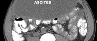
- The presence or absence of fluid in the abdominal cavity.
- The presence or absence of gallstones.
- Aortic diameter.
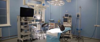
At the multidisciplinary clinic “Zdorovye”, ultrasound of the abdominal organs is performed using modern equipment. Expert class equipment has high resolution. This allows doctors to accurately determine the location of pathological changes in the area being examined.
Our team consists of the best professionals in the city. Doctors have many years of experience and regularly improve their skills in specialized courses. Diagnosticians detect diseases with high accuracy even in the early stages of development.
Possibilities of the technique
Ultrasound examination is prescribed for almost all patients with suspected benign or malignant disease in the stomach . High-frequency waves pass well through human tissue and are partially reflected from them (depending on the density indicator). They are then captured by a sensor located on the front wall of the abdomen, processed by a computer and displayed as an image on the screen.
Ultrasound is a routine test if there are symptoms of dyspepsia, decreased appetite or pain in the upper abdomen. In addition to the stomach, the condition of the pancreas, liver, gallbladder, spleen, lymph nodes and large vessels of the abdominal cavity is examined.
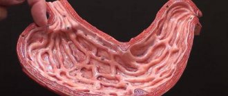
With high-quality preparation of the patient, ultrasound allows one to clearly visualize the walls of the antrum and body of the stomach . The bottom of the organ in most patients is poorly visible due to the presence of gases in it (even a small amount). If the study is carried out by an experienced specialist, he can also approximately estimate the volume of gastric juice that is in the cavity.