Foreign bodies of the esophagus
The reasons for the entry of foreign bodies into the esophagus may be the habit of holding various objects in the mouth (in small children, in workers of certain professions), negligence in food preparation and hasty eating, and the deliberate swallowing of various objects by mentally ill people. In more than 50% of cases, the foreign body freely passes through the esophagus and through other parts of the digestive tract and exits naturally. Sharp foreign bodies (needles, nails, fish and meat bones, etc.) get stuck in the initial part of the esophagus or in other parts of the digestive tract. Large objects are retained in places of physiological narrowing of the esophagus (at the level of the trachea bifurcation, above the cardia). The retention of a foreign body in the esophagus is facilitated by spasm of the esophageal muscles in response to irritation of the mucous membrane by the foreign body.
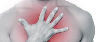
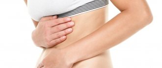
Clinical picture and diagnosis. Symptoms depend on the type of foreign body, the level of its retention in the esophagus, and the degree of damage to the esophageal wall. After suddenly swallowing a foreign body, patients experience a feeling of fear, pain in the throat, in the area of the jugular fossa or behind the sternum, which intensifies when swallowing saliva or liquid.
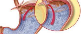
If a large foreign body enters the oropharynx at the same time as the esophagus, the entrance to the larynx is blocked, so instant death from asphyxia is possible. Prolonged stay of a foreign body in the esophagus causes traumatic esophagitis, ulcerations, perforation of the esophageal wall with the subsequent development of complications in the form of mediastinitis, purulent pleurisy, etc.
The diagnosis is based on medical history and x-ray examination. In this case, metallic foreign bodies are easily detected; less contrasting foreign bodies are detected during examination of the esophagus with a water-soluble contrast agent and esophagoscopy. In case of perforation of the esophagus, leakage of the contrast agent beyond its contours and the presence of mediastinal emphysema are noted. Esophagoscopy, which clarifies the nature of the foreign body and its location, is of great diagnostic importance. An esophagoscope can be used to remove a foreign body.
Treatment. If a foreign body is suspected in the esophagus, the patient must be urgently sent to a surgical hospital. Removal is carried out using a fiberoptic or rigid esophagoscope and a set of special tools for grasping a foreign body. If it is impossible to remove it through an esophagoscope, surgery is indicated to remove the foreign body. Surgical access to the esophagus depends on the level of location of the foreign body. After removing the stuck object, the wound in the esophageal wall is sutured.
Foreign body in the gastrointestinal tract
Definition, etiology and pathogenesistop
Ingestion and foreign body entrapment in the gastrointestinal tract is more common in children. In adults, the most common reason for intervention is pieces of food, dentures or implants stuck in the esophagus - most often in diseases of the central nervous system that occur with dysphagia, along with post-inflammatory stenosis, eosinophilic esophagitis or tumor (cancer) of the esophagus; less common causes are pharyngeal diverticula (including Zenker's diverticulum), Schatzky's ring and complications of surgery (eg, narrowing of the anastomosis in patients after partial resection of the esophagus or stomach) and radiation therapy.
Clinical picturetop
1. Foreign body in the esophagus: almost always causes symptoms (dysphagia, odynophagia, chest pain, esophageal obstruction, nausea and vomiting). Respiratory symptoms, such as choking, shortness of breath, and stridor (guttural whistling), occur as a result of aspiration of saliva or pressure of a foreign body on the trachea. Drooling and inability to swallow indicate complete closure of the esophagus.
2. Foreign body of the alimentary canal below the esophagus: usually does not cause symptoms, but can lead to significant complications such as perforation of the gastrointestinal tract. Symptoms of perforation: fever, tachycardia, peritoneal symptoms, subcutaneous emphysema and neck swelling.
Diagnosticstop
The history should include information about the time of the event and the type of foreign body ingested, as well as the time after which symptoms began.
Additional research methods
1. RG: a stuck piece of food does not require this examination. In the case of other foreign bodies, perform X-ray of the neck, chest and/or abdominal cavity, preferably in 2 projections (front and side). Classic X-ray has limited effectiveness (≈50% false negative results), mainly due to the poor absorption of X-rays by many foreign bodies, such as a piece of food, fish or chicken bones, wood, plastic, glass. Avoid the use of liquid barium contrast agents due to the risk of aspiration and potential endoscopic intervention.
2. CT: indicated in case of doubt, as well as in case of suspected severe complications such as perforation.
3. Endoscopy: a study confirming the presence of a foreign body in the gastrointestinal tract, as well as the main therapeutic intervention. If a piece of food gets stuck in the esophagus for no apparent organic reason, biopsies should be taken for histological examination to exclude eosinophilic esophagitis.
treatmenttop
Conservative therapy
Possible in the case of asymptomatic, small and blunt objects located below the esophagus (usually eliminated from the body 1–2 weeks after passing into the stomach). Perform weekly abdominal X-ray examination to assess foreign body movement along the gastrointestinal tract. In case of delay in the stomach after 3-4 weeks. endoscopic intervention is indicated.
Pharmacological treatment has limited use and should not delay endoscopic or surgical treatment; There are several reports of the usefulness of glucagon in the treatment of a stuck foreign body in the esophagus.
Endoscopic treatment
Necessary in 10–20% of patients. The time of endoscopic intervention depends on the type and location of the foreign body in the gastrointestinal tract:
1) urgent intervention (within several hours) - sharp foreign bodies and batteries in the esophagus, as well as foreign bodies that completely close the lumen of the esophagus;
2) accelerated intervention (within 24 hours) - small, blunt foreign bodies in the esophagus and magnets, batteries, sharp or long objects (>5-6 cm; may become wedged in the duodenal flexure) in the stomach;
3) planned intervention (within 72 hours) - medium-sized foreign bodies in the stomach (>2–2.5 cm in diameter [may become wedged in the pylorus or in the area of the ileocecal valve] and <5–6 cm in length).
If a foreign body gets stuck in the esophagus, it is enough to carefully push it with the tip of the endoscope into the stomach. In other cases, treatment consists of intercepting the foreign body using an appropriate instrument (forceps, loop, mesh, Dormia basket) and removing it from the gastrointestinal tract. To protect the airway and esophageal walls from damage, it is recommended to use additional accessories such as tubes or latex caps installed at the end of the endoscope. In cases where the risk of aspiration is high, tracheal intubation is indicated before endoscopic surgery.
Surgery
Necessary in ≈1% of cases, mainly after unsuccessful endoscopic treatment. Indications for emergency surgery: gastrointestinal perforation, bleeding that cannot be controlled endoscopically, and small bowel obstruction by a foreign body.
In clinical practice, unusual foreign bodies (FBs) of the esophagus and stomach, swallowed by patients intentionally, are rarely encountered.
A large number of tools have been proposed for extracting FB from the gastrointestinal tract (forceps, loops, baskets). However, it is not always possible to remove FB from the stomach using a flexible endoscope [1]. In addition, there is no consensus among clinicians regarding the choice and sequence of use of rigid and flexible esophagoscopes, depending on the complexity of the FB and the suspected complication [2].
In 2007, S. Gulati et al. A rare gastric FB has been described - a toothbrush, which was removed using a rigid esophagoscope [3]. The peculiarity of such FBs is their shape and significant size, which does not allow them to migrate beyond the duodenum. Attempts at endoscopic extraction of such FBs are not always successful, so open operations are required.
We present a clinical observation.
Patient M.
, 27 years old, entered the Research Institute of SP named after. N.V. Sklifosovsky 08/05/14 with complaints of dull pain in the epigastric region. According to the patient, since 2010 he has been observed in a psychoneurological clinic for schizophrenia. He was repeatedly treated in a psychiatric hospital. 3 hours before entering the institute, against the background of imperative, verbal hallucinations, he swallowed a kitchen knife. He had not made any previous suicide attempts.
Upon examination, the patient’s condition is satisfactory, he is conscious and available for productive contact. No particularities in organs and systems.
Mental status: accessible to contact, oriented in place, time and self, mood background is smooth, no criticism. He reported that he heard “voices” that ordered him to swallow a knife. Cannot help but submit to verbal hallucinations.
The patient underwent an X-ray examination of the abdominal cavity (Fig. 1, a). Radiographs revealed no free gas in the abdominal cavity. In the middle, in the posterior sections at the level of the VIII-XII thoracic vertebrae, a large foreign body of metallic density is detected - a knife blade with the tip upward. The length of the metal foreign body is 12 cm. When marking the esophagus and stomach with a water-soluble contrast agent during a polypositional study, the upper part of the foreign body for 8 cm is located in the lumen of the lower third of the esophagus, and the lower part passes into the lumen of the stomach (Fig. 1, b). No disruption of the contrast medium's patency or leakage beyond the contours of the esophagus and stomach was detected.
Rice. 1. Radiographs. a - straight; b - oblique projection. Explanation in the text.
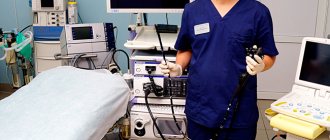
Taking into account the fact that the patient has a highly traumatic foreign body located partly in the esophagus and partly in the stomach, it was decided to perform a combined esophagoscopy (using a rigid and flexible esophagoscope) under general anesthesia.
Tube No. 1 of a rigid Brunnings esophagoscope was passed into the esophagus, through which a fibrogastroscope was inserted. The latter is carried to the lower third of the esophagus, where the proximal part of the IT is visualized - a knife blade with serrated sharpening (Fig. 2, a). The esophageal mucosa in the middle and lower third was not changed; no traumatic injuries to the esophageal mucosa were detected at the level of the foreign body or proximally. The IT is captured by a loop passed through the channel of the fibrogastroscope, by the proximal part of the blade. With constant air insufflation, the FB is pulled into the upper third of the esophagus and inserted into the lumen of the rigid esophagoscope tube (Fig. 2, b), after which the fibrogastroscope is removed, and the FB is captured by a rigid crocodile forecept and removed along with the rigid esophagoscope tube (Fig. 3 ).
Rice. 2. Photographs during esophagoscopy. a - foreign body in the lumen of the esophagus; b — foreign body in the tube of the Brunnings esophagoscope.
Rice. 3. Removed foreign body.
During control fibroesophagoscopy, the mucous membrane of the oropharynx is not changed, and in the area of the entrance to the esophagus there are single pinpoint submucosal hemorrhages. The mucosa distal to the located parts of the esophagus and the area of the cardioesophageal junction is also not changed. During a control X-ray examination of the esophagus with a barium suspension, the act of swallowing was not impaired, the esophagus was freely passable, and no leakage of the contrast agent was detected. No gas was detected in the soft tissues of the neck and peri-esophageal tissue.
After removal of the FB, the patient experienced moderate pain when swallowing for 2 days, which went away on its own on the 3rd day. After 6 days, the patient was discharged in satisfactory condition under the supervision of a doctor at a psychoneurological dispensary.
Thus, the above clinical observation shows that combined esophagoscopy (using a rigid and flexible esophagoscope), performed under anesthesia, allows you to remove a large foreign body of the esophagus and stomach of increased trauma (knife) without damaging the walls of the esophagus and oropharynx and avoid open surgery.