Diseases of the stomach and intestines - the organs that make up the gastrointestinal tract - occupy 1st place among all diseases occurring in people of different ages. These pathologies bring patients many unpleasant moments - from an uncomfortable state to excruciating pain. But the most dangerous thing is that it is gastrointestinal diseases that cause a huge number of complications - perforated ulcers, severe inflammation and cancerous tumors, leading to disability and even death. That is why it is recommended for every person to undergo a gastrointestinal ultrasound periodically, even if nothing hurts yet.
And your gastrointestinal tract is healthy: stomach health is a matter of time
The content of the article
Let's look at medical statistics on diseases of the stomach and intestines. Alas, it is frightening, even without taking into account hidden patients who have not been examined and residents of the poorest countries where there is no access to medical services.
According to statistics:
- Almost 90% of the population of developed countries suffers from gastritis of varying degrees of neglect.
- 60% of the world's inhabitants are infected with Helicobacter pylori, a bacterium that causes inflammation of the mucous membrane of the stomach and intestines, and is the cause of gastritis and stomach ulcers.
- In Western countries, up to 81% of citizens, according to statistics, periodically experience heartburn, which is a symptom of gastroesophageal reflux disease - a disease of the esophagus that leads to disruption of the gastrointestinal tract.
- About 14% of people have stomach ulcers.
At the age of over 60 years, the quality and length of life depends on the condition of the stomach and intestines, but it is possible to get rid of existing pathology only in the initial stages of the disease. That is why it is so important to be attentive to your health and not bring the problem to a chronic stage.
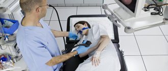
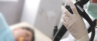
Gastroscopy at the Central Clinical Hospital of the Russian Academy of Sciences (in a dream)
At the Central Clinical Hospital of the Russian Academy of Sciences, modern equipment is used to perform gastroscopy; the examination is carried out by a highly qualified endoscopist. When performing gastroscopy under anesthesia, the patient's condition is monitored throughout the entire study by an experienced anesthesiologist. After completing the procedure, the patient spends several hours in a comfortable day hospital at the clinic - this is necessary to monitor his condition while recovering from anesthesia.
Advantages of visiting the RAS clinic:
- reliable protection of personal data;
- highly qualified endoscopists;
- consultations with doctors of the highest qualification category;
- attentive and caring nursing staff;
- modern equipment;
- quick receipt of examination results;
- the possibility of performing gastroscopy in a dream.
After undergoing gastroscopy, a consultation with a gastroenterologist is necessary. At the Central Clinical Hospital of the Russian Academy of Sciences, consultations are conducted by doctors with extensive experience in clinical practice - professors, doctors of medical sciences. The study of modern therapeutic and diagnostic methods allows our specialists to detect diseases of the stomach and other digestive organs at the earliest stages and begin treatment on time. This allows us to achieve positive results in the treatment of most gastrointestinal pathologies: both common and rare.
You can make an appointment with a gastroenterologist or for gastroscopy at the Central Clinical Hospital of the Russian Academy of Sciences on the website or by phone.
How to check the stomach and intestines quickly, cheaply and informatively?
There are several types of examination of the intestines and stomach, but only ultrasound diagnostics has a full range of advantages, which doctors consider invaluable and very effective in making a diagnosis.
- An ultrasound can be done urgently for any patient’s condition. The examination will take a maximum of 15-30 minutes.
- Ultrasound diagnostics are carried out painlessly, without causing psychological discomfort. Unfortunately, other methods of examining the gastrointestinal tract require very unpleasant procedures - swallowing tubes, inserting sharp instruments into the anus, sometimes to a considerable depth, ingesting liquids that cause vomiting, etc.
- Ultrasound is completely safe. The method is based on echolocation and does not require the use of X-ray and MRI equipment.
- This is one of the cheapest examinations. An examination of the gastrointestinal tract along with the rest of the abdominal organs will cost around 1 thousand rubles.
With all this, this technique is sometimes even more informative than other methods of examining the stomach and intestines. For example, unlike the endoscopic diagnostic method (using probes that are inserted inside), ultrasound reveals intestinal inflammation, thickening and protrusion of the walls, stenosis (expansion of the lumen), abscesses, fistulas, congenital anomalies (Crohn's disease), neoplasms in the early stages of development diseases.
Preparation for gastroscopy under anesthesia (in a dream)
Gastroscopy is prescribed by the patient's attending physician, usually a gastroenterologist. The procedure may be recommended after an MRI, in case of certain patient complaints, or in case of negative test results.
The study is usually carried out in the morning and is performed on an empty stomach. Eating is possible no later than 7 hours before the examination. You must refrain from smoking for at least 2 hours: it irritates the gastrointestinal mucosa, which negatively affects the accuracy of the study. During the examination, the patient lies on his left side, with a cushion placed under his head. When performing gastroscopy without anesthesia, immediately before the procedure, the patient receives local anesthesia: the oral cavity and pharynx are treated with lidocaine in the form of an aerosol. When performing gastroscopy under general anesthesia, intravenous anesthesia is used. With this type of examination, the patient is asleep, so he does not experience any discomfort when the doctor inserts a probe into the oral cavity, esophagus and stomach. To perform endoscopy under anesthesia, hospitalization for several hours in a day hospital is required. The duration of an endoscopic examination depends on its type and purpose. As a rule, the procedure takes from 5–7 minutes in the absence of serious pathological changes to 15–30 minutes in the presence of multiple disorders or the need to perform any therapeutic manipulations.
Specifics of the gastrointestinal tract examination: why the stomach and intestines need to be examined in detail
Despite the close relationship between the stomach and intestines, the doctor examines both organs in detail, since they not only have similar diseases. For example, ulcers can be localized in any part of the gastrointestinal tract or form in all parts at once. The same applies to oncological tumors, inflammation and other processes.
Depending on the patient’s complaints, the specialist examines the intestines and stomach separately. Having received data indicating dangerous processes, the doctor refers the patient for additional diagnostics.
Along with an ultrasound, it is recommended to simultaneously take a breath test for Helicobacter pylori. This analysis is also not traumatic - the patient will only need to exhale air a few times. A complex ultrasound plus analysis for Helicobacter will allow literally in 15-20 minutes to identify the cause of heartburn, pain and cramps in the abdomen, diarrhea or constipation, bloating and other symptoms, establish the extent of the processes and prescribe treatment without resorting to unpleasant diagnostic methods.
Gastroscopy
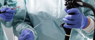
Gastroscopy is an endoscopic examination of the esophagus and stomach using a video camera attached to a flexible probe.
The device is inserted through the patient's mouth. Gastroscopy of the stomach is prescribed as an independent diagnostic procedure or performed during esophagogastroduodenoscopy (EGDS) - it depends on the capabilities of the equipment used. The procedure is generally painless, but many patients experience great fear and are unable to swallow the endoscope. In such cases, doctors use sedation. The patient is put into light medicated sleep, which significantly reduces stress levels and allows the doctor to carefully examine all areas of the stomach.
You can have a gastroscopy in Moscow at the Alfa Health Center clinic. We create the most comfortable conditions for each patient, using anesthesia if necessary. We guarantee high accuracy of results and reasonable prices for gastroscopy in a dream. The price list for services is posted on our website.
How to examine the intestines: Ultrasound plus additional techniques
The intestine has three sections: the large, small intestine and rectum, and the study of each of them has its own characteristics and nuances.
- Ultrasound of the colon
helps detect cancer at a very early stage. To make sure, the patient is prescribed a contrast X-ray and colonoscopy. Irrigoscopy, an X-ray examination using contrast fluid, will also be very effective. The method allows you to “see” areas that are invisible to colonoscopy and difficult to distinguish with ultrasound, for example, areas of bends or accumulations of mucus. - Ultrasound of the small intestine
is hampered by tortuosity and deep burial, as well as the accumulation of gases that distort the image on the monitor. A special curved sensor and the latest high-precision equipment help to examine the small intestine. Ultrasound evaluates wall thickness, visualization of layers, patency, wall expansion, and peristalsis. - An ultrasound of the duodenum
is performed together with an examination of the stomach. Allows 100% diagnosis of stomach ulcers, cancer, gastroduodenitis.
Depending on the area being examined, the doctor uses a sensor with certain characteristics.
What can be replaced?
If the patient has contraindications to FGDS, the doctor prescribes an examination using alternative methods. It is considered advisable to combine several diagnostic procedures to obtain more accurate data and prevent errors at the diagnostic search stage.
Sometimes patients themselves look for an alternative to gastroscopy, experiencing discomfort or fear of the study. In such cases, it is better to coordinate your actions with your doctor and choose a diagnostic method together . After all, to make a diagnosis in each case, a different amount of examination may be required. Below we list the main alternative studies and briefly discuss each of them.
Ultrasound machines for intestinal examination
The intestines are examined using two types of sensors: transabdominal (through the abdominal wall) and endorectal. To study the colon, a 2D device is sufficient, which produces a flat two-dimensional image. Such an examination already provides reliable information about the patient’s health status. The endorectal method is more informative because the sensor is inserted into the anus and examines the organ from the inside.
The doctor decides which sensor to choose depending on the patient’s complaints. In special cases, both methods are used.
- In 15% of cases, the transabdominal sensor “does not see” the rectum, as well as the area of the anal canal. The endorectal method is not possible with stenosis of the terminal gastrointestinal tract (abnormal narrowing).
- The endorectal probe usually examines the distal parts of the rectum. A rectal examination requires preparation.
Gastroscopy without swallowing the probe
This type of examination also includes transnasal fibrogastroscopy, in which a probe is passed through the nasal passages and descends into the stomach along the back wall of the pharynx. This is a gentle method intended for particularly sensitive patients exposed to stress. For example, in case of hypertension, inserting a fiberscope through the mouth can provoke a surge in pressure or even a hypertensive crisis. With transnasal FGS, these undesirable consequences can be avoided, since negative emotions from the manipulation are minimized.
Gastroscopy of the stomach without swallowing a probe becomes possible in its pure form thanks to capsule endoscopy. The patient is asked to swallow a small capsule, inside of which there is a built-in video camera and a video signal transmitter for it. There are different companies that produce capsules, they may have specific features, for example, there are capsules intended for the small or large intestine, for the stomach. Along with the capsule, the patient receives a signal receiver. Then, from this receiver, the doctor will take the data obtained during capsule endoscopy, but at this stage the patient can leave the hospital and return home. Later, the capsule will naturally leave the gastrointestinal tract, having previously captured the entire picture throughout the gastrointestinal tract. It is disposable and it is not necessary to control its output.
If you experience discomfort, changes in bowel habits or pain, you should immediately contact your doctor. A relative disadvantage of this study is that it is only diagnostic in nature; there is no possibility of conducting therapy or taking material for analysis.
Price issue
The cost of studying the gastric mucosa correlates with the complexity of the study. On average, prices for classic fibrogastroscopy vary from 2 to 4 thousand. With additional manipulations (biopsy, pH measurements) carried out during the study, the price can increase to 10 thousand.
Transnasal gastroscopy is limited to an average of 4 thousand rubles, so it is impossible to carry out additional actions.
Video capsule gastroscopy is the most expensive study, since the most expensive materials and modern technological developments are used to create a disposable capsule (20-50 thousand).
*Prices are indicated in rubles.
Preparation and performance of intestinal ultrasound
Preparation for the procedure begins 3 days in advance, the patient refuses food that causes constipation or flatulence (legumes, sweets, flour products, smoked and spicy foods).
The day before, from 18.00, the patient completely refuses any food, having first taken a laxative (Guttalax, Regulax, Duphalac, Bisacodyl). If there are problems with peristalsis, the patient is given an enema, and in special cases, a special cleansing enema is performed using a Bobrov apparatus (a glass vessel for introducing a large amount of liquid inside).
In the morning, the patient goes for an ultrasound examination until 11.00 am. This is due to the fact that the procedure is carried out only on a well-cleansed intestine and a completely empty stomach, while long breaks in food intake are contraindicated.
In the ultrasound diagnostic room, the patient lies on the couch on his side with his back to the machine, having first removed his clothes below the waist and lowered his underwear. The legs are tucked with the knees to the chest. Ultrasound begins in the direction from the lower parts to the higher ones. In parallel with this, the doctor moves the probe in such a way as to examine the intestine in the transverse, longitudinal and oblique planes. When the echogenic picture is not entirely clear, the doctor asks the patient to change position (lean on his knees and elbows, stand up).
Ultrasound of the large intestine is performed using a transabdominal probe. A contrast liquid (barium sulfate solution) is first injected into the empty intestine. Thanks to this, a clear picture is obtained on the monitor screen.
To examine the rectum, 3.5-5 MHz sensors are used. Ultrasound of a given length passes through the soft tissue of the intestine, being reflected back. The built-in receiving sensor picks up the signal and transmits it in processed form to the monitor screen. Various compactions, neoplasms and erosions are expressed in the form of white, black or mixed areas of varying echogenicity. An experienced doctor does not make a diagnosis immediately, but correlates the data obtained with the results of tests and other studies.
Description of the procedure
Planned FGDS is prescribed in the morning. The procedure is carried out in an equipped endoscopy room.
Before manipulation, it is necessary to remove objects squeezing the body:
- belts;
- chains;
- tight clothes.
How does the procedure work?
Step-by-step process of the procedure:
- An hour before gastroscopy, the patient is given premedication - 1 ml of atropine and 1 ml of trimeperidine are administered intramuscularly.
- The person is placed on the couch on his left side. You can bend your legs slightly and wrap your arms around yourself at elbow level. Place a tray in front of your face. During the examination, you need to behave correctly - you cannot move, turn your head, swallow saliva, or talk.
- The patient is given a silicone insert to clamp between the teeth, through which the probe is inserted. It is necessary to prevent damage to the gastroscope. If it is impossible to perform the procedure due to the person’s strong tension or a pronounced gag reflex, anesthesia is performed - then it will be painless.
- Next, the doctor inserts a tube into your throat. You need to swallow it while taking a deep breath. When the tube passes through the pharyngeal ring, the discomfort stops.
- The endoscopist examines the internal organs. Sometimes it is possible to go through the intestine completely and examine the place of its transition to the next section.
- After this, the doctor carefully removes the gastroscope tube.
Photo gallery
Full insertion of the probe
Examination directly through a gastroscope
On-screen inspection
Gastroscopy with anesthesia
Is it painful to swallow a tube and tube?
The gastroscope probe has a diameter of 1.5-2 cm, so it does not cause pain when swallowing. Only discomfort appears from the presence of a foreign object in the throat.
The thickness of the tube for small children is even smaller - 5-10 mm. Therefore, there is no need to worry about pain, because the diameter of the esophagus is much larger and the tube will pass through it absolutely freely.
How often can you do it
Initially, healthy people do not often need to undergo gastroscopy. FGDS is carried out only according to indications, on which the regularity of the manipulation will depend. It is prescribed when appropriate symptoms appear; the further number of examinations depends on the established diagnosis.
If you have uncomplicated gastrointestinal diseases, the test can be taken once a year. Severe pathologies - with complications, rapidly progressing - require gastroscopy every 3-6 months. In most cases, the frequency of FGDS is determined individually.
Interpretation of intestinal ultrasound results
A healthy intestine has two membranes. The outer one is muscle tissue with low echogenicity, the inner mucous membrane is in contact with gas, and therefore is visualized as a hyperechoic layer.
During an ultrasound examination, the following parameters are assessed:
- Dimensions and shape
. The wall thickness is 3-5 mm. The picture is distorted in the event of the formation of gases that deform the ultrasound, and insufficient filling of the intestines with liquid. - The location of the intestine
relative to other organs. - Wall structure (echogenicity)
. The outer layer is hypoechoic, while the inner wall is characterized by hyperechogenicity. The contours are smooth, the intestinal lumen should not have expansions or contractions. Peristalsis is noticeable. - Length and shape of various sections.
The thermal section is 5 cm, the middle section is 6-10 cm, the middle ampullary section is 11-15 cm. - Lymph nodes.
Should not be visualized.
Deviations from the norm indicate various pathologies:
- Enteritis (inflammation of the small intestine): dilation of the intestine, increased peristalsis, accumulation of contents of varying echogenicity;
- Hirschsprung's disease (congenital pathology of an increase in certain sizes of the intestine): significant expansion of the lumen, uneven contours, heterogeneous wall thickness, noticeable places of thinning, lack of peristalsis;
- If it is impossible to determine the layers of the intestine, we can talk about acute mesenteric thrombosis - a consequence of myocardial infarction, expressed in thrombosis of the mesenteric artery;
- Uneven internal contours (which causes ulcerative lesions of the mucosal surface), weak echogenicity, thickening of the wall - all this indicates nonspecific ulcerative colitis;
- Chronic spastic colitis: areas of high echogenicity against the background of a hypoechoic surface, thickening of the walls;
- Ischemic colitis: inability to visualize layers, uneven thickening, reduced echogenicity;
- Acute appendicitis: the monitor screen shows a vermiform appendix 7 mm in diameter, the layers of the appendix do not differ from each other, the walls of the appendix are thickened asymmetrically, free fluid is visualized, increased echogenicity indicates an abscess;
- Diverticulitis (protrusion of the intestinal walls): at the site of the diverticulum, ultrasound “sees” a thickening of the wall more than 5 mm above normal, echogenicity indicates an abscess, the contours are uneven;
- Mechanical damage to the intestine: in addition to severe tension in the abdominal muscles, the patient’s echogenicity at the site of the hematoma is reduced, the walls at the site of damage are thickened;
- Oncology (cancerous or precancerous tumor): the external contours are uneven, the lumen is narrowed, peristalsis is impaired at the site of the tumor, lymph nodes of reduced echogenicity are visualized.
Of course, only a doctor can make a diagnosis. In this case, the results of other studies should be taken into account, for example, a blood test showing the degree of inflammation and the presence of parasites in the gastrointestinal tract, ultrasound of the liver and pancreas, etc.
What are the advantages and disadvantages of ultrasound of the intestinal gastrointestinal tract?
Ultrasound diagnostics of the intestine is used for initial examination in case of suspected pathology, as well as in cases where the endoscopic method is contraindicated due to the patient’s health condition (perforation (damage) of the intestine, inflammatory process).
Ultrasound examination of the intestines has a number of advantages:
- The patient does not experience psychological discomfort.
- The doctor receives information about the size of the organ, its structure, thickness, number of layers, without penetrating inside the organs.
- Ultrasound allows you to examine the inflamed intestines and clearly sees the upper gastrointestinal tract.
- Peristalsis is visualized in real time and intestinal obstruction is determined.
- On an ultrasound of the intestines, the specialist will see even small compactions or changes in the echostructure of tissues.
- Ultrasound allows you to do screening (endorectal method), completely confirm or refute oncology.
Despite the large number of advantages, diagnosing this organ with ultrasound has some disadvantages, the main one of which is the impossibility of making an accurate diagnosis without additional examination.
Also, the disadvantages of the method include the following:
- Only functional disorders in the functioning of the organ are detected.
- Structural changes are determined without defining the parameters of the changes.
- It is not possible to assess the condition of the internal mucous surface; if structural changes are detected, colonoscopy is prescribed - an endoscopic method
Tests and studies that complement intestinal ultrasound
As mentioned above, intestinal ultrasound is not 100% confirmation of a particular diagnosis, although in many ways the method is informative and accurate. Depending on the preliminary diagnosis, in addition to ultrasound, the patient is prescribed:
- Capsule examination
. The patient swallows a capsule with a sensor inside, which conducts video surveillance and transmits the image to the monitor screen. The method allows you to see areas inaccessible to the endoscope. Significant advantages also include the absence of trauma (the intestinal walls are not scratched) and radiation (unlike X-rays).
The disadvantages of the capsule technique include the low prevalence of capsule examination, because the method was first tested in the USA in 2001, and today it is still not widespread. Its cost is very high, and this limits the circle of clients. Other disadvantages include the inability to conduct a capsule study in case of intestinal obstruction, infections, and peritonitis. The method has age restrictions associated with the peculiarities of peristalsis.
- Colonoscopy
. This is an endoscopic method that allows you to examine the internal mucous membrane for polyps, colitis, tumors, Crohn's disease, inflammation and other pathologies. The disadvantage of this method is the risk of intestinal trauma and perforation (punctures of the walls). Colonoscopy also does not see tumors between the intestinal walls. - Irrigoscopy
. This is a special method aimed at identifying hidden tumors located between the inner and outer lining of the intestine. In addition, the method, unlike colonoscopy, sees areas on the folds of the intestine and its remote areas.
Irrigoscopy involves the introduction of a liquid solution of barium sulfate through the anus, which allows a clear contrast image to be obtained upon contact with air. The advantages of irrigoscopy are the ability to examine structural changes in tissue (scars, diverticula, fistulas). The method is used for diarrhea or constipation, mucus in the intestines, pain in the anus.
“In 30-40% of cases, screening colonoscopy reveals precancerous changes”
— At what age should a colonoscopy be done?
- If there are indications, then it is performed even on children and infants. After all, there are congenital pathologies. If we talk about screening colonoscopy, and now a colorectal cancer screening program has been launched in Belarus, then it should be done starting from the age of 50 for people who are not worried about anything. That is, these are relatively healthy people who have no vomiting, no nausea, no constipation, no diarrhea, but they are over 50 years old. In 30−40% of such cases, during screening colonoscopy, we identify various precancerous changes.
— What is the difference between colonoscopy and ileocolonoscopy?
— The difference is in the depth of insertion of the device. Ileocolonoscopy is a colonoscopy, that is, an examination of the large intestine, plus an examination of a section of the small intestine. Ileocolonoscopy is only needed in a limited number of patients when some inflammatory and degenerative bowel disease, such as Crohn's disease, is suspected. But not every patient can undergo ileocolonoscopy.
- Why?
— There are anatomical features of the structure of the body in almost any person. The anatomy that is taught in institutes is actually represented in perhaps 10% of people. Everything else is a variant of the norm.
All people have hands, and their hands have fingers. But some fingers are long, some are short, some are thick, and others are thin. It’s exactly the same inside: one person’s intestines are turned upside down in a loop, another has sharp corners. But the thinner the patient, the more difficult it is for him to do a colonoscopy. In a thin and overweight patient, the length of the intestine is the same, but the volume of the abdominal cavity is different, and in a small volume the intestine is strongly shifted.
In addition to anatomical nuances, there are features that arose as a result of the treatment of certain diseases. Let’s say a patient has undergone surgery on the abdominal organs, and there are already adhesions and anastomoses there...
— To what depth is the endoscope inserted during a colonoscopy?
— The length of the endoscope is 1 meter 60 cm, and in some cases it is inserted to this depth. But it all depends on how the colon is laid out. And it can be laid 60 cm, or maybe 1 meter 60 cm.
— Does a person feel pain during a colonoscopy or is it rather discomfort?
“It’s discomfort, but some people perceive it as pain. Each person has their own pain sensitivity threshold. We have been performing colonoscopy without anesthesia for many years, and I must say: if you make every effort, then for 80% of patients the examination proceeds quite comfortably.
Of course, anesthesia makes it possible to conduct the examination more calmly for both the patient and the doctor, but one must understand that pain indicates that something is wrong with the examination itself. When the patient experiences pain, it means that the intestine is overstretched somewhere or its mesentery is very stretched. And when the patient is under anesthesia, he doesn’t feel anything, and we don’t know it. That is, in fact, there are pros and cons.
In my opinion, colonoscopy should be attempted without anesthesia, but we must be prepared to use it if necessary. This would be ideal.
— What to do if a person is afraid to have a colonoscopy, but he should?
— When a doctor prescribes any test, you need to ask why it is prescribed. When the patient goes for a study and knows for what purpose it is prescribed, then issues with fear are resolved on their own. But in a number of cases we see excesses in the prescription of invasive studies. Perhaps, sometimes it is necessary to do a non-invasive radiation study to determine the diagnosis. After all, as a result of colonoscopy there can be complications, for example, perforation of the colon. This is a hole in the colon that will require surgical repair.
- Does this happen rarely?
— It’s rare, but it happens, and you need to know about this, because in most cases our patients do not know what complications may arise as a result of colonoscopy. Or maybe bleeding, intestinal volvulus, intestinal obstruction... Our whole life is a scale. And if we put what is beneficial on one cup and what is harmful on the other, then which cup outweighs? This should not only be decided by the doctor. The patient must also take some part in his destiny.
Ultrasound of the stomach is an important part of the gastrointestinal tract examination using ultrasound.
For a long time, ultrasound diagnostics was not used in the study of the stomach. This is due to the fact that the stomach is a hollow organ, and the air does not allow full use of a conventional ultrasound sensor - special sensors are needed to examine the back walls. In addition, accumulated gases distort the displayed results. However, medicine does not stand still, and modern techniques already provide sufficient information to make an accurate diagnosis.
Sensors for studying the stomach appeared relatively recently, in the late 2000s. However, the speed and safety of scanning makes ultrasound examination of the stomach increasingly popular.
During an ultrasound examination, the doctor evaluates the organ according to the main indicators:
- Stomach volume.
It is a hollow muscular organ that resembles a pouch. The volume of an empty stomach is 0.5 liters, and when full it stretches to 2.5 liters. The height of the stomach reaches 18-20 cm, width - 7-8 cm. When filled, the stomach stretches up to 26 cm in length and up to 12 cm in width. - Structure.
Near the heart is the cardiac region, in which the esophagus passes into the stomach. On the left you can see the bottom of the organ, where air entering with food accumulates. The body of the stomach is the largest part, rich in glands that produce hydrochloric acid. The pyloric zone is the transition from the stomach to the intestine. There, partial absorption of substances received from food occurs. - Structure.
The walls of the stomach have a muscular layer that is responsible for contracting and promoting the food coma. The serosa is intermediate between the muscular and mucous layers. Lymph nodes and blood vessels accumulate in it. The mucous layer is covered with the finest villi, which secrete gastric juice produced by the glands. - Blood supply.
The circulatory system covers the entire organ. The organ is supplied with venous blood by three main vessels: the left, hepatic and splenic. The venous network runs parallel to the arterial network. Various bleeding occurs when the gastric mucosa is damaged (ulcers, tumors).
In what cases are there no analogues?
Among the indications for gastroscopy, there are several particularly important ones, in the presence of which it cannot be avoided :
- gastrointestinal bleeding (vomiting "coffee grounds" or black stools);
- bleeding from varicose veins of the esophagus (bloody vomiting);
- foreign bodies in the esophagus or stomach.
This is due to the fact that in such cases FGDS is performed not only for diagnostic purposes, but also for therapeutic purposes. it is not possible to eliminate the problem using other methods , it is impossible to replace the procedure.
It is also important to perform gastroscopy in patients with suspected gastric cancer and the need for a biopsy . After all, the patient’s management tactics depend on the results of the study.
How is an ultrasound of the stomach performed?
Preparation for an ultrasound of the stomach is similar to an ultrasound of the intestines: the patient adheres to a strict diet for 3 days, and the night before does not eat any food from 18.00. If there is a tendency to form gas, the patient drinks 2 capsules of Espumisan before bed. In the morning, half an hour before the procedure, you should drink a liter of water so that the walls of the stomach straighten.
There is also a method of ultrasound examination with contrast. Water is an excellent conductor of ultrasound, and without it, scanning an organ is somewhat difficult.
The procedure is carried out on an empty stomach. The doctor assesses the condition and thickness of the walls on an empty stomach, looking for the presence of free fluid. Then he asks the patient to drink 0.5-1 liter of liquid, and uses an ultrasound machine to evaluate changes in the expanded stomach. A third ultrasound scan is performed 20 minutes later when the stomach begins to empty. The doctor evaluates the motility of the organ and the rate of fluid loss. Normally, a glass of water (250 ml) comes out of the stomach in 3 minutes.
The patient lies on the couch on his side, the specialist applies gel to the peritoneal area and moves the sensor over the surface. Periodically, he tells the patient to change position or change his posture slightly. The doctor pays attention to the following indicators:
- position of the stomach and its size
- Has the mucous surface of the stomach expanded?
- is there thickening or thinning of the walls
- what is the state of the circulatory system of the stomach?
- contractility of the stomach
- are there inflammations and neoplasms?
The entire examination takes a maximum of 30 minutes and does not cause discomfort or pain. Ultrasound, unlike FGDS, is much easier to tolerate for children and the elderly.
Advantages and disadvantages of ultrasound of the stomach when examining the gastrointestinal tract
The doctor prescribes an ultrasound examination of the stomach to the patient as a primary auxiliary diagnostic method.
The advantages of ultrasound are as follows:
- the exit section most susceptible to diseases is examined;
- ultrasound “sees” any foreign bodies in the cavity;
- Ultrasound accurately assesses the thickness of the walls of the organ;
- thanks to the method, venous blood flow is clearly visible;
- using diagnostics, benign and malignant tumors of minimal size are identified;
- stomach ulcers are well assessed;
- the degree of inflammation of the gastric mucosa varies;
- the method allows you to see reflux disease - the reflux of the contents of the lower sections back into the stomach;
- the organ is examined from different points and in different sections, which is impossible with x-rays;
- Ultrasound sees what is happening in the thickness of the stomach wall;
- thanks to the echo structure, ultrasound can easily distinguish a polyp from an oncological neoplasm;
- in addition to diagnosing the stomach, ultrasound diagnostics reveals concomitant pathologies of other organs (usually with gastritis, diseases of the biliary tract and pancreas develop);
- Ultrasound is performed on newborns and small children for whom it is impossible to undergo an FGDS or x-ray.
The main advantage of ultrasound over FGDS is the ability to detect forms of cancer developing in the thickness of the organ wall (infiltration forms), which cannot be detected using fibrogastroscopy.
Despite all the advantages, ultrasound has some disadvantages that do not allow the method to become widespread as an independent examination of the stomach.
The disadvantages include the following:
- Unlike endoscopic examination, ultrasound does not allow tissue samples to be taken for further study (for example, gastric juice;
- scraping of the mucous membrane, tissue biopsy);
- Ultrasound cannot assess the degree of changes in the mucous membrane;
- limitation of the areas studied (it is possible to examine only the outlet zone of the stomach).
Fibrogastroscopy
Fibrogastroscopy is the most common way to examine the gastric mucosa by inserting a fibrogastroscope into the organ cavity. This procedure is indicated for patients who are suspected of having gastritis, stomach ulcers, tumors, or polyps. Sometimes FGS is prescribed to identify possible causes of allergies or neuroses; the range of diseases is quite wide. Now the study is carried out in two versions of the probe: through the mouth and through the nose.
FGS with transoral administration
The procedure lasts only a few minutes, and the doctor can announce preliminary results immediately after the manipulation.
The patient is in a reclining position on his stomach, holding a special plastic mouthpiece in his mouth. The gastroenterologist endoscopist passes a probe through it and asks the patient to swallow the tube. Since light anesthesia was administered in advance, his gag reflex is weakened, and the patient does not feel the urge to gag, only discomfort and a sensation of a foreign body.
The advantages of this method include:
- short duration of the study (only 2-5 minutes);
- quick receipt of visual observation results;
- the ability to manipulate a video camera under eye control to study areas of particular interest;
- the possibility of carrying out therapeutic measures (biopsy, coagulation of bleeding vessels, removal of polyps);
- minimal risk of complications.
The disadvantages of fibrogastroscopy include:
- long period of preparation for the study, dietary restrictions;
- discomfort during gastroscopy;
- a large number of contraindications.
FGS with transnasal administration
This test is not yet widely used; transnasal testing means passing a flexible probe through the nose, along the back of the throat and down the esophagus. Since the fiberscope does not affect the root of the tongue and the uvula of the soft palate, the patient is not bothered by the gag reflex. The patient no longer requires local anesthesia or sedation. If there are allergic reactions to the anesthetic, this will be a separate point in favor of the transnasal method.
Obviously, the tube in this case will be much thinner than with fibrogastroscopy through the mouth. The diameter of the tube should not exceed half a centimeter, which means that the additional capabilities of gastroscopy will be significantly limited (you cannot take a biopsy through a thin channel, you cannot perform coagulation during bleeding). Such a tube is easier to insert, and the quality of the study itself does not deteriorate at all.
Scheme of passing the tube through the nose.
In addition, with the transnasal route of administration, the patient’s verbal functions are preserved; he can immediately report any unpleasant sensations to a specialist, which significantly reduces fear and anxiety before the procedure.
But, like any procedure, transnasal FGS has its drawbacks. Some patients report the appearance of nosebleeds after gastroscopy through the nose.
For fibrogastroscopy, regardless of the method of administration, there are a number of contraindications that reduce the versatility of the method. Relative contraindications are temporary; when reduced body functions are restored, they are removed, and FGS becomes possible. In addition, with the development of life-threatening conditions and the need for urgent gastroscopy, you can turn a blind eye to some of these indications. Such restrictions include:
- inflammatory diseases of the ENT organs;
- last trimester of pregnancy;
- burns of the esophagus and oral cavity;
- angina pectoris and advanced arterial hypertension.
Absolute contraindications limit gastroscopy indefinitely. In the presence of varicose veins, severe narrowing, scars of the esophagus, aortic aneurysm and curvature of the spine, FGS is strictly contraindicated.
What does ultrasound of the stomach reveal when examining the gastrointestinal tract?
The ultrasound method is not the most popular when examining the gastrointestinal tract, but it makes it possible to obtain very important information.
The stomach is an extension of the digestive canal in the form of a bag. It is a hollow organ whose walls have an outer muscular layer and an inner mucous layer. The mucous membrane is rich in glands that produce gastric juice and hydrochloric acid, as well as enzymes. With their help, incoming food is softened and treated with a natural antiseptic. The stomach is separated from the esophagus by the sphincter, and from the duodenum by the pylorus.
The organ is examined by ultrasound in two ways:
- Transabdominal (through the walls of the peritoneum). It is carried out with different sensors, but the results always require additional confirmation.
- Probe (sees the stomach from the inside). Used extremely rarely.
When conducting a study using a sensor, the specialist pays attention to the following:
- thickness, folding, structure of the mucous membrane (are there any neoplasms, bulges, or irregularities on it);
- thickness of the muscle layer (expansion or thinning indicates pathology);
- the integrity of the gastric wall (are there any perforations, ulcers or neoplasms);
- amount of free fluid (indicates inflammation);
- peristalsis, motility and contractility of the stomach;
- transitional sections of the stomach (sphincter and pylorus, their features
- functioning).
It is worth noting that ultrasound of the stomach and duodenum is significantly inferior in its informative value to the more popular method known as FGDS. But in some cases, other research methods are unacceptable for the patient due to health conditions or fear of a traumatic procedure.
Transabdominal examination identifies three layers of the gastric wall: hyperechoic mucosal layer (1.5 mm), hypoechoic submucosal layer (3 mm) and hyperechoic muscular layer (1 mm). With the probe method of research, 5 layers up to 20 mm thick are determined.
Ultrasound diagnostics of the stomach allows us to identify the following pathologies
| Symptoms | Possible disease |
| Swelling of the antral mucosa | Acute pancreatitis, nephrotic syndrome (kidney damage) |
| Thickening of the stomach wall, uneven rounded neoplasm, rich in blood vessels, no boundaries between layers, no peristalsis | Carcinoma (malignant tumor) with distant metastases |
| Lack of boundaries between layers, narrowing of the pyloric lumen | Pyloric stenosis (narrowing of the pylorus due to scarring caused by an ulcer) |
| Changes in the echostructure of the stomach walls, the walls are expanded, the contours are uneven | Neuroma (tumor developing from the tissues of the peripheral nervous system), leiomyoma (benign tumor of the smooth muscles of the stomach), adenomatous polyp |
| Expansion of the abdominal region (compared to the norm) after filling the stomach with water, splitting of the echo signal, the presence of hypoechoic inclusions, stagnation of fluid in the cardiac region | Gastroesophageal reflux (reflux of intestinal contents back into the esophagus) |
| Small amount of fluid, rapid release of fluid from the stomach, changes in the contour of the stomach | Diaphragmatic hernia |
| Dense hyperechoic formations with a clear structure, the boundaries between the layers are clearly visible, the echogenicity of the mucous and muscle layers is not changed | Cystic formations |
| Uncertain changes recorded by ultrasound | Affected hollow organ syndrome. This diagnosis requires mandatory confirmation by other types of research (CT, MRI, FGDS, X-ray). |
| Anechoic crater-like areas on the inner wall of the stomach | Stomach ulcer |
Ultrasound scanning of different parts of the stomach
Thanks to ultrasound, the doctor assesses the condition of the following areas of the organ:
Bulbar or duodenal bulb
. This part of the organ is located in the area where the stomach exits, and controls the flow of contents processed by gastric juice into the intestinal lumen. With intestinal diseases, ulcers and sites of inflammation form on the bulb. The main reasons for duodenal ulcers are increased acidity and the bacterium Helicobacter pylori, which begins to actively multiply under such conditions.
The study is carried out in real time with a linear or convex sensor with a frequency of 3.5-5 MHz. To detail the condition of the walls, sensors with a frequency of 7.5 MHz are used, but they are ineffective for obese patients with developed subcutaneous fat.
If a patient is diagnosed with a gastric and duodenal ulcer, then in most cases the walls of the bulb are affected. On ultrasound, this is reflected by anechoic areas, because, unlike healthy walls, the ulcer does not reflect ultrasound.
The diagnosis of “stomach and duodenal ulcer”, if areas of anechoicity are identified on ultrasound, is made conditionally. Additionally, the condition of the walls of the bulb is assessed (they have a mucous structure with longitudinal folds). The normal thickness should be no more than 5 mm, and in the antrum (the transition of the stomach into the duodenum) - up to 8 mm. With thickening, we are not talking about an ulcer, but about an oncological neoplasm. The patient will need additional research: endoscopic with sampling of material for biopsy.
Due to the fact that ultrasound is not able to establish an accurate diagnosis, the patient is given a preliminary diagnosis of “anechoic areas”, and then he is sent for fibrogastroduodenoscopy. It is this method that makes it possible to take tissue from the wall of the bulb to determine the nature of the pathology. FGDS also allows you to assess the condition of the organ’s vessels.
Pyloric canal or pylorus of the stomach.
This is a slight narrowing at the junction of the bulb and the duodenum. It consists of smooth muscle walls 1-2 cm long, located both in the annular and transverse directions. Normally, there is some curvature of the canal. Ultrasound can detect diseases such as polyps, stenosis (narrowing), ulcers, and pyloric spasm.
Sphincter (cardia)
- This is the border between the peritoneum and the esophagus. Normally, the sphincter opens only after eating, and remains closed the rest of the time. Due to its functional significance, the sphincter has a stronger muscular layer than the stomach, which allows it to open and close like a valve. When eating, the sphincter closes the exit from the stomach, allowing food to be digested. But as a result of increased acidity and other pathologies, the organ ceases to function normally, and the contents of the stomach enter the esophagus.
Who is gastroscopy indicated for?
It is recommended to check the stomach with an endoscopic capsule for people who cannot swallow a tube due to narrowing and spasm of the esophagus or are afraid or do not want to do this.
But the symptoms of the disease require clarification of the diagnosis. Tubeless gastroscopy is recommended if the patient is concerned about:
- heartburn;
- nausea and vomiting;
- difficulty swallowing;
- choking and coughing while eating;
- constant bloating;
- weight loss;
- pain in the epigastric area, associated or not associated with food;
- the appearance of clots or fresh blood in stool and vomit;
- anemia (anemia) with weakness, dizziness.
Fibrogastroscopy is certainly performed:
Why does my stomach hurt after drinking alcohol?
- if it is necessary to take material for morphological examination of the mucosa (taken from five sections);
- to remove polyps in the stomach;
- at tank analysis of mucus for the presence of Helicobacter;
- to accurately determine the acidity of the patient’s gastric juice;
- if it is necessary to stop bleeding from an ulcer;
- to monitor the effectiveness of the course of treatment.
There are contraindications to fibrogastroscopy; in these cases, only an endoscopic capsule can replace it. It is difficult or impossible to pass the probe:
- for tumors that have significantly narrowed the lumen in the esophagus and stomach;
- pronounced defects of the chest and spine;
- existing diverticula in the wall of the esophagus;
- mental disorders of the patient;
- aortic aneurysm, significant myocardial hypertrophy;
- diseases of the hematopoietic organs with impaired coagulation and a tendency to bleeding;
- severe form of bronchial asthma.
Some experts believe that it is possible to examine children as early as one and a half years old, but a gastroscope will be required to insert the capsule into the stomach.
Gastroenterologists with long experience working with video capsules distinguish absolute and relative indications
Absolute indications include mandatory examination conditions: long-term causeless constant weakness, blood in the stool, positive Gregersen reaction (for occult blood), weight loss, symptoms of vitamin deficiency with adequate nutrition.
With these signs, patients are suspected of:
- anemia - with blood hemoglobin below 100 g/l, the indicator should be especially alarming in male patients, since in women it is often associated with heavy menstruation;
- hidden gastrointestinal bleeding - manifested by signs of anemia, registration of a positive Gregersen reaction at least twice, absence of detected pathology on fibrogastroscopy and colonoscopy, examination is necessary to exclude bleeding ulcers of the small intestine or tumor;
- acute gastrointestinal bleeding in the absence of an obvious source, when a standard examination shows that there are no erosions, ulcers, tumors, diverticula, but there is blood in the stool;
- Crohn's disease with damage to the small intestine - the used colonoscopy makes it possible to examine the lower part of the small intestine (approximately an area of 10-15 cm before connecting to the large intestine), and with Crohn's disease the changes may be located higher, therefore hidden from the standard examination;
- hereditary syndromes accompanied by polypous growths - they are usually suspected in children, adolescents, if they already exist in the family, in close relatives.
Relative indications (desirable, but not mandatory) include: suspicion of celiac disease - impaired digestion is caused by damage to the villous layer of the small intestine by foods containing gluten protein (gluten from wheat, barley, oats, rye), timely diagnosis allows you to prescribe the correct diet and fully restore patient's digestion.
Also contraindications are the need to assess the extent of intestinal damage in Crohn's disease and the results of treatment, prolonged patient complaints of abdominal pain with unclear causes or ineffectiveness of prescribed therapy, the same applies to diarrhea that is incomprehensible to a gastroenterologist, if loose stools continue up to several times a day.
In such cases, video capsule probeless endoscopy helps to identify a previously unknown cause of damage to the stomach and small intestine.
Today the best video capsules are considered to be Israeli.
Pathology detected: should it be rechecked?
Ultrasound of the stomach and intestines is very informative, but it is impossible to make a diagnosis based on the data obtained. If problems are detected, the patient undergoes additional examination. The most popular methods for examining the gastrointestinal tract include:
- FGDS. This is an endoscopic method that allows you to see bleeding, tumors in the stomach and intestines.
- Probing. It involves taking the contents of the stomach for further laboratory testing.
- Gastropanel. This is an innovative method, according to which the patient is drawn from a vein, and a possible ulcer, atrophy, or cancer is detected using certain markers.
- CT scan. They take cross-sectional images in different projections and identify the location of tumors, hematomas, hemangiomas, etc.
- MRI. This is the most expensive and effective research method. Allows you to visualize not only the organ itself, but also nearby lymph nodes and blood vessels.
- Endoscopy. Used when collecting material for biopsy.
- X-ray. Reveals incorrect location of the stomach and intestines relative to other organs, pathology of shape, and various neoplasms.
- Parietography. Translucent the walls of the stomach and intestines thanks to the injected gas.
- Laboratory tests (blood, urine, stool tests).
After undergoing additional diagnostics, the doctor decides on treatment methods. It is important to understand that treatment of the gastrointestinal tract cannot be done in a “mono” mode - it is always a set of measures related to restoring health and preventing relapses and complications. You can also monitor the quality of treatment using ultrasound, comparing previous results of a gastrointestinal examination with new ones.
If you find an error, please select a piece of text and press Ctrl+Enter