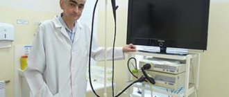
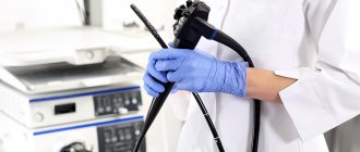
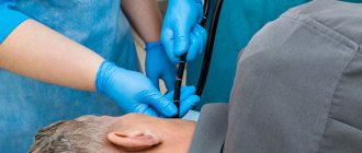
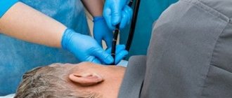
The combination of fibrogastroduodenoscopy (FGS, FGDS) with fine-needle biopsy is a procedure performed using endoscopic equipment. This is a device that allows you to see the condition of these organs using a miniature video camera inserted into the esophagus and stomach. The doctor can see the condition of the mucous membranes and tissues on the monitor screen, and in a many times enlarged form.
This allows the doctor to visually highlight those areas of the stomach, esophagus or duodenum that are affected by any disease. The inserted thin probe is also adapted to allow the collection of biological material - a small amount of tissue that is suitable for morphological analysis.
There may be erosions, polyps, ulcers, and areas of inflammation on the gastric mucosa. There may also be areas of degenerating cancer cells. All these zones are visible during the examination, and the doctor has the opportunity to take tissue from these zones. In addition, with FGDS, sources of intragastric bleeding are visible, and this bleeding can even be stopped during the examination procedure itself.
Indications for gastric biopsy
Endoscopic examination of the stomach with biopsy sampling is indicated in the following cases:
- Any neoplasms of a tumor nature.
- Sluggish and long-term non-healing ulcers of the mucous membrane.
- Gastritis that is difficult to treat.
- Gastritis with an unclear type of secretion.
- Stomach ulcer.
- Visually detectable changes in the mucous membrane (suspicion of intestinal metaplasia).
- A combination of dyspeptic symptoms and unmotivated weight loss, especially in people with a family history of cancer.
- The presence of polyps of the mucous membrane.
- Previous surgical interventions on the stomach for diagnosed cancer.
Performing a gastric biopsy can serve several purposes:
- Study of the morphological structure of the affected area (differentiation between benign and malignant processes is carried out).
- Determination of inflammatory activity.
- Determination of the type of mucosal epithelial dysplasia.
- Detection of Helicobacter pylori.
- Monitoring the effectiveness of treatment carried out after the diagnosis of stomach cancer.
Indications for gastroscopy with biopsy
Any organic disease of the gastrointestinal tract and long-term, especially steadily progressing, complaints of gastric discomfort may be grounds for endoscopy.
Individual screening - regular preventive examination for early signs of cancer is advisable if there is a high probability of the disease, for example, with atrophic gastritis in combination with other risk factors and “bad” heredity.
Clinical studies have not provided objective evidence of the benefits of mass screening of the European population for stomach cancer, but in Japan and South Korea, where there is a high incidence of the disease, FGDS is included in the screening performed for people over 40 years of age every 2 years.
Contraindications to gastric biopsy
Gastric biopsy has a number of contraindications, both absolute and relative. The first group includes:
- Acute somatic pathologies requiring emergency medical care (stroke, myocardial infarction, bronchial asthma attack).
- Cicatricial narrowing of the lumen of the esophagus, making it impossible to advance the endoscope.
- Aortic aneurysm.
Relative contraindications for gastric biopsy include the following conditions:
- Inflammatory process in the nasopharynx.
- Feverish condition.
- Epilepsy.
- Blood clotting disorders, hemorrhagic diathesis.
- Mental illnesses in the acute stage.
- Heart failure and hypertensive crisis.
Preparation for the procedure
No specific preparation is required for the biopsy. It is enough to follow the general rules shown before performing gastroscopy. First, the doctor explains to the patient the purpose and course of the procedure and obtains his consent. In case of emotional lability, before endoscopic examination of the gastric mucosa and taking a biopsy, mild sedatives may be prescribed to calm the patient. In addition, it is necessary to carefully collect anamnesis to identify allergic reactions to drugs used during endoscopy with biopsy (especially anesthetics).
It is recommended to exclude hot and spicy foods and alcohol from the diet a week before the examination, as they can irritate the mucous membranes and distort the picture. You should also reduce your consumption of foods that cause increased gas formation. You should not eat food 6 hours before the procedure, drink 2 hours before. In some cases, it is recommended to rinse the stomach immediately before the biopsy. For example, with pyloric stenosis, the evacuation of food from the stomach into the duodenum may be slowed down.
Biopsy sampling methods
A tissue sample from the gastric mucosa can be obtained by two methods. The first method is surgical. It is carried out directly during an operation on an organ, when the question arises about the volume of tissue to be removed. A small section of tissue is removed with a scalpel and sent urgently to the laboratory. The doctor examines the drug under a microscope and makes a conclusion after 10 - 15 minutes. During this time, the operating team does not perform any actions. After deciphering the biopsy, the operation resumes and the necessary adjustments are made to the extent of the surgical intervention.
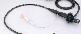
The more common method is endoscopic. For this purpose, a flexible probe is used - a fiberscope, at one end of which there is a camera lens, an LED flashlight, holes for air and water supply, and a mount for attachments. At the other end of the tube there is a handle with a control unit and an eyepiece. Nowadays, targeted biopsy is mainly used because it minimizes the risks of the procedure.
Procedure
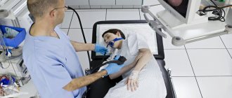
A gastric biopsy can be performed on an outpatient basis, under local anesthesia: the pharynx and pharynx are irrigated with a 10% lidocaine solution to suppress the gag reflex. The subject should be in a lying position on his left side. The endoscope is inserted with gentle movements, and all areas of the mucous membrane along the esophagus and stomach are examined one by one. To improve visualization, air is injected, which straightens the folds of the hollow organ.
A piece of tissue is pinched off from the area of mucosa being examined with a special biopsy instrument (biopsy forceps) inserted into the instrument port of the fiberscope. To increase the accuracy of histological examination, material is collected from several points of the mucosa (at least 4), and the biopsy sample is taken from the border between the pathologically changed and normal areas.
An additional technique of chromatogastroscopy is also used, which simplifies the determination of the boundaries between different areas of the surface under study. A dye is sprayed over the gastric mucosa, which is unevenly absorbed (depending on the functional and organic state of the mucosa). For staining you can use Lugol's solution, methylene blue, Congo red. The area that is stained more intensely is most often pathologically changed, and the biopsy is performed on the periphery of this area (if there is an ulcer or tumor, then in the center too). The duration of the procedure can vary from 10 to 40 minutes and depends on the scope of the study.
After a gastric biopsy, it is advisable not to eat for 2 hours. After this period, you should avoid eating hot foods and drinks for some time. As a rule, there is no pain either during or after the procedure, but there may be slight discomfort in the epigastric region. In rare cases, there may be slight bleeding that stops on its own.
Is a biopsy always informative?
In most cases, analysis of pieces of tissue taken from the pathologically altered mucous membrane gives a positive result, that is, it allows one to make an accurate diagnosis or reject the disease.
In the infiltrative form of gastric cancer, when the malignant tumor is located under the unchanged mucous membrane, standard tissue sampling does not always confirm the diagnosis, which means a false negative result. In such cases, deep penetration of tweezers into the gastric wall and additional x-ray examination are resorted to.
During diagnostic laparoscopy for established cancer, a biopsy is also performed from areas of the peritoneum suspicious for metastases.
How the material is examined
The piece of tissue taken during the biopsy is placed in an airtight container with a preservative. The container is signed (full name of the person being examined and the date of the procedure) and sent to the laboratory.
In the histological laboratory, the tissue is fixed in formalin and embedded in paraffin; if an urgent examination is performed, the tissue is frozen. Then the laboratory assistant uses a microtome to make thin sections of the tissue being examined. The specimen is then placed on a glass slide, stained, dried, and examined under a microscope.
After studying the drug, the pathologist issues a conclusion in which he indicates:
- thickness of the gastric mucosa;
- the nature of the epithelial tissue;
- the presence of dysplasia or metaplasia of the epithelium;
- degree and nature of inflammation;
- the presence of atypical cells, their maturity and location;
- presence of H. pylori, degree of contamination.
A possible additional analysis may be the polymerase chain reaction of a biopsy specimen, which gives the most accurate result in terms of diagnosing Helicobacter pylori.
Morphologically, the presence of the following signs speaks in favor of stomach cancer: abnormal cell shape (polymorphism not typical for this area), cell size (usually tumor cells are larger), size and fragmentation of nuclei, multinucleation, pathological inclusions in the cytoplasm.
The results of a gastric biopsy, if it is not urgent, are given to the patient and the attending physician 10 to 14 days after the procedure. An urgent study is performed on the same day, but its accuracy is somewhat inferior to a planned study.
Carrying out cell analysis
To conduct a differentiated analysis of cells taken from a patient, several different laboratory techniques are used:
- sowing on nutrient media;
- preparing a smear for microscopy;
- polymerase chain reaction (PCR);
- urease test with dye.
In order to analyze the nature of changes in the foci of neoplasms, a targeted biopsy is taken - at least 4-5 fragments are excised, which makes it possible to increase the information content of the test.
Possible complications and consequences of a biopsy
With proper preparation and a biopsy performed by a qualified doctor using modern equipment, the risk of complications is significantly reduced. However, in practice the following situations may rarely occur:
- acute bleeding from the area where the material was taken;
- perforation of the gastric mucosa. It occurs extremely rarely, usually when a “blind” biopsy is performed by an inexperienced specialist);
- the occurrence of allergic reactions to the anesthetic;
- damage to teeth and oral mucosa.
In addition, after the procedure, long-term consequences may occur, which, as a rule, are facilitated by concomitant pathology, as well as some anatomical features:
- bleeding from a vessel damaged during biopsy;
- aspiration pneumonia due to violation of the technique of inserting and removing the fiber gastroscope or vomit entering the respiratory tract;
- inflammatory diseases of the upper digestive system.
A gastric biopsy is in most cases a painless and low-traumatic procedure, which, despite its relative simplicity, can provide a lot of important information about the nature and course of the pathological process.
Book a consultation 24 hours a day
+7+7+78
How to prepare for the examination
Endoscopic examination can be done when necessary, so if gastric bleeding is suspected, FGDS is the leading diagnostic method, during which they try to stop the bleeding.
To identify gastric disease or cancer screening, FGDS is performed routinely on an empty stomach, preferably 8 hours after a meal, and you should not smoke for at least 3 hours.
In Moscow, the cost of a biopsy depends on the volume - the more pieces taken for analysis, the higher the price. The cost is affected by additional manipulations during FGDS, for example, the use of a contrast agent to clarify the location of tissue collection, and special morphological studies to make an accurate diagnosis.
The Medicine 24/7 diagnostic center offers to perform a biopsy of the stomach (polyp, ulcer or mucous membrane) and conduct an analysis at a competitive price in Moscow. You can get information about the cost of a gastric biopsy and a detailed consultation by phone
The material was prepared by oncologist, endoscopist, chief surgeon of the Medicine 24/7 clinic, Konstantin Yuryevich Ryabov.
