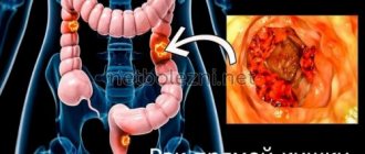
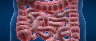
Intestinal tuberculosis is an infectious disease caused by tuberculosis bacteria (Koch bacillus). The disease is expressed in the form of granuloma (inflammation), most often localized at the transition point of the small intestine to the large intestine - the ileocecal region. According to medical statistics, the probability of contracting an infection is 1/2000.
There are 3 forms of the disease:
- primary - the lesion affects the intestinal lymph nodes;
- secondary - inflammation of the walls of the ileum is observed;
- ileocecal tuberculosis is a mixed type disease (combines the features of forms 1 and 2).
Prevention and treatment of intestinal tuberculosis
It is generally accepted that most often Koch's bacillus affects the respiratory organs - the bronchi or lungs. Medical statistics say that this is far from true; one of the most common forms of the disease is intestinal tuberculosis. It is difficult to determine the disease without special research - the signs are often confused with other, less dangerous ailments. It is better not to try to diagnose the disease yourself and try to eliminate it - treatment of intestinal tuberculosis should be carried out only under the supervision of a doctor.
Treatment
Intestinal tuberculosis responds well to treatment, which is carried out in a clinical hospital. There are 2 ways to get rid of the disease:
- medicinal - eliminating the infection with the help of drugs that stop the vital activity of Koch's bacillus, as well as chemotherapy. Among the drugs aimed at combating tuberculosis, the following have proven themselves to be excellent: Phtivazid; PASK; Streptomycin.
- surgical - used in the presence of an ulcer, stenosis of the colon or fistula.
During treatment, the patient must follow a special diet. Doctors recommend eating mostly non-solid foods: porridge cooked in water, fresh fruits, vegetables and specialized dietary supplements (for example, fiber). You should avoid foods that create a favorable environment for fermentation. These include:
- canned food;
- smoked meat;
- dairy products;
- carbonated drinks.
Most patients diagnosed with intestinal tuberculosis have a high chance of recovery. The development of the disease can be stopped in the early stages, and therefore it is much easier for patients with mildly expressed degenerative changes in the intestinal walls to eliminate the consequences.
Advanced forms of the disease are difficult to treat. The only way to get rid of tuberculosis is extensive resection (removal) of the affected intestinal tissue.
Causes of intestinal tuberculosis
Doctors call secondary infection one of the main causes of intestinal tuberculosis. The development of the disease usually begins against the background of pulmonary tuberculosis. The infection can enter the intestines through the blood and lymph. Swallowing large amounts of sputum, which contains bacteria, is also considered a cause of intestinal tuberculosis.
What causes intestinal tuberculosis, and what factors contribute to its development? Especially often, the disease begins to progress when the body’s immune strength decreases. Inflammatory processes progressing in internal organs also contribute to the spread of infection.
How is an X-ray done to diagnose tuberculosis?
X-rays of light
- the procedure is simple. To perform it, you will need to undress to the waist and stand next to the device in accordance with the doctor’s instructions. Jewelry and other objects that may interfere with scanning are removed, and the abdomen and reproductive organs are covered with radiation protection: a leaded rubber apron. Then, at the command of the radiologist, you need to draw air into your lungs and hold your breath for a few seconds. During this time a picture is taken.
Symptoms of intestinal tuberculosis
The disease has several signs that, with some observation, can be determined independently, which should be the reason for an immediate visit to the doctor. Symptoms of intestinal tuberculosis include:
- profuse cold sweat;
- fever, intense increase in body temperature;
- prolonged diarrhea (often confused with intestinal upset);
- painful sensations in the lower abdomen, aggravated by walking or physical activity.
Signs of intestinal tuberculosis also include blood clots in the stool. Defecation may be accompanied by severe abdominal pain.
A doctor cannot diagnose intestinal tuberculosis on the basis of an oral description of symptoms; one will have to undergo a series of studies that will confirm suspicions of a serious disease. An x-ray of the peritoneal area and chest is recommended. Additionally, an ultrasound and tuberculin test are performed. Stool tests are required to detect occult blood.
Pulmonary tuberculosis on x-ray
The description of x-rays for tuberculosis can be very varied, since this disease has many forms. However, the main sign of tuberculosis of the lung on an x-ray is a violation of the structure of the organ, depending on the duration of the disease. To exclude other diseases affecting the lungs, it is necessary to supplement x-rays with tests and other diagnostic methods. In some cases, MRI and CT are prescribed to diagnose tuberculosis, but x-ray remains the most accessible.
How to treat intestinal tuberculosis
Doctors recommend studying the basic rules of therapy before treating intestinal tuberculosis. Treatment consists not only of the use of medications, but also of additional measures. A prerequisite is diet correction. You should exclude sweets and dishes rich in animal fat from the menu. It is recommended to introduce fermented milk products (kefir, natural yoghurts without sweet additives) and vegetable dishes into the diet.
How to cure intestinal tuberculosis, and what drugs are used to continue therapy? Antibacterial drugs (Streptomycin) and chemotherapy drugs (Tubazid, para-aminosalicylic acid) are usually prescribed. The dosage, number of doses, and duration of treatment are calculated only by a specialist. The doctor takes into account the form of intestinal tuberculosis, the general condition of the patient, and additional complications.
Treatment of intestinal tuberculosis with folk remedies is not recommended, although with the permission of a doctor, herbal decoctions can be used as an auxiliary therapy. Closely monitor the body's reaction to herbal formulations - the appearance of side effects or irritation should be an immediate reason to refuse to continue the course.
Features of diagnosis and treatment of tuberculous peritonitis
V.F. CHIKAEV1, Yu.V. BONDAREV2, K.M. ZIYATDINOV3, D.M. PETUKHOV2
1Kazan State Medical University, 420012, Kazan, st. Butlerova, 49
2City Clinical Hospital No. 7, 420103, Kazan, st. Chuikova, 54
3 Branch of the RCTB - Kazan Tuberculosis Hospital, 422700, RT, Vysokogorsky district, Kamenka village, st. Nagornaya, 3
Chikaev Vyacheslav Fedorovich - Doctor of Medical Sciences, Professor of the Department of Traumatology, Orthopedics and ChES, tel. +7-927-434-48-29, e-mail1
Bondarev Yuri Viktorovich - Candidate of Medical Sciences , Head of Surgical Department No. 3, tel. +7-917-269-91-12, e-mail: [email protected]
Ziyatdinov Kamil Mukhametovich - candidate of medical sciences , phthisiatrician surgeon, tel. +7-960-052-29-02, e-mail3
Petukhov Denis Mikhailovich - surgeon, tel. +7-917-276-89-76, e-mail2
The article presents the results of an analysis of the features of diagnosis and treatment of tuberculous peritonitis. In the Russian Federation, abdominal tuberculosis among extrapulmonary forms accounts for 2-3% of all specific lesions. The complexity of diagnosing abdominal tuberculosis is determined by the variety of pathogenetic mechanisms, morphological and clinical manifestations. As a rule, patients are admitted to the general surgical department with complicated secondary peritonitis and require complex therapy taking into account the underlying pathological process.
Key words: tuberculosis, peritonitis, diagnosis, treatment.
VF CHIKAYEV1, Yu.V. BONDAREV2, KM ZIYATDINOV3, DM PETUKHOV2
1Kazan State Medical University, 49 Butlerov St., Kazan, Russian Federation, 420012
2City Clinical Hospital No. 7, 54 Chuikov St., Kazan, Russian Federation, 420103
3Kazan Tuberculosis Hospital (branch of Republican Clinical Tuberculosis Dispensary), 3 Nagornaya St., Kamenka settlement, Russian Federation, 422700
Diagnosis and treatment of tuberculous peritonitis
Chikayev VF - D. Med. Sc., Professor of the Department of Traumatology, Orthopedics and Urgent Surgery, tel. +7-927-434-48-29, e-mail1
Bondarev Yu.V. — Cand. Med. Sc., Head of Surgery Department No. 3, tel. +7-917-269-91-122
Ziyatdinov KM - Cand. Med. Sc., surgeon-phthisiatrician, tel. +7-960-052-29-02, e-mail: [email protected]
Petukhov DM - surgeon, tel. +7-917-276-89-76, e-mail2
The article presents the results of analyzing the diagnosis and treatment of tuberculous peritonitis. Abdominal tuberculosis presents 2-3% of extrapulmonary specific lesions in the Russian Federation. Complexity of abdominal tuberculosis diagnosing is determined by the variety of its pathogenic mechanisms, morphological and clinical manifestations. Patients are usually admitted to general surgery with complicated secondary peritonitis, which requires complex therapy due to the basic pathological process.
Key words: tuberculosis, peritonitis, diagnosis, treatment.
The problem of peritonitis remains relevant, despite all the achievements of scientific and technological progress. This is evidenced by summary data, according to which average mortality rates remain at the level of 20-30%, and in the most severe forms, for example, postoperative peritonitis, they reach 40-50% [1].
In primary peritonitis, the inflammatory process develops without violating the integrity of the hollow organs, and it is the result of spontaneous hematogenous translocation of microorganisms into the peritoneal cover or transudation of a specific monoinfection from other organs. The following types of primary peritonitis are distinguished: spontaneous peritonitis in children, spontaneous peritonitis in adults, tuberculous peritonitis.
Since the discovery of Mycobacterium tuberculosis by Robert Koch to the present day, tuberculosis has remained a pressing problem. Today, about 2 billion people are infected with tuberculosis in the world. At the turn of the 20th-21st centuries, the epidemic situation regarding tuberculosis in Russia is assessed as unfavorable. According to operational records, in 2012, 89.7 thousand cases of active tuberculosis were detected for the first time. In the Russian Federation, the incidence of tuberculosis increased by 2.4 times, mortality by 2.3 times [2-5]. Forms of infection with multidrug resistance and combined with infection with the immunodeficiency virus (HIV) have emerged. In 2012, the number of cases of active tuberculosis decreased by 5%, and the number of cases caused by HIV increased by 12.5%. In recent years, the incidence rate per 100 thousand people has tended to decrease, but remains at a high level. Especially many cases of active tuberculosis are registered among young and middle-aged men. Thus, in the peak year of 2000, for every 100 thousand men aged 18-34 years, 212 sick people were identified. Among extrapulmonary forms, abdominal tuberculosis occupies a special place, accounting for 2-3% of all extrapulmonary specific lesions in the Russian Federation [6-9]. Over the last decade, the share of newly diagnosed abdominal tuberculosis of the total number of patients with extrapulmonary forms in some regions of the Russian Federation ranges from 4.4 to 8.3% [10].
According to statistical data, in the structure of the abdominal form of tuberculosis, mesenteric lymph nodes are affected in 70%, and the peritoneum in 12%. Tuberculous peritonitis (peritoneal tuberculosis) is classified primarily as a primary tuberculosis infection as a consequence of the lymphohematogenous spread of the process, or it is a complication of specific damage to the lymph nodes of the abdominal cavity, intestines, genitals, spine, spreading by contact. Particularly severe peritonitis develops when a tuberculous intestinal ulcer perforates into the abdominal cavity or breaks through the caseous lymph nodes of the mesentery. During the period of secondary tuberculosis, the spread of the process from the mesenteric nodes, intestines and genital organs often leads to the development of a dry form of peritonitis with damage to limited areas of the peritoneum. Regardless of the genesis, the picture of peritonitis has a dominant position in the general symptomatology of the disease. In patients with abdominal tuberculosis, concomitant nonspecific pathology of the digestive organs leads to difficulties in diagnosing the underlying disease. In the vast majority of cases, tuberculosis of the gastrointestinal tract, both in Russia and abroad, is detected either during surgical interventions for its complications, or at a section [11014].
In tuberculous peritonitis, depending on the depth of damage to the peritoneum, tubercular, exudative, exudative-adhesive, adhesive, tumor-like or caseous-ulcerative forms are distinguished. The most common are adhesive, chronic forms of tuberculous peritonitis. The complexity of diagnosing abdominal tuberculosis is determined by the variety of pathogenetic mechanisms, morphological and clinical manifestations, and the absence of characteristic signs of gastrointestinal lesions at various levels of the gastrointestinal tract [15]. Abdominal tuberculosis has no specific symptoms; its diagnosis can only be made by histological or bacteriological methods. Bacteriologically specific abdominal lesions are rarely detected - Mycobacterium tuberculosis is found in ascitic fluid only in 2.6% of cases, in feces - in 7.3% of cases. Specific tuberculin tests are positive less than 50%.
The clinical picture of abdominal tuberculosis is polymorphic, there are no pathognomonic symptoms and clear diagnostic criteria, therefore, as a rule, it occurs under the guise of other diseases of the abdominal organs and is detected only in a small part of patients, while in the majority it remains undiagnosed. According to the recommendation of R. Seth et al. (1974), that in any patient with unclear abdominal symptoms tuberculous peritonitis should be suspected. Tuberculous peritonitis is characterized by a triad of symptoms:
— vague dull or cramping pain in the abdomen in the right half in the projection of the ileocecal angle, vomiting, diarrhea;
- fever, low-grade body temperature;
- ascites (isolated ascites is observed only in TVS peritonitis).
Radiation methods are informative in diagnosis, but their data must be differentiated from UC, Crohn's disease, and cancer. During plain radiography of the abdominal organs, the identification of calcified lymph nodes almost always indicates the presence of tuberculous mesadenitis [16].
Ultrasound can reveal segmental lesions of the intestine, enlarged regional lymph nodes and encysted ascites [17, 18].
Data from CT and magnetic resonance imaging, as a rule, do not have independent significance and can only be taken into account in conjunction with the results of classical X-ray examination and endoscopy.
Endoscopic instrumental diagnostic methods are more informative. In case of intestinal tuberculosis during colonoscopy, the characteristic signs are segmental lesions with a change of affected and unaffected areas, and in case of UC, the process involves large sections of the intestine and passes to normal structures without a sharp boundary [19-21].
The information content of laparoscopy with peritoneal biopsy remains at a high level [11, 22, 23].
Secondary tuberculous peritonitis is characterized by the presence of a primary focus of tuberculous damage to the lungs or other organs. Once pulmonary tuberculosis is detected, the diagnosis becomes much easier. In such patients, tuberculin tests are usually positive.
The first operation in Russia for the exudative form of tuberculous peritonitis was performed by N.A. Velyaminov (1884). Indications for surgical treatment are: exudative peritonitis, complicated forms of tuberculous peritonitis, progressive intestinal obstruction, perforation of hollow organs, disintegrating pseudotumor process, purulent peritonitis [24, 25]. With tuberculous peritonitis, even laparotomy alone usually leads to an improvement in the patient's condition. This is explained by the fact that during laparotomy the effusion from the abdominal cavity is evacuated, and the air entering the abdominal cavity actively affects the tuberculosis process. Each laparotomy for tuberculous peritonitis makes it possible to accurately establish the diagnosis by taking a biopsy [25].
An alternative at the present stage is laparoscopic sanitation of the abdominal cavity and biopsy of nodules [26].
Since tuberculous peritonitis is quite rare among all surgical diseases, in most cases patients admitted to surgical departments are usually operated on with a diagnosis of an acute surgical disease (appendicitis, acute peritonitis of unknown etiology, acute intestinal obstruction). The etiology and form of peritonitis are revealed only on the operating table.
The purpose of the study is to analyze the features of diagnosis and treatment of tuberculous peritonitis.
Materials methods
An analysis of the literature data on the diagnosis and treatment of tuberculous peritonitis at the present stage was carried out. The medical documentation of patients hospitalized in the emergency surgery department of the State Emergency Hospital No. 1 in Kazan was studied.
Results and discussion
Over the past 5 years, 4 male patients aged 38-72 years with secondary tuberculous peritonitis have been hospitalized at the clinic. All patients in the emergency surgery department were hospitalized on an emergency basis. In three cases, patients were admitted from an anti-tuberculosis dispensary. In one case, the patient was brought from home. All received complex treatment taking into account the underlying pathological process.
In three cases, patients were hospitalized with a diagnosis of acute intestinal obstruction, peritonitis. It was characteristic that all patients were malnourished and had pale skin. Plain radiography of the abdominal cavity of Kloiber's thicket with ultrasound of the abdominal cavity shows free fluid. Moreover, one of them does not have a primary diagnosis of tuberculosis. After conservative therapy due to non-resolving obstruction, all patients underwent emergency surgery. Laparotomy revealed a productive phase of inflammation with the development of adhesive peritonitis, the formation of adhesions between the peritoneum and adjacent abdominal organs. There is a massive adhesive process in the abdominal cavity, which involves intestinal loops and the omentum. Serous exudate was detected between the loops of the small intestine. Millet-like rashes were detected along the peritoneum. Separation of adhesions, sanitation of abdominal exudate, and biopsy of nodules for analysis were performed. Histological analysis subsequently confirmed tuberculosis. The operation was completed by Abbott-Miller intubation of the jejunum; the probe could not be inserted below due to a pronounced adhesive process. In the postoperative period, complex therapy was carried out in collaboration with phthisiatricians. A feature of the postoperative period was the persistence of low-grade fever. Patients were discharged with a recommendation for examination and treatment by a phthisiatrician.
In one case, a 46-year-old patient was delivered with generalized peritonitis from an anti-tuberculosis dispensary, where he was being treated for pulmonary tuberculosis. The patient's condition is serious, gray complexion, low nutrition, dry tongue, the abdomen is painful in all parts, the symptom of peritoneal irritation is positive. After preoperative preparation, the patient was operated on as an emergency. Laparotomy revealed perforation of the small intestine and widespread diffuse peritonitis. In the abdominal cavity there is a pronounced planar adhesive infiltrative process. The perforation hole was sutured and the abdominal cavity was sanitized by washing with ECHAR-anolyte. The rationale for its use is the high oxygen content in the solution. A programmed relaparotomy and sanitation of the abdominal cavity was planned, the operation was completed with laparostomy. When sanitation of the abdominal cavity, the use of ozonated solutions is probably justified.
On the second day after the operation, the abdominal cavity was sanitized and the abdominal wall was sutured. On the 3rd day, repeated multiple perforations of the small intestine were performed, relaparotomy was performed, suturing of the perforated operations was performed, resection of the small intestine was not performed due to a pronounced adhesive productive process. Subsequently, the patient developed progressive peritonitis and multiple organ failure. The treatment ended in death. At the autopsy - intestinal tuberculosis, mesadenitis.
Thus, the literature analysis and our own observations allow us to draw the following conclusions:
Taking into account the growth of general tuberculosis, one should remember the possibility of inflammation of the peritoneum by a specific process.
As a rule, this category of patients is admitted to emergency surgery with secondary complications.
Surgical tactics for tuberculous peritonitis depend on the etiology, form, and extent of the pathological process.
In the postoperative period, complex therapy for tuberculous peritonitis is carried out in collaboration with phthisiatricians.
LITERATURE
1. Savelyev V.S. Guide to emergency abdominal surgery. - Moscow, 2004. - 640 p.
2. Kononenko V.G., Shkurupiy V.A. Epidemiology of pulmonary tuberculosis and parenteral chemotherapy / V.G. Kononenko. - Novosibirsk, 2002. - 164 p.
3. Krasnov V.A. On the state of morbidity and anti-tuberculosis care for the population of the Siberian Federal District // Bulletin of the interregional association “Health of Siberia”. - 2002. - No. 4. - P. 82-83.
4. Onishchenko G.G. Epidemiological situation in the Russian Federation and measures to stabilize it // Problems of tuberculosis and lung diseases. - 2003. - No. 11. - P. 4-9.
5. Khegai L.N. Pathomorphological assessment of the dynamics of tuberculosis in Tashkent // Problems of tuberculosis and lung diseases. - 2004. - No. 9. - P. 16-17.
6. Abramovskaya A.K. Current problems of extrapulmonary tuberculosis // Minsk, 1995. - pp. 67-68.
7. Vasiliev A.V. Current problems of care for patients with extrapulmonary tuberculosis // Extrapulmonary tuberculosis - an urgent problem of healthcare. - St. Petersburg, 1997. - P. 10.
8. Garbuz A.E. Current state of the problem of extrapulmonary tuberculosis // Problems of tuberculosis. - 1998. - No. 2. - P. 32-34.
9. Galkin V.B., R.K. Yagafarova, V.M. Hokkanen et al. // Epidemiological and clinical aspects of extrapulmonary tuberculosis in the north-west of Russia // Problems of tuberculosis. - 1998. - No. 2. - P. 36-38.
10. Skopin M.S., Batyrov F.A., Kornilova 3.X. Tuberculosis of the abdominal organs and features of its detection // Problems of tuberculosis and lung diseases. - 2007. - No. 1. - P. 22-26.
11. Lomachenkov V.D. Tuberculosis of the abdominal organs, diagnosed by laparoscopy // Problems of tuberculosis. - 1998. - No. 1. - P. 53-55.
12. Monkemuller K.E. Massive rectal bleeding from colonic tuberculosis // Am. J. Gastroenterol. — 1996 Jul. - Vol. 91, Issue 7. - P. 1439-1441.
13. Jain V.K. Ileocaecal tuberculosis associated with adenocarcinoma of the Caecum and Colon // J. Indian Med Assoc. - 1996. - No. 94 (1). - P. 36.
14. Kaushik R., Sharma R., Attri AK Coexisting tuberculosis and carcinoma of the colon: a report of two cases and a review of the literature // Trop. Gastroenterol. — 2003, Jul. Sep. - Vol. 24(3). — P. 137-139.
15. Barinov V. S. Abdominal tuberculosis // Phthisiology: national manual / ed. M.I. Perelman. - M.: GEOTAR-Media, 2007. - P. 329-321.
16. Vengerov B.B. et al. The use of ultrasound research in recognizing pathological changes in the biliary system of patients with pulmonary tuberculosis // Tuberculosis. - Kyiv: Health, 1989. - Issue. 21. - pp. 26-28.
17. Ivanov A.K. Ultrasound examination of the liver in patients with pulmonary tuberculosis / A.K. Ivanov, K.A. Dzodzuashvili // Clinical medicine. - 1990. - No. 1. - P. 100-102.
18. Malik A. Ultrasound in abdominal tuberculosis / A. Malik, NC Saxena // Abdom. Imaging. 2003, Jul.-Aug. - Vol. 28(4). - P. 49.
19. Volobuev N.N. Diagnosis and treatment of intestinal tuberculosis / N.N. Volobuev // Surgery. - 1994. - No. 4. - P. 43-45.
20. Baraga J. at al. Endoscopic appearance of colonic tuberculosis // Endoscopy. — 2003 Mar. - Vol. 35(3). - P. 256.
21. Sato S., Yao K., Yao T. et al. Colonoscopy in the diagnosis of intestinal tuberculosis in asymptomatic patients // Gastrointestinal Endosc. — 2004 Mar. - Vol. 59(3). — P. 362-368.
22. Semenovsky A.V., Barinov V.S., Kocharova M.N. and others. Laparoscopy in the complex diagnosis of abdominal and genital tuberculosis // Problems of tuberculosis. - 1999. - No. 3. - P. 36-39.
23. Matrosov V.M. et al. Laparoscopy in the diagnosis of tuberculosis of the abdominal organs // Tuberculosis in Russia, year 2007: materials of the YTII Russian Congress of Phthisiologists. - M.: Idea, 2007. - P. 348-349.
24. Savina T.A. et al. Surgical treatment of patients with abdominal tuberculosis // Problems of tuberculosis. - 2001. - No. 9. - P. 58-59.
25. Batyrov F.A. et al. Surgical interventions for complicated forms of tuberculosis of the abdominal organs // Surgery. 2005. - No. 1. - P. 51-53.
26. Medzhidov R.T. et al. Videolaparoscopic diagnosis and treatment of abdominal tuberculosis // Tuberculosis in Russia, year 2007: materials of the YIII Russian Congress of Phthisiatricians. - M.: Idea, 2007.
REFERENCES
1. Savelyev VS Guidelines for emergency abdominal surgery. Moscow, 2004. P.640.
2. Kononenko V. G, Shkurupiy VA Epidemiology of pulmonary tuberculosis and parenteral chemotherapy. Novosibirsk, 2002. P. 164.
3. Krasnov VA On the state of disease and TB care to the population of the Siberian Federal District. Bulletin of the Interregional Association “Siberian Healthcare”, 2002, no. 4, pp. 82-83.
4. Onischenko GG Epidemiological situation in the Russian Federation and stabilizing measures. Problems of Tuberculosis and Lung Disease, 2003, vol. 11, pp. 4-9.
5. Hegay LN Pathological assessment of the dynamics of tuberculosis in Tashkent. Problems of Tuberculosis and Lung Disease, 2004, vol. 9, pp. 16-17.
6. Abramovskaya. AK Actual problems of extrapulmonary tuberculosis. Minsk, 1995. Pp. 67-68.
7. Vasilyev AV Actual problems of patient care with extrapulmonary tuberculosis. Extrapulmonary tuberculosis as an urgent public health problem. Saint Petersburg, 1997. P. 10.
8. Garbuz AE State of the extrapulmonary tuberculosis problem. Problems of Tuberculosis. 1998, vol. 2, pp. 32-34.
9. Galkin VB, yagafarova VG, Hokkanen VM Epidemiological and clinical aspects of extrapulmonary tuberculosis in Northwest Russia. Problems of Tuberculosis, 1998, vol. 2, pp. 36-38.
10. Skopin MS, Batyrov FA, Kornilova Z.Kh. Abdominal tuberculosis and features of identifying. Problems of Tuberculosis and Lung Disease, 2007, vol. 1, pp. 22-26.
11. Lomachenkov VD Abdominal tuberculosis diagnosed at laparoscopy. Problems of Tuberculosis. 1998, vol. 1, pp. 53-55.
12. Monkemuller K. E. Massive rectal bleeding from colonic tuberculosis. Am. J. Gastroenterol. 1996, Jul., vol. 91, issue 7, pp. 1439-1441.
13. Jain V. K. Ileocaecal tuberculosis associated with adenocarcinoma of the Caecum and Colon. J. Indian Med Assoc., 1996, no. 94(1). P. 36.
14. Kaushik R., Sharma R., Attri A. K Coexisting tuberculosis and carcinoma of the colon: a report of two cases and a review of the literature. Trop. Gastroenterol., 2003, Jul.–Sep., no. 24 (3), pp. 137-139.
15. Barinov VS Abdominal tuberculosis. Phthisiology: national guideline. Ed. MI Perelman. Moscow: GEOTAR-Media, 2007. Pp. 329-321.
16. Vengerov BB et al. Using ultrasonic method research in recognition of pathological changes in the biliary system of patients with pulmonary tuberculosis. Tuberculosis. Kiev: Health 1989, vol. 21, pp. 26-28.
17. Ivanov AK, Dzodzuashvili KA Ultrasound examination of the liver in patients with pulmonary tuberculosis. Clinical Medicine, 1990, vol. 1, pp. 100-102.
18. Malik A., Saxena NC Ultrasound in abdominal tuberculosis. Abdom. Imaging. 2003, Jul.-Aug., No. 28 (4). P. 49.
19. Volobuev NN Diagnosis and treatment of intestinal tuberculosis. Surgery, 1994, vol. 4.Pp. 43-45
20. Baraga J. at all.Endoscopic appearance of colonic tuberculosis. Endoscopy, 2003, Mar 35 (3). P. 256.
21. S. Sato, K. Yao, T. Yao et al. Colonoscopy in the diagnosis of intestinal tuberculosis in asymptomatic patients. Gastrointestinal Endosc. 2004, Mar., no. 59(3). pp. 362-368.
22. Semenovsky AV, Barinov VS, Kocharova MN et al. Laparoscopy in the complex diagnosis of abdominal and genital tuberculosis. Problems of Tuberculosis, 1999, vol. 3, pp. 36-39.
23. Matrosov VM et al. Laparoscopy in the diagnosis of abdominal tuberculosis. Tuberculosis in Russia Year 2007: Proceedings of the YTII Congress of Russian phthisiologists. Moscow: OOO “Idea”, 2007. Pp. 348-349.
24. Savina TA et al. Surgical treatment of abdominal tuberculosis. Problems of Tuberculosis. 2001, no. 9, pp. 58-59.
25. Batyrov FA et al. Operative intervention in complicated forms of abdominal tuberculosis. Surgery, 2005, vol. 1, pp. 51-53.
26. Medzhidov RT et al. Videolaparoscopic diagnosis and treatment of abdominal tuberculosis. Tuberculosis in Russia Year 2007: Proceedings of the YTII Congress of Russian phthisiologists. Moscow: OOO “Idea”, 2007. Pp. 349-350.
Stages of intestinal tuberculosis
There are several degrees of intestinal tuberculosis, each of which has its own characteristics. The first has no special signs and is practically asymptomatic. A dangerous disease can only be detected by special studies - a reaction to the Mantoux test or the Diaskin test.
The second stage of intestinal tuberculosis is manifested by a number of signs that are characteristic of the disease. At this stage, treatment of intestinal tuberculosis is not particularly difficult - the spread of Koch's bacillus can be suppressed with drug therapy in just a few months.
At the third stage of development of the disease, damage to internal organs and systems begins; the sick person poses a threat to others, releasing bacteria when coughing into the air. Often the third stage of the disease ends in death - even powerful pharmaceutical drugs can be powerless.
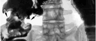
Diagnostics
Tuberculous lesions of the ileocecal region of the intestine are detected through x-ray examination of the abdominal organs . The image clearly shows characteristic symptoms:
- scarring;
- a large number of ulcerations localized on the mucous membrane of the colon;
- less often, defects in the filling of the organ with feces can be identified.
An intestinal biopsy (excision of intestinal tissue for later study) can also show the presence of Koch's bacillus.
The simplest way to diagnose the disease is the Mantoux test, which determines the presence of an immune response to tuberculin injected under the skin of the suspected patient. 48 hours after the injection, the test result is checked: if a papule has formed at the patient’s injection site, the size of which exceeds 10 mm, then there is a high probability of infection in his body.
An ultrasound method for examining the abdominal organs also makes it possible to identify degenerative changes in the intestinal walls, which are considered a characteristic feature of the disease.
A TB doctor can determine the presence of the disease by relying on the results of the following tests :
- blood;
- feces;
- a scraping taken from the inner walls of the intestine.
Prevention of intestinal tuberculosis
Doctors warn that vaccination is considered the most effective and efficient rule for preventing intestinal tuberculosis. It is especially important to administer the vaccine to people who often come into contact with a sick person or live in the same room with him.
Another measure to prevent intestinal tuberculosis is taking the drug Isoniazid. Doctors prescribe the drug to people at risk who have weak immunity.
Treatment of intestinal tuberculosis is a complex process, therapy can last for several months, so it is recommended to undergo regular medical examinations, which will help to identify the disease in a timely manner. It should be remembered that only in the early stages will treatment take place without any particular difficulties. Severe forms of the disease that develop in the absence of drug therapy can result in death.
Prevention
Statistics show that tuberculosis is directly related to the livelihoods of the population and its living conditions. In order to minimize the risk of developing an epidemic, it is necessary to follow a number of preventive measures:
- providing patients with medicines;
- regular medical examinations for the detection of Koch's bacillus, which are mandatory;
- mandatory vaccination of newborns;
- keeping infected people in specialized places (clinics, dispensaries, hospitals), where they will be provided with professional medical care.
Source: https://www.gastromap.ru/