- home
- —
- Clinic
- —
- Diagnostics
- —
- Biopsy
- —
- Stomach biopsy
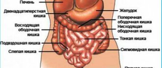
A gastric biopsy is the intravital removal of a tissue fragment for histological or cytological examination, that is, the study of tissue or cellular characteristics. This makes it possible to identify pathological processes, find out their nature, and in the case of malignant processes, not only detect them, but also recognize the type and variety of the tumor, which is necessary to determine the further course of treatment.
Hospitalization of cancer patients. Daily. Around the clock
9,500 patients trust us annually.
Call me back!
A biopsy of the gastric mucosa is mandatory for oncology, and is also used for other diseases: gastritis, slow-healing ulcers and peptic ulcers, and dyspeptic symptoms. A sharp decrease in body weight, polyps, suspicion of intestinal metaplasia (tissue degeneration), the presence of cancer in close relatives, and previous operations for tumors may also require a biopsy examination.
Interpretation of FGDS with biopsy of the antrum and body of the stomach
Share information with your Facebook friends
VK
Question
Hello, Andrey Viktorovich! Help decipher the biopsy.
1. Two fragments were taken from the mucous membrane of the antrum of the stomach: the mucous membrane is moderately thinned, the architectonics of the location of the glands is disrupted, and focal hyperproliferation of the integumentary-pit structures is noted. In the lamina propria there is a moderate diffuse focal infiltration of lymphocytes and plasma cells, the stroma is diffusely fibrotic, expanding the glands of the lamina propria with a slight decrease in their number. Groups of glands are identified in which cylindrical cells alternate with a large number of goblet cells.
2. Two fragments from the mucous membrane of the body of the stomach: the pit structures are hyperplastic, excessively tortuous, and randomly located. Stroma with pronounced fibrosis and diffuse focal lymphoid infiltration. The glands of the lamina propria are separated by strands of connective tissue, some of them are cystically dilated. Groups of glands are identified in which cylindrical cells alternate with a large number of goblet cells.
Conclusion: 1. The morphological picture corresponds to moderate chronic minimally atrophic diffuse reactive gastritis with foci of intestinal metaplasia of the glandular epithelium. 2. Moderate chronic diffuse inactive gastritis. Hyperplastic polyp with signs of chronic inflammation, with foci of intestinal metaplasia of the glandular epithelium. N.bacter is negative. No treatment prescribed. They recommended doing an FGDS in six months. Please tell me what does this mean?
Answer
Greetings! I apologize for the delay, I participated in the conference. 1. “The architecture of the location of the glands is disrupted, focal hyperproliferation of integumentary-pit structures is noted” - these are signs of long-term chronic inflammation and tissue irritation either by chemical agents or active substances of neutrophils and lymphocytes that accumulate in the tissue during inflammation. 2. “The stroma is diffusely fibrotic, pushing apart the glands of the lamina propria with a slight decrease in their number” - the effect of inflammation, leading not to true atrophy, but to apparent atrophy, due to the spreading of tissues in different directions. 3. “Goblet cells” are signs of small intestinal metaplasia; in the antrum this may not cause significant concern. 4. “The glands of the lamina propria are separated by strands of connective tissue, some of them are cystically dilated” - apparently, on this basis they made a conclusion about a hyperplastic polyp, which is not entirely true. In the body, such a picture will most likely correspond to a polyp of the fundic glands. Histology alone is not enough for me. We need a description of the protocol, photos and video materials. According to histology - nothing terrible. Control after 3-5 years.
©
print version
Methods for removing biopsy material
When prescribing a procedure, various goals can be pursued:
- detection of Helicobacter pylori infection;
- determination of the benignity or malignancy of the process, study of morphological features;
- determining the degree of inflammation activity;
- for dysplasia - determination of its type;
- after cancer treatment, monitoring its effectiveness.
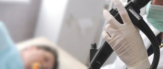
There are actually 2 methods used for gastric biopsy - surgical and endoscopic. The first is used during operations - the biomaterial is taken with a scalpel and urgently sent to the laboratory. The operation is paused until the results are available, which usually takes about 15 minutes. In accordance with the conclusion of the study, the volume of surgical intervention and the parts of organs removed are adjusted. This type of biopsy for stomach cancer allows you not to delay treatment, since literally every day is important.
More often, an endoscopic method is used, in which tissue sampling is combined with gastroscopy (FGDS, EGDS). For this, a flexible probe (fiberscope, endoscope) with instruments is used. If earlier manipulations were carried out “blindly,” now a camera and a flashlight are installed on the device, and water irrigation and air supply are used to smooth out the mucous membrane. The doctor has the opportunity to examine the epithelium and has complete control over the process.
Our expert in this field:
Ivanov Anton Alexandrovich
Medical director, oncologist-surgeon, candidate of medical
sciences Call the doctor
Call the doctor
Procedure for performing a gastric biopsy
No lengthy preparation for endoscopy is required. It is important not to eat or drink much fluid at least 8-10 hours before it starts. If a biopsy is included in the procedure, you should stop smoking and drinking liquids rich in coloring substances (tea, coffee, juices, carbonated drinks).
The biopsy is performed as follows:
- The patient is placed on his left side.
- The larynx and oral cavity are treated with an antiseptic.
- A plastic mouthpiece is placed in the mouth.
- An endoscope is inserted and air is injected to inflate the walls of the esophagus and stomach to ensure a good view.
- Based on the image from the device’s camera, the doctor looks for suspicious areas and takes tissue samples with a special device.
Endoscopy with biopsy is completed by removing the endoscope and sending the resulting samples for detailed histological analysis. The entire procedure lasts 20-40 minutes, the results of the biopsy analysis will be ready in a few days.
Our prices
- Repeated appointment with an oncologist* RUB 3,000.
- Primary appointment with a leading specialist/candidate of medical sciences (oncology)* RUB 4,250.
- Appointment with an oncologist DMN* 5,000 rub.
- Repeated appointment with a leading specialist/candidate of medical sciences (oncology)* RUB 4,000.
- Preventive appointment with an oncologist* RUB 1,490.
- Primary appointment with an oncologist-chemotherapist* RUB 4,600.
- Repeated appointment with an oncologist-chemotherapist* RUB 4,000.
- Consultation with an oncologist on palliative care* RUB 2,000.
Preparation rules
This procedure does not require special preparation measures, but doctors still give a number of recommendations that should be followed. What should the patient do:
- Stop taking medications, any course of treatment.
- The procedure is carried out on an empty stomach, which is why it is so important not to eat anything before it, but you can drink water.
- If a brush biopsy of the cervix is performed, exclude any sexual contact for 2-3 days. Also, you should not treat with vaginal suppositories or use tampons.
- It is also important to give up bad habits – alcohol and smoking – within 2-3 days, and when interviewed at a doctor’s appointment, report allergies and existing chronic diseases.
After the brush therapy procedure, despite its gentle mode of implementation, the patient may need help, which is why it is so important to inform doctors about any change in your condition.
Book your room today
6 seats
1 local ward
- 4 meals a day
- Bathroom in the room
- Anti-decubitus mattresses
- Nurse
6700 rub.
Book
13 places
2 local ward
- 4 meals a day
- Bathroom in the room
- Anti-decubitus mattresses
- Nurse
4200 rub.
Book
2 seats
VIP chamber
- a guest room
- 4 meals a day
- Bathroom in the room
- Anti-decubitus mattresses
- Nurse
16200 rub.
Book
When will the results be ready?
Taking into account the scope of the study and the method, the timing can be very different.
- Histological examination.
In histology, biological material is examined under a microscope when a tissue section is placed on a laboratory glass, first placed in paraffin, and then stained. The staining itself is necessary for clearer visibility of the cells - results are obtained after 4-14 days. If urgent diagnostic results are needed, the biomaterial is frozen, sectioned and examined, obtaining data after 40-45 minutes. - Cytological studies
make it possible to determine the cell structure of the collected biomaterial - it is used if it is impossible to collect and study a piece of tissue. In this case, a smear is taken and applied to a laboratory glass for examination, and results are obtained after 1-3 hours
14.11.2017
Reviews
23.04.2021
“I was surrounded by care, love and relieved of pain!”
16.04.2021
Feedback from a patient at the NACFF clinic after surgery
14.04.2021
Mass in the transverse colon
05.03.2021
Colon and rectosigmoid cancer
View all reviews
Hospital, hospitalization, emergency medical care - 24/7, 7 days a week
+7 (495) 259-44-44
So, methods of conducting internal biopsy can be of the following types:
- Collection using a puncture
- this manipulation is carried out using thin needles and this method is used more likely for lesions located close to the surface of the skin. To avoid errors and inaccuracies, and to quickly and accurately collect biomaterial, external control is carried out using ultrasound. The process involves puncturing the skin with a needle and inserting it directly into the affected area of the body and collecting samples. 2-3 similar punctures are made in order to obtain more accurate and reliable results. - Aspiration
– special tubes are used in the process of collecting biomaterial; most often this method is used to examine the uterine cavity. - Trepanation
- in this option, biomaterials are taken directly from the bone and bone marrow and involves drilling the externally hard membranes.
Advantages and capabilities of the method
FGDS with biopsy is prescribed if it is necessary to conduct a microscopic examination of mucosal fragments.
The combined technique allows:
- detect pathological changes in tissues, inflammatory processes, neoplasms;
- assess the location, stage of development, nature of tumors.
Thanks to clear images and a high degree of detail, when combined with FGD and biopsy, it is possible to detect even minor abnormal changes in tissue structures. The method is low-traumatic and does not cause complications.
Where is FGDS with biopsy done in Moscow?
To diagnose diseases of the gastrointestinal tract, the Central Clinical Hospital of the Russian Academy of Sciences uses high-precision equipment that provides excellent quality video recording and data transmission. The examination is performed by experienced, highly qualified doctors. You can do an FGD with biopsy at an affordable price at a time convenient for you by appointment.
To make an appointment for examination and treatment at the Central Clinical Hospital of the Russian Academy of Sciences and clarify the cost of services, you can call or use a special form on the website.
How is the examination carried out?
The procedure is carried out using an endoscopic device with a mini video camera. Tissue collection is performed using forceps built into an elastic probe. A thin tube is inserted into the esophagus. For the patient, this is the most unpleasant moment of the procedure, when a gag reflex may appear. To make the insertion of the probe easier, the root of the tongue and the oral cavity are pre-treated with lidocaine to temporarily deprive the receptors of sensitivity. In critical cases, general anesthesia is used.
During the study, a video camera records all changes in the condition of the mucosa. A medical report is issued to the patient a few days after receiving the results of the tissue analysis.
Endoscopic examination with biopsy: indications and contraindications
Indications for the procedure are:
- polyps of the gastric mucosa;
- stomach bleeding;
- peptic ulcer;
- Crohn's disease;
- acute and chronic gastritis;
- infection with the bacterium Helicobacter pylori.
The study is also used for early diagnosis of precancerous conditions and tumor processes.
Contraindications to gastroscopy: decompensated condition with any diseases of internal organs, exacerbation of chronic diseases, inflammatory processes in the larynx, pharynx or nasal cavity, severe mental illness.