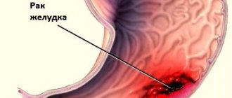
The stomach is a hollow muscular organ, the widest part of the gastrointestinal tract (GIT), where food accumulates and its initial digestion and partial absorption of nutrients and water begin. How does the stomach work, how is gastric juice formed and why are its protective functions disrupted?
Anatomical structure of the stomach
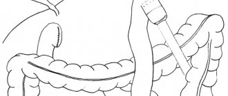
Food from the esophagus enters the stomach, which at its other end is connected to the duodenum. Usually the stomach is shaped like the letter “J”, but it can change depending on the position of the body, food intake, etc. Due to the fact that the stomach is a muscular organ, its walls can stretch when eating food and water. The volume of the stomach also depends on age and, of course, dietary habits. Anatomically, the stomach has 4 parts:
- the part that is adjacent to the esophagus is the cardiac part;
- adjacent to the duodenum - pyloric or pyloric;
- between these sections is the body of the stomach;
- and the part that is located to the left of the cardiac part is the bottom.
The stomach is separated from the esophagus by the lower esophageal sphincter, and from the duodenum by the pyloric sphincter. Their main task is to prevent the backflow of contents from the stomach into the esophagus, from the duodenum into the stomach. Although failures do happen.
The structure of the stomach wall
The structure of the human stomach wall can be divided into four main layers. The inner part is the mucous membrane, which is covered with single-layer columnar epithelium. Below is the submucosa, and after that is the muscle layer, in the structure of which several sublayers of smooth muscle can be distinguished. The peculiarity of these muscles is that their contraction is not controlled by a person; all movements occur unconsciously, since this is the “jurisdiction” of the autonomic nervous system. And the outer layer of the stomach wall is the serosa. Between the submucosa and the muscular layer is the submucosal plexus, which regulates the secretory function of epithelial cells.
Features of the structure of the gastric mucosa
The mucous membrane is formed by a single-layer cylindrical layer, under which muscular plates are located. They form folds, fields and gastric pits. The excretory ducts of the gastric glands are concentrated here. This layer of the mucosa contains many gastric glands, which consist of several protective cells of the stomach:
- parietal cells, their main task is the production of hydrochloric acid;
- the main cells produce the enzymes pepsin and pepsinogen;
- additional - secrete protective mucus, which protects the walls of the stomach from hydrochloric acid.
The entire surface of the gastric mucosa is covered with a thin layer of mucus. Its chemical composition forms a mucobicarbonate barrier of the stomach, protecting it from its internal aggressive factors. Antimicrobial agents are concentrated in this mucus: lysozyme, lactoferrin and other components.
Gastric juice and digestion
The entire surface of the stomach has a pitted structure. This is precisely what ensures minimal contact of its walls with hydrochloric acid. Therefore, the acidity on the surface of the epithelium is close to neutral. The cells that produce hydrochloric acid are located in the superficial layer of the stomach walls, so the path it travels is quite short. And it is precisely the rapid nature of the production of digestive juices that protects the stomach from damage. Under the influence of hydrochloric acid, proteins and fats, such as milk, are digested in the stomach. However, salivary enzymes continue to operate in the digestive bolus, digesting carbohydrates and sugars. The production of gastric juice, like saliva, occurs reflexively. The stimulus for the production of digestive juice is not only the direct consumption of food, but also the sight and smell of food.
Motility and digestion of food in the stomach
Peristaltic contractions constantly occur in the stomach, even on an empty stomach they last on average 15-20 seconds.
During food intake, the anterior part of the stomach expands, and this process in medicine is called accommodation. It provides the ability to fit the entire volume of food into the stomach. As soon as food has entered the stomach, due to peristalsis, the food bolus (chyme) is mixed with hydrochloric acid. Evacuation of food from the stomach is carried out due to a pressure gradient. But its speed depends on the consistency of the food, for example, liquid food is evacuated literally immediately, but dense food can remain in the stomach for 4-6 hours, but protein foods leave the stomach much faster compared to fatty foods. The motor-evacuation function of the upper gastrointestinal tract and stomach in particular is carried out by the sympathetic and parasympathetic nervous system. Text: Yulia Lapushkina.
Histology.RU
Material taken from the site www.hystology.ru
The stomach performs secretory, mechanical and endocrine functions. There are single-chamber and multi-chamber stomachs.
Unichamber stomach . Its wall is built of mucous, muscular and serous membranes (Fig. 265).
The mucous membrane consists of an epithelial layer, a basic lamina, a muscular lamina and a submucosa.
Rice. 265. A - diagram of the microscopic structure of the fundus of the stomach:
1 - single-layer cylindrical glandular epithelium; 2 - gastric dimple; 3 - own fundic glands of the stomach; 4 - lamina propria of the mucous membrane; 5 - muscular plate of the mucous membrane; 6 - submucosa (a - blood vessel, b - fat cell); 7 - muscular layer; 8 - intermuscular nerve plexus; 9 - serous membrane; 10 - mucous membrane; 11 - oblique layers of the muscularis propria; 12 - circular layer and 13 - longitudinal layer of the muscular layer. B - diagram of the electron microscopic structure of the mucous cells of the superficial epithelial layer of the stomach (according to Ito): 1 - microvilli; 2 — granules of mucous secretion; 3 - mitochondria; 4 - Golgi complex; 5 - granular endoplasmic reticulum; 6 - basement membrane.
Rice. 266. Glands of the fundus of the stomach:
I - neck of the gland; II - body of the gland; III - bottom of the gland; 1 - single-layer glandular epithelium; 2 - gastric pit; 3 — own plate; 4 - additional cells; 5 - parietal cell; 6 - main cells; 7 - muscular plate.
Its surface is in the form of an uneven contour, which is facilitated by the loose connection of the mucous and muscular membranes.
The mucous membrane forms folds, fields and pits. All layers of the mucosa take part in the formation of folds, and the formation of fields involves the epithelial layer and the main lamina, in which the glands are located in groups delimited by connective tissue. Gastric dimples are formed as a result of the immersion of the epithelium into the thickness of the main lamina (A - 2).
The epithelial layer is represented by a single-layer columnar glandular epithelium. Its cells are characterized by pronounced polar differentiation: the basal pole contains an oval nucleus and numerous mitochondria; Above the nucleus is the Golgi complex. The apical pole contains secretory granules and drops of mucoid secretion (B). The surface epithelial layer produces mucus, which protects the mucosal tissue from mechanical damage from the rough part of the feed and the negative effects of gastric juice. The lamina propria is built from loose connective and reticular tissue. Here, adjacent to each other, lie simple tubular unbranched (branched) glands. Their excretory ducts open into the gastric pits. The structure and function of the glands of different zones of the stomach wall vary, and therefore they are divided into fundic, pyloric and cardiac. On this basis, parts of the stomach are usually called fundic, pyloric, cardiac.
Simple, tubular fundic glands have an unbranched or weakly branched terminal section and a short excretory duct that opens into a relatively shallow gastric fossa. The gland is divided into a neck, body and bottom (Fig. 266). The neck is the excretory duct, the body and bottom are the secretory section.
The gland has a very narrow, barely noticeable lumen; it consists of chief, parietal (lining), mucous, cervical, endocrine (argyrophilic) cells (see color table XI).
Most of the bottom and body of the gland are built from the main cells. The cell has basal and apical poles. The first of them is characterized by basophilia, which is caused by the localization of the protein-synthesizing system of the cell, that is, the granular endoplasmic reticulum. The formation of the proenzyme pepsinogen is associated with this zone of cells. The second pole is filled with granules of protein secretion; its plasmalemma forms short microvilli. In the central part of the main cell there is an oval nucleus (Fig. 267).
Parietal (parietal) cells, adjacent to the basement membrane, lie outside the main and mucous cells. They are round in shape and larger in size than the main ones. The rounded nucleus lies in the center of the cell, the cytoplasm is oxyphilic. Inside the parietal cell there is a system of intracellular tubules with numerous microvilli. They pass into intercellular tubules located between the cells of the gland and in contact with the lumen of the gland (B). The cytoplasm is rich in mitochondria. Parietal cells produce chlorides, from which hydrochloric acid is formed. In its presence, pepsinogen is converted into pepsin, an enzyme in gastric juice. Its action is aimed at breaking down the protein part of the feed.
The basal pole of the mucous cell contains a flattened? core. The apical pole contains mitochondria, the Golgi complex, and numerous round mucus granules. The cells are localized in the body of the gland.
At the base of each gland located in the neck is a flattened or triangular nucleus. In its apical part there are drops of secretion, well stained with mucicarmine. Cervical cells are characterized by high mitotic activity. They are considered not only ferrous, but also have regenerative ability.
Endocrine cells are found in the body and bottom of the gland. These cells secrete biologically active substances like hormones that stimulate the secretory function of the glands.
Simple tubular cardiac glands have a highly branched terminal section and a wide lumen of the excretory duct. The cells of the terminal section are cylindrical or cubic in shape, they have a nucleus pushed towards the base and light cytoplasm. The cells secrete amyloid enzymes that break down starch. Chief, parietal and mucous cells may be found in the glands. The cardiac glands are located in the main plate near the esophagus (Fig. 268).
The pyloric glands are tubular, simple, with short and highly branched terminal sections, with wide lumens. In the main plate they lie more loosely. Glandular cells are similar in structure to the mucous cells of the fundic glands. The cells are cylindrical in shape with a light cytoplasm containing mucus and flattened nuclei pushed towards the basal pole. There are cervical cells and no parietal cells
Rice. 267. Scheme of the electron microscopic structure of the main cell of the gland of the fundus of the stomach (A):
a - mitochondria; b - granular endoplasmic reticulum; c — Golgi complex; d — pepsinogen grains; d - microvilli; e - basement membrane; g - core; B - diagram of the electron microscopic structure of the parietal gland of the fundus of the stomach: 1 - tubule; 2 - mitochondria; 3 - core; 4 - lysosome; 5 - Golgi complex.
Rice. 268. Three types of stomach glands:
A - bottom; B - pyloric; B - cardiac gland; a - integumentary epithelium; b - isthmus; c - body; g - bottom of the gland; d - transverse and oblique sections of individual branches of the gland; e - main; g - parietal and h - accessory cells.
cells. The gastric pits are deeper than other glands.
The muscle plate is built from bundles of smooth muscle cells arranged circularly and longitudinally. It consists of two longitudinal and one internal circular layers. The contraction of muscle cells causes the formation of folds of the mucous membrane, which improves the secretion of secretions from the lumen of the glands,
The submucosa is built of loose connective tissue and contains the choroid and nerve plexuses, a network of lymphatic vessels. This structure determines the mobility of the mucous membrane.
The muscularis propria consists of three layers of smooth muscle cells: inner, outer and middle. The inner layer is oblique, the middle layer is circular, the outer layer is longitudinal. Between the layers of muscles are the ganglia of the intramural myenteric plexus and many lymphatic vessels.
The serous membrane is built of loose connective tissue and is covered on the outside with mesothelium (single-layer squamous epithelium).
In animals of different species, differences in the structure of single-chamber stomachs come down mainly to different ratios of the area of the glandular and glandular parts, the ratio of the length of the terminal sections and excretory ducts, which is due to the nature of the food taken, and in connection with this, the specific need for enzymes.
The multi-chamber stomach of ruminants (cattle, sheep, goats) consists of three proventriculuses: the rumen, book, mesh and glandular stomach - abomasum. Unlike abomasum, there are no glands in the mucous membrane of the proventriculus. Here, mechanical processing and decomposition of feed takes place with the participation of bacteria and protozoa. The mucous membrane of the proventriculus is covered with stratified keratinizing epithelium, which is a continuation of the epithelial layer of the esophagus. In the epithelium of the proventriculus, the stratum lucidum is not expressed; there is a granular and stratum corneum; a system of cracks is intensively developed between the cells.
Scar (Fig. 269 - A). Its mucous membrane forms protrusions (papillae) of the main plate of various sizes and shapes, covered with stratified squamous epithelium. The muscular plate consists of separate bundles lying at the base of the papillae. Individual muscle cells are found in the main plate of the papilla. The muscular coat is built from smooth muscle cells, among which there are individual fibers of striated muscle tissue. There are two layers; the inner one is circular and the outer one is longitudinal.
The scar is covered with a serous membrane, represented by loose connective tissue and mesothelium.
Grid (B). The main lamina of the mucous membrane forms a large number of folds, which, like the cells lying between them, are covered with stratified squamous epithelium. The muscular plate is not expressed. Single smooth muscle cells are scattered in the connective tissue base. The muscular layer is represented by two layers of smooth muscle cells that form spirals and intersections. The muscularis tunic is connected to the muscular lining of the esophagus and the esophageal groove. On the outside, the mesh is covered with a serous membrane. The esophageal gutter is similar in structure to the mesh. The muscular layer of the mucous membrane is more developed, which forms a continuous layer near the mesh. At the base of the lips of the groove there is a longitudinal smooth muscle layer that has not lost connection with the muscular membrane of the mesh. In addition, there is an inner circular layer of smooth muscle cells and an outer longitudinal layer of striated muscle fibers extending from the esophagus.
Book (B). The main and muscular plates are involved in the formation of leaflets. At the apex, the muscular plate thickens, forming a longitudinal layer of the leaf margin. The large layers are penetrated by muscle cells of the annular layer of the muscularis propria. The latter also consists of smooth muscle cells, forming a thinner longitudinal layer and a thicker
Rice. 269. Scheme of the histological structure of the proventriculus of cattle:
A - scar; B - mesh; B - book; a — epithelial layer; b - base of the mucous membrane; c — muscular plate; d — submucosa of the mucous membrane; e - annular and longitudinal layers of the muscularis propria; e - serous membrane; g - ingrowth of connective tissue into the epithelium; h — papillae of the rumen mucosa; and - large fold of the mesh; j - its muscle bundles; l - large sheet of book; m - lamellar process of the annular layer of the muscular layer. At high magnification: n - scar papilla; o - mesh fold; p - a piece of book (according to Tehver).
circular. On the outside lies a serous membrane consisting of loose connective tissue and mesothelium.
Thus, a common morphological feature of the proventriculus of ruminants is the presence of connective tissue papillae covered with squamous stratified epithelium. The forestomachs are abundantly supplied with blood. The papilla includes several arterial vessels, which form a dense network of capillaries in it.
Abomasum. The wall of the glandular stomach of ruminants, like other animals, is built of mucous, muscular and serous membranes. The main lamina of the mucosa contains the cardiac, fundic and pyloric glands. Simple tubular fundic glands have a longer neck and a relatively short secretory section. These glands occupy most of the stomach wall. The zone of cardiac glands is insignificant. It is located next to the entrance of the book into the abomasum. Compared to other animals, the pyloric glands are longer.
Reviews (0)
Add a review