What is a biochemical blood test?
Biochemical blood test (biochemistry) is currently one of the most universal methods of laboratory diagnostics, as it allows you to assess the condition of all systems of the human body. You can take this test and get the result in one to two business days. Using this method, doctors evaluate whether the liver, kidneys, pancreas and other internal organs are functioning normally, identify active inflammatory and rheumatic processes, as well as impaired water-salt metabolism and an imbalance of microelements. A biochemical blood test is of great importance for making a diagnosis, prescribing and adjusting treatment, as well as determining the stage of the disease.
Detailed laboratory examination of the pancreas
A comprehensive blood test that allows you to identify the main disorders of various etiologies in the functional state of the pancreas.
The research results are provided with a free doctor’s commentary.
What is this analysis used for?
- For the diagnosis of pancreatic diseases.
When is the test scheduled?
- For clinical symptoms of pancreatic diseases;
- if pancreatic dysfunction is suspected.
English synonyms
Pancreatic panel.
Research method
- C-peptide – competitive solid-phase chemiluminescent enzyme-linked immunosorbent assay;
- plasma glucose – enzymatic UV method (hexokinase);
- alanine aminotransferase (ALT) – UV kinetic test;
- lipase – enzymatic colorimetric method;
- gamma-glutamyl transpeptidase (gamma-GT), total serum amylase, serum cholinesterase, total bilirubin - kinetic colorimetric method;
- C-reactive protein, quantitative (highly sensitive method) - immunoturbidimetry;
- CA 19-9 – chemiluminescence immunoassay (“sandwich” method).
Units
- C-peptide – ng/ml (nanograms per milliliter);
- plasma glucose – mmol/l (millimoles per liter);
- alanine aminotransferase (ALT), lipase – IU/l (international unit per liter);
- gamma-glutamyl transpeptidase (gamma-GT), total amylase in serum - U/l (unit per liter);
- C-reactive protein, quantitative (highly sensitive method) – mg/l (milligrams per liter);
- CA 19-9, serum cholinesterase - U/ml (unit per milliliter);
- total bilirubin – µmol/l (micromoles per liter).
What biomaterial can be used for research?
Venous blood.
How to properly prepare for research?
Do not smoke for 30 minutes before the test.
General information about the study
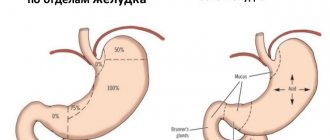
The pancreas is an organ of the gastrointestinal tract located behind the stomach and performs important exocrine and endocrine functions. Digestion of proteins and fats in the small intestine is carried out due to the synthesis and secretion of digestive enzymes by the exocrine part of the gland. In addition to proteo- and lipolytic enzymes, it releases bicarbonates, neutralizing the hydrochloric acid of gastric juice in the duodenum. The endocrine function of the pancreas is provided by islet tissue, in which the hormones insulin, glucagon, somatostatin and pancreatic polypeptide are synthesized and then secreted into the blood. Insulin and glucagon regulate blood glucose levels and its transport in tissues. Pathology of the pancreas primarily leads to digestive disorders, and in chronic diseases contributes to the development of endocrine disorders (diabetes mellitus).
The causes of pancreatic diseases are different: genetic and autoimmune disorders, infections (usually viral), injuries, toxic lesions, taking certain medications (estrogens, furosemide, azathioprine, etc.), neoplasms. Most often, pathology of the pancreas occurs against the background of liver dysfunction, diseases of the biliary tract (cholelithiasis with choledocholithiasis), due to impaired outflow of bile and pancreatic juice. Another common cause of pancreatic diseases is alcohol abuse.
Clinical manifestations of pancreatic diseases depend on the etiology, degree of dysfunction and activity of the process. Acute inflammatory changes, trauma to the gland, as well as chronic diseases during exacerbation in most cases are accompanied by pain and burning in the epigastric region with irradiation to the back, nausea, vomiting, and increased body temperature. Chronic diseases of the pancreas lead to pancreatic insufficiency, weight loss, and the development of ascites due to impaired digestion and absorption of nutrients from the intestine.
An increase in the activity of pancreatic enzymes (amylase and lipase) and the level of C-reactive protein in the blood are signs of active inflammation of the organ - acute pancreatitis. Changes in the level of glucose and C-peptide indicate a violation of the endocrine function of the pancreas and are an indirect sign of damage to pancreatic islet tissue, which can occur with chronic pancreatitis. A sharp increase in the tumor marker CA 19-9 against the background of changes in biochemical indicators of gland function most often indicates pancreatic cancer.
An increase in the concentration of alanine aminotransferase, gamma-glutamyl transpeptidase, cholinesterase, bilirubin and the enzymes amylase and lipase indicates the simultaneous involvement of the liver and pancreas in the pathological process, which usually occurs with stones of the common bile duct and reactive pancreatitis.
If the indicators of this complex analysis change, it is necessary to perform additional laboratory and instrumental studies to clarify the causes and mechanisms of the development of the disease and select therapy.
What is the research used for?
- To assess the functional state of the pancreas and the severity of damage;
- for differential diagnosis of pancreatic diseases;
- for monitoring a patient with chronic diseases of the hepatopancreabiliary zone (cholelithiasis, cholelithiasis, chronic pancreatitis);
- to monitor the effectiveness of treatment of pancreatic diseases.
When is the study scheduled?
- For symptoms of possible damage to the pancreas (girdling pain and/or burning in the upper abdomen, nausea, vomiting, change in color, quantity and consistency of stool);
- when the structure and size of the pancreas changes according to instrumental research methods;
- when examining persons who abuse alcohol;
- if there is a family history of pancreatic diseases;
- when monitoring patients with chronic diseases of the liver, pancreas and biliary tract;
- during a preventive examination.
What do the results mean?
Reference values
C-peptide: 0.9 - 7.1 ng/ml.
Plasma glucose
| Age | Reference values |
| Less than 3 years | 3.3 - 5.5 mmol/l |
| 3 – 16 years | 3.3 - 5.5 mmol/l |
| More than 16 years | 4.1 - 5.9 mmol/l |
Alanine aminotransferase (ALT)
| Floor | Age | Reference values |
| Male | Less than 1 day | 13 – 45 IU/l |
| More than 1 day | 0 – 50 IU/l | |
| Female | Less than 1 day | 13 – 45 IU/l |
| More than 1 day | 0 – 35 IU/l |
Gamma-glutamyl transpeptidase (gamma-GT)
| Floor | Age | Reference values |
| Female | Less than 3 months | 15 – 74 U/l |
| 3-6 months | 9 – 28 U/l | |
| 6 months – 1 year | 4 - 17 U/l | |
| 1-13 years | 3 - 22 U/l | |
| 13-18 years old | 2 - 42 U/l | |
| Over 18 years old | 0 - 38 U/l | |
| Male | Less than 3 months | 15 – 74 U/l |
| 3-6 months | 9 – 28 U/l | |
| 6 months – 1 year | 4 - 17 U/l | |
| 1-13 years | 3 - 22 U/l | |
| 13-18 years old | 2 - 42 U/l | |
| Over 18 years old | 0 - 55 U/l |
Lipase
| Age | Reference values |
| Less than 1 year | 0 – 8 IU/l |
| 1 – 10 years | 5 – 31 IU/l |
| 10 – 18 years | 7 – 39 IU/l |
| Over 18 years old | 21 – 67 IU/l |
Total amylase in serum: 28 - 100 U/l.
C-reactive protein, quantitative (highly sensitive method): 0 - 1 mg/l.
CA 19-9: 0 - 35 U/ml.
Total bilirubin
| Age | Reference values |
| Less than 2 days | 58 – 197 µmol/l |
| 2-6 days | 26 - 205 µmol/l |
| 6-30 days | 5 – 21 µmol/l |
| More than a month | 5 – 21 µmol/l |
Serum cholinesterase
| Floor | Reference values |
| Female | 3.93 - 11.5 U/ml |
| Male | 4.62 - 11.5 U/ml |
Serum total amylase
Reasons for the increase:
- Acute or reactive pancreatitis
- Pancreas cancer
- Alcoholism
- Biliary obstruction (cholelithiasis)
- Diabetic ketoacidosis
- Hyperthyroidism
- Inflammation of the salivary glands
- Parotitis
- Perforation of a stomach or intestinal ulcer
- Peritonitis
- Pregnancy
- Reasons for the downgrade:
- Cirrhosis of the liver
- Hepatitis
- Cystic fibrosis
- Severe burns
- Severe thyrotoxicosis
Serum cholinesterase
Reasons for the increase:
- Hyperlipidemia type VI
- Nephrotic syndrome
- Obesity
- Diabetes
- Chorea
- Neurosis
Reasons for the downgrade:
- Liver diseases (hepatitis, cirrhosis with jaundice, liver failure, massive tumor or liver metastases)
- Organophosphate poisoning
- Congestive heart failure
- Acute infection, tuberculosis
- Acute myocardial infarction
- Poor nutrition
- Anemia
- Dermatomyositis
- Congenital cholinesterase deficiency
Lipase
Reasons for the increase:
- Acute pancreatitis
- Chronic pancreatitis, pancreatic trauma or cancer, pancreatic duct obstruction
- Cholecystitis
- Hemodialysis
- Peritonitis
- Primary biliary cirrhosis
- Strangulated intestinal obstruction, intestinal infarction
- Chronic renal failure
- Reasons for the downgrade:
- Decreased thyroid function
- Cystic fibrosis
Plasma glucose
Reasons for the increase:
- Endocrine diseases (diabetes mellitus, Cushing's syndrome, hyperthyroidism, acromegaly, pheochromocytoma, pituitary adenoma, glucagonoma)
- Acute emotional or physical stress
- Obesity
- Hemochromatosis
- Acute or chronic pancreatitis, pancreatic cancer
- Chronic kidney disease
- Pregnancy
- Reasons for the downgrade:
- Alcoholic liver disease, liver toxicity, liver necrosis, cirrhosis, liver failure
- Insulinoma
- Adrenal insufficiency
- Malabsorption
- Starvation
- Overdose of insulin drugs
- Physical stress, intense physical activity
C-reactive protein
Reasons for the increase:
- Acute pancreatitis
- Appendicitis
- Bacterial infection
- Burns
- Inflammatory bowel diseases
- Systemic lupus erythematosus
- Lymphomas
- Myocardial infarction
- Pelvic inflammatory diseases
- Polymyalgia rheumatica
- Rheumatoid arthritis
- Sepsis
- Tuberculosis
- Surgery – the first 3 days after it
Alanine aminotransferase (ALT)
Reasons for the increase:
- Acute infectious or toxic hepatitis (increase in rate by 30-50 times)
- Alcoholic liver disease, cirrhosis (moderate increase)
- Metastatic liver damage (moderate increase)
- Impaired bile outflow due to biliary cirrhosis, cholelithiasis, choledocholithiasis, pancreatic cancer (moderate increase)
- Pancreatitis (possibly slightly enlarged)
- Infectious mononucleosis
- Myocardial infarction, heart failure
- Polymyositis
- Severe burns, skeletal muscle injuries
- Severe shock
Gamma-glutamyl transpeptidase (gamma-GT)
Reasons for the increase:
- Biliary obstruction (cholelithiasis, choledocholithiasis, cholecystitis, cholangitis)
- Acute or chronic hepatitis
- Alcoholic liver disease
- Metastases in the liver
- Cirrhosis of the liver
- Acute pancreatitis
- Pancreas cancer
- Kidney cancer
- Congestive heart failure, myocardial infarction
- Systemic lupus erythematosus
Total bilirubin
Reasons for the increase:
- Choledocholithiasis
- Cholelithiasis
- Viral hepatitis
- Sclerosing cholangitis
- Biliary cirrhosis of the liver
- Pancreatic head cancer
- Dubin–Johnson syndrome
- Rotor syndrome
- Biliary atresia
- Alcoholic liver disease
- Pregnancy
- Cirrhosis
- Infectious mononucleosis
- Toxic hepatitis
- Liver echinococcosis
- Liver abscesses
- Metastases or massive liver tumors
- Blood diseases accompanied by hemolysis (hemolytic, pernicious, megaloblastic, sickle cell anemia, erythremia, thalassemia, congenital microspherocytosis)
- Gilbert's syndrome
- Crigler–Najjar syndrome
- Post-hemotransfusion reaction, transfusion of incompatible blood groups
- Malaria
- Myocardial infarction
- Sepsis
- Hemorrhagic pulmonary infarction
- Hemorrhage into tissue
- C-peptide
Reasons for the increase:
- Insulinoma, islet tissue tumor
- Diabetes mellitus type 2 (non-insulin dependent)
- Pancreas transplant
- Kidney failure
- Taking oral hypoglycemic drugs
- Reasons for the downgrade:
- Diabetes mellitus type 1
- Pancreatectomy
- Hypoglycemia when using insulin
CA 19-9
Reasons for the increase:
- Pancreatic cancer (significant increase)
- Lung cancer (significant increase)
- Gallbladder cancer (significant increase)
- Colorectal cancer (moderate increase)
- Stomach cancer (moderate increase)
- Cholecystitis (slight increase)
- Cholelithiasis
- Liver pathology, cirrhosis
- Cystic fibrosis (cystic fibrosis)
- Pancreatitis
What can influence the result?
Factors distorting the result:
- study with intravenous administration of a contrast agent 24 hours before analysis;
- pregnancy;
- concomitant pathology (diseases of other parts of the gastrointestinal tract, malignant neoplasms or acute inflammation of other localizations, endocrine pathology, blood diseases);
- infectious mononucleosis, adenovirus infection, mumps and other acute infections affecting the pancreas;
- taking medications that affect certain test indicators.
Important Notes
- Considering the fact that each individual biochemical indicator of the study is not a strictly specific marker of pancreatic function, changes identified by the analysis require the exclusion of possible pathology of other organs and systems, as well as clarification of the etiology of the pathological process.
- Test results should be analyzed taking into account clinical symptoms, disease duration and comorbidities. When interpreting them, a careful differential diagnosis is necessary, which is carried out by a qualified physician. Sometimes instrumental studies (ultrasound, CT scan of the abdominal cavity) can be used.
Also recommended
- Laboratory diagnosis of pancreatitis
- Laboratory examination of the liver
- Bilirubin and its fractions (total, direct and indirect)
- Laboratory diagnosis of hepatitis - biochemical markers
- Lipidogram
- Total amylase in daily urine
- Pancreatic amylase
- Serum calcium
- Insulin-like growth factor
- Serum elastase
- Glycated hemoglobin (HbA 1c)
- C-peptide in daily urine
- Glucose tolerance test
- Glucose in urine
- Antibodies to insulin
- Antibodies to pancreatic islet cells
- Insulin
- Proinsulin
- Coprological elastase
- Gastrin
- Complete blood count (without leukocyte formula and ESR)
- Erythrocyte sedimentation rate (ESR)
- General urine analysis with sediment microscopy
- Coprogram
- Serum albumin
- Alpha fetoprotein (alpha FP)
- Carcinoembryonic antigen (CEA)
- Antibodies to smooth muscle
- Antinuclear factor on HEp-2 cells
- Anti-mitochondrial antibodies (AMA)
Who orders the study?
Gastroenterologist, therapist, general practitioner.
Deadline
Until 12:00 the next day. If tests are taken before 12:00, the result can be received by email until 24:00 of the same day
The complex includes
- Serum total amylase
- Plasma glucose
- Lipase
- Serum C-peptide
- CA 19-9
Units
ng/ml (nanograms per milliliter), mmol/l (millimoles per liter), IU/l (international unit per liter), unit/l (unit per liter), mg/l (milligram per liter), unit/ ml (unit per milliliter), µmol/l (micromole per liter)
Preparing for the study
- Do not eat for 12 hours before the test.
- Avoid physical and emotional stress for 30 minutes before the test.
- Do not smoke for 30 minutes before the test.
Type of biomaterial and methods of collection
| Type | At home | In the center | On one's own |
| Deoxygenated blood | Yes | Yes |
At home: biomaterial can be collected by a mobile service employee.
At the Diagnostic Center: collection or independent collection of biomaterial is carried out at the Diagnostic Center.
Independently: collection of biomaterial is carried out by the patient himself (urine, feces, sputum, etc.). Another option is for the doctor to provide samples of biomaterial to the patient (for example, surgical material, cerebrospinal fluid, biopsies, etc.). After receiving the samples, the patient can either independently deliver them to the Diagnostic Center or call a mobile service at home to transfer them to the laboratory.
Literature
- Nazarenko G.I., Kishkun A. Clinical assessment of laboratory research results. – M.: Medicine, 2000. – 533 p.
- Fischbach FT, Dunning MB A Manual of Laboratory and Diagnostic Tests, 8th Ed. Lippincott Williams & Wilkins, 2008: 1344 p.
- Wilson D. McGraw-Hill Manual of Laboratory and Diagnostic Tests 1st Ed. Normal, Illinois, 2007: 666 p.
Indications for biochemical blood test
Most often, a biochemical blood test is prescribed during a comprehensive examination of the body. Even if the patient does not have obvious manifestations of the disease, a biochemical analysis will help determine which organ is not coping with its task and is not working as it should. Any change in the biochemical composition of the blood indicates an unfavorable situation and the need for urgent intervention. Doctors of almost all specialties use the results of biochemical blood tests in their practice. Let's name the most common areas: gastroenterology, urology, cardiology, gynecology and many others. A biochemical blood test is a set of tests, each of which is prescribed to the patient depending on the complaints and clinical picture. Your doctor may order a standard panel of tests or just 1-2 tests. If you have a need to donate blood for biochemistry, but you do not know what indicators you need, seek help from a doctor.
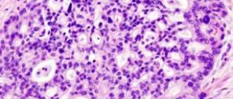
CT and MRI in the diagnosis of pancreatic cancer
HOW TO DIAGNOSE PANCREAS CANCER
The pancreas is a very important organ that produces pancreatic juice necessary for digestion, and also takes part in the production of hormones, including insulin. Tumor diseases of the gland are common. Their peculiarities include the fact that there are no early symptoms of pancreatic cancer as such. At an early stage, neoplasms of the pancreatoduodenal zone do not manifest any symptoms, which is why they are diagnosed late. Thus, the patient may not be aware of cancer for a long time. Symptoms appear only when the tumor grows into neighboring organs, when its size increases (for example, when it compresses the papilla of Vater, into which the bile ducts open). With this variant of the course of the disease, obstructive jaundice appears. This forces the patient to undergo diagnostic testing (CT, MRI, ultrasound), which reveals the oncological process. In other cases, the tumor may be an incidental finding during studies performed for some other reason. More often, a tumor is discovered by chance during a preventive ultrasound.
HOW TO TEST YOUR PANCREAS FOR CANCER
Today, there are several methods for accurately diagnosing this oncology, both X-ray and non-X-ray. In this article we will take a detailed look at what pancreatic cancer looks like on various images and how to find it using each of these methods. We will also provide diagnostic signs of pancreatic cancer, as well as consider clinical examples illustrating this dangerous disease.
Today, scientists have proven that the most informative way to check the pancreas for cancer and to identify oncopathology as early as possible is magnetic resonance imaging (MRI). This study makes it possible to clearly visualize the tumor, determine its structure and structure, distinguish between the soft tissue and cystic components, identify growth into the parapancreatic tissue (that is, the tissue surrounding the gland, from the Latin word “pancreas”), into neighboring organs (into the spleen, into the renal fascia , into the duodenum, into other parts of the intestine, into the omentum). In this case, it is necessary to take into account the fact that MRI must be performed on a high-field device (with a field strength of not <1.5 Tesla), because only such a device provides the required quality of diagnostic images. If necessary, the study is supplemented with the introduction of a contrast agent. In addition, it should be borne in mind that image analysis should be carried out by an experienced radiologist with good experience in diagnosing pathology of the pancreaticoduodenal zone. To avoid a diagnostic error, you can re-analyze the MRI images and get a Second Opinion from an experienced specialist (more on this at the bottom of the article).
Get an MRI of the pancreas in St. Petersburg
Also, a formation in the pancreas can be detected by ultrasound. This is a simple and accessible method that evaluates changes in the structure of the organ, changes in size (thickening), the presence of a cystic (liquid) component, the degree of expansion of the pancreatic duct, and signs of cancer germination (invasion) into neighboring tissues. The disadvantage of ultrasound is its low tissue resolution, and in doubtful cases, tomographic methods are prescribed - computed tomography and MRI.
A space-occupying pancreas formation can also be suspected during FGDS (fibrogastroduodenoscopy), a study of the organs of the digestive system using a flexible fiber-optic probe. In this case, the endoscopist may note a deformation (persistent, permanent) of the duodenum and suspect some kind of volumetric process in the pancreaticoduodenal zone. If there is such a suspicion, a computer or magnetic resonance imaging scan is required.
CT DIAGNOSIS OF PANCREAS CANCER
The most widely used X-ray method for visualizing pancreatic volumetric processes is computed tomography. The study can be either native (without contrast) or with contrast. Contrast CT helps to better differentiate normal and tumor tissue, as well as clearly identify the pathology of the vessels surrounding the gland. In addition, contrast enhancement on CT scans is mandatory if the doctor wants to exclude the spread of the process to the liver (liver metastases) or surrounding lymph nodes.
SIGNS OF A NEOPOLOGY (CANCER) OF THE PANCREAS DETECTED BY CT
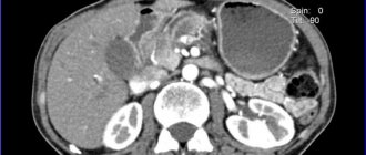
1) Local thickening (tail, body and head are thickened evenly). Thickening can also be diffuse (affecting all departments) - in this case it is customary to talk about “pseudotumorous pancreatitis” and not about cancer. Sometimes it is difficult to distinguish between these two conditions on CT or MRI, and consultation with an experienced radiologist is necessary. With local thickening, the size of the affected area is usually measured on axial sections and assessed according to the TNM system (T - lat., "tumor" - tumor, N - "nodus" - lymph node, M - "metastasis" - the presence of metastases in distant organs, more often total liver).
On the images: local thickening of the pancreas head due to a soft-tissue (solid) neoplasm that practically does not accumulate contrast (red arrow). The dilated duct of Wirsung is highlighted with a yellow arrow.
The tail of the pancreas is sharply thickened (yellow arrow), its structure is disturbed: multiple poorly contrasted areas (necrosis and decay) are visible. In addition, there is ascites (fluid in the abdominal cavity). Metastasis to the perinephric tissue (retroperitoneal) is highlighted with a red asterisk.
The same patient. When analyzing the remaining scans, it becomes clear that the volumetric process is not limited only to the tail, but also extends to the body and head. The prognosis here is unfavorable; life expectancy with such a prevalence of oncological process is usually short. The photo in the top row shows a soft tissue formation with a focus of decay in the center, the images in the bottom row (left) show pathologically altered regional lymph nodes - in the gates of the liver, as well as in the tissue near the aorta and retroperitoneally, in the tissue near the gates of the left kidney. The red arrow also highlights a large solid-cystic area in the liver (metastatic in nature). Green arrows indicate fluid in the abdominal cavity.
2) The presence of a formation that has a heterogeneous structure, with uneven edges (lumpy). With computed tomography, you can evaluate its structure, determine the predominant component (solid, soft tissue; or liquid, cystic), the presence of areas of necrosis, purulent melting, calcifications, hemorrhages, etc.
What does pancreatic cancer look like? Photo (CT). The yellow circle reveals pathological tissue in the head region (with uneven edges, located near the wall of the duodenum).
3) Increased density of parapancreatic fiber. The “turbidity” of the fiber may indicate its tumor infiltration and/or an associated infection, or autolysis (self-digestion) by released enzymes (as a result of the destruction of pancreatic tissue) and the development of pancreatitis.
Please note: near the pancreatic head, the fiber loses its usual structure, its density is higher, the edges of the head are blurred and indistinct. All these are signs of infiltration of fiber by cancer cells (contact metastasis) or signs of pancreatitis (secondary).
Get a CT scan of the pancreas in St. Petersburg
4) Dilation of the pancreatic (Wirsung) duct above the site of the lesion. Cancer of the head causes expansion of the pancreatic duct in the area of the body and tail (normally it has a width of 1-2 mm, with tumor lesions it can expand to 0.5 cm or more).
On the images: a tumor of the head of the gland (in the yellow circle), yellow arrows indicate the dilated pancreatic (Wirsung) duct as a result of a violation of the outflow of secretions. Native (without contrast) computed tomography.
5) Uneven accumulation of contrasted blood by the tumor (it has a lower density compared to unchanged tissue). The solid (soft tissue) component accumulates contrast; areas of necrosis and cystic restructuring do not accumulate it, because they do not have blood vessels in their structure and remain hypodense (low density).
6) Germination of the tumor into the nearest organs (into the spleen, into the duodenum, into the superior vena cava, into the portal vein, into the splenic vein, into the hepar, into the omentum, into Gerota’s fascia, separating the retroperitoneal space from the abdominal cavity) are extremely unfavorable signs - markers of neoplasm inoperability (T4 according to TNM).
In this case, with a tumor of the head of the pancreas, surgery is not indicated, because germination of a blood vessel occurs and hemorrhage occurs in the hepatic parenchyma (hematoma is indicated by a red arrow), a space-occupying lesion is indicated by a yellow circle. According to the TNM classification, pancreatic cancer corresponds to T4 (tumor of any size with invasion into surrounding tissues, including blood vessels).
A mass formation of the tail of the gland growing into the spleen (inoperable, TNM T4), on the left - before contrast, on the right - after contrast administration. A large cystic lesion (O) was detected in the tail area (and also partially in the body area), with a predominant fluid component, with multiple zones of necrosis, close to the spleen (C), also adjacent to the anterior renal fascia on the left. Metastases (distant) are marked with red asterisks.
The same patient. CT images reformatted in the coronal plane allow assessment of the extent of the lesion. A volumetric process with germination into the spleen is clearly visible, and fluid along the edge of the spleen is also visible. Hypodense secondary lesions in liver tissue.
7) The presence of altered and enlarged lymph nodes in the parapankeratic tissue, in the para-aortic tissue, in the hilum of the liver and (or) spleen indicates lymphogenous metastasis of the neoplasm and makes it possible to set N1 according to the TNM system (N - Latin “nodus”, lymph node). TNM N1 means damage to regional (located near the organ) lymph nodes and is an unfavorable sign, but does not exclude surgical intervention.
Tumor of the head (in the yellow circle) with metastases to the lymph nodes near the porta hepatis, as well as to the para-aortic lymph nodes (green arrows). Contrast-enhanced CT images (arterioparenchymal phase contrast enhancement) are shown. According to the TNM classification, the neoplasm belongs to stage N1M1 - the presence of enlarged regional nodes with a pathologically altered structure and distant secondary foci.
 Presence of liver metastases. The neoplasm can metastasize not only through the lymphogenous route, but also through the hematogenous route. The organ that is affected first is the liver, while the elimination of tumor cells occurs through the portal vein system. Detection of metastases to distant organs in pancreatic cancer is an unfavorable symptom (according to the TNM system, M1 is set - “distant metastases detected”). If distant metastases are found with a pancreatic tumor, surgery is not indicated. There are, however, modern methods of treating liver metastases (chemoembolization, ultrasound ablation, etc.), carried out in advanced oncology hospitals.
Computed tomography with contrast. Stage of the process is M1 according to the TNM classification (with the presence of symptoms of jaundice - due to compression of the common bile duct - and distant metastases). On the left, red arrows highlight multiple hypodense (low density), weakly accumulating contrast agent areas (1-3 cm in diameter), round in shape, diffusely distributed over the entire section area. On the right, under the blue arrow, is the part of the volumetric process that does not accumulate contrast (which contains a small number of arterial vessels), under the green arrow is the soft tissue part of the formation, and under the red arrow is the unchanged part of the organ (body and tail).
Pancreatic tail cancer. CT symptoms. Yellow arrows indicate a formation that has a heterogeneous structure, with multiple hypodense areas of necrosis and decay. There is a complication - massive ascites, i.e. accumulation of fluid in the abdominal cavity (fluid is marked with yellow stars). Distant metastasis is highlighted with a green arrow.
SECOND OPINION IN CANCER
Without a doubt, a pancreatic tumor is a dangerous diagnosis, which in some cases has an unfavorable prognosis. Patient survival, life expectancy, success of a particular treatment method, and choice of surgical tactics strictly depend on the stage of the tumor process. Therefore, the most important condition for successful treatment is not only the timely detection of oncopathology, but also its accurate staging using the TNM scale. In particular, it is very important for oncologists to know whether the tumor is spreading into surrounding organs and tissue, vascular damage, metastasis to the lymph nodes and liver. Therefore, it is necessary not only to undergo a modern diagnostic examination, such as CT or MRI, but also to correctly analyze the images in order to identify or exclude all of the above signs.
To be confident in the accuracy of the diagnosis, today you can order a review of CT and MRI results from specialists in the radiological diagnosis of oncological diseases. Such expert analysis is carried out in institutions that specialize in oncological and surgical pathology. This allows us to increase the accuracy of diagnosis and describe the disease according to modern standards. The resulting expert opinion is an accurate guideline for the attending physicians - oncologist surgeons.
You can get a Second Opinion on a CT scan of the pancreas in the National Teleradiological Network (NTRS) system. This service receives complex and controversial diagnostic cases from all Russian regions. Specialists from the Moscow Institute of Surgery named after. Vishnevsky and other specialized centers will conduct a remote review of your CT or MRI. It is enough to download CT or MRI images from a disk on the NTRS website and receive a qualified report signed by a doctor within 24 hours.
Vasily Vishnyakov, radiologist
Read more about Second Opinion
Read more about telemedicine
Pavel Popov
Candidate of Medical Sciences, Member of the European Society of Radiologists
Decoding the results of a biochemical blood test
For all indicators of a biochemical blood test, there are no strictly defined “normal” values. They speak of normality if they are in the interval between the minimum and maximum permissible values. In addition, due to the use of different reagents or slightly different technologies, the absolute values for each indicator of a biochemical blood test may vary depending on the laboratory in which the study was performed. Therefore, the main attention should be paid to whether the value “falls” within the range from the minimum to the maximum permissible value. Based on the symptoms of the disease and the results of the analysis, doctors will make a diagnosis, conduct a consultation and prescribe treatment.
The value and range of normal values of biochemical analysis indicators
Total protein
The total concentration of blood serum proteins, it consists mainly of the concentration of 2 main protein fractions: albumins and globulins. Proteins are involved in maintaining the pH and osmotic pressure of the blood, in coagulation and immune reactions (immunoglobulins, acute phase proteins), and also transport lipids, bilirubin, steroid hormones to organs and tissues (transport proteins). The concentration of total protein is an important diagnostic parameter for many diseases, especially those associated with metabolic disorders.
Normal: 64–84 g/l.
An increase in concentration may indicate an infectious disease, autoimmune diseases (rheumatoid arthritis, rheumatism, systemic lupus erythematosus) or cancer. It also increases with dehydration, that is, loss of water (prolonged diarrhea, especially in children, persistent vomiting, extensive burns).
A decrease in concentration can occur due to insufficient protein intake from food, diseases of the liver, intestines, kidneys or cancer, thyrotoxicosis, as well as due to prolonged physical activity.
Glucose
The most important component of blood, the main participant in carbohydrate metabolism. Our body receives more than half of its energy in glucose oxidation reactions. Glucose enters our body with food, is formed in the cells of the body from amino acids and lactate, excess glucose is stored by the body “in reserve” in the liver in the form of glycogen. The concentration of glucose in the blood is determined, primarily for the diagnosis of diabetes mellitus types 1 and 2. It is recommended that all people over 45 years of age have this test (even if there are no symptoms) because changes in glucose levels occur several years before clinical signs of diabetes appear. Measuring glucose concentration is also of great importance in the diagnosis of diseases of the thyroid gland, adrenal glands, pituitary gland, as well as in obese patients and pregnant women.
Normal: 3.30–5.50 mmol/l.
An increase in glucose concentration is observed in type 1 or type 2 diabetes, impaired glucose tolerance, hormonal disorders, acute or chronic pancreatitis, chronic liver and kidney diseases.
A decrease in concentration occurs with pancreatic tumors, some endocrine diseases, severe liver damage (cirrhosis, tumors), etc.
Urea
The main product of protein breakdown, its content in the blood depends on the nature of the diet (the predominance of meat foods leads to its increase, plant foods – a decrease), age (increases in older people).
Normal: 2.5–8.3 mmol/l.
An increase in serum urea concentration may be a consequence of poor kidney function, heart failure, tumor process, bleeding, intestinal obstruction or urinary tract obstruction. Short-term increases in urea levels may occur after intense training or physical activity.
A decrease in concentration is observed in liver diseases (urea synthesis is impaired); malabsorption in the intestine, during pregnancy.
Creatinine
Creatinine is the end product of creatine phosphate metabolism, which is involved in the energy metabolism of muscles and other tissues. It is excreted by the kidneys, so its concentration is an indicator of kidney function.
Normal (depending on muscle mass): men – 62–115 µmol/l, women – 53–97 µmol/l.
An increase in concentration, as a rule, indicates renal failure or hyperthyroidism, and is also observed with massive damage to muscle tissue (trauma, surgery, long-term compartment syndrome).
A decrease in creatinine concentration occurs during fasting, a significant decrease in muscle mass, and during the 1st and 2nd trimester of pregnancy.
Total cholesterol (cholesterol)
A component of fat metabolism, it is involved in the construction of cell membranes, the synthesis of sex hormones and vitamin D.
Normal: 3.5–6.5 mmol/l.
Elevated cholesterol levels are a risk factor for atherosclerosis and coronary heart disease (CHD).
An increase in cholesterol concentration also occurs in diseases of the liver and pancreas, kidneys, hypothyroidism, diabetes mellitus, gout, and alcoholism. A diet rich in carbohydrates and fats leads to an increase in cholesterol levels.
A decrease in cholesterol concentration can be observed in case of malabsorption in the intestine, severe acute diseases, including infectious diseases, chronic obstructive diseases and pulmonary tuberculosis, etc.
Bilirubin
Yellow-red pigment, one of the main components of bile, is present in the blood serum in the form of two fractions: direct (protein-bound, or conjugated) and indirect (free, or unbound) bilirubin, which together constitute total bilirubin. In serum, As a rule, the concentration of total and direct bilirubin is determined; the difference between these indicators is the value of free (unconjugated, indirect) bilirubin. The determination of this indicator is of great importance for the differential diagnosis of jaundice of various origins.
Normal (total bilirubin): 5–20 µmol/l. When its concentration increases to values above 27 µmol/l, jaundice occurs.
An increase in the concentration of total bilirubin occurs with liver diseases (cancer, hepatitis, poisoning, cirrhosis), cholelithiasis, cholestasis, as well as against the background of hemolytic anemia, lack of vitamin B12.
Alanine aminotransferase ALT (ALT)
An enzyme that is predominantly found in liver cells and is therefore used to assess liver function. It is also found, but in smaller quantities, in the cells of the kidneys, heart, pancreas, and skeletal muscles. It enters the bloodstream when the cells of these organs are destroyed. Determination of ALT activity in serum plays an important role in the diagnosis of viral hepatitis; the degree of increase in the activity of this enzyme depends on the severity of the disease and can exceed the norm by 5–10 times or more. The level of ALT activity, and most importantly, the ratio of this indicator to the activity of another enzyme, aspartate aminotransferase, is of great importance for the diagnosis and prognosis of the course of myocardial infarction.
Norm: men – up to 41 U/l, women – up to 31 U/l.
High ALT activity in the blood indicates damage to the heart or liver and makes one suspect the presence of serious diseases such as viral hepatitis, cirrhosis, liver cancer, myocardial infarction, heart failure or myocarditis.
Aspartate aminotransferase AST (AST)
The cellular enzyme, like ALT, is found in the cells of the heart, liver and kidneys. Participates in amino acid metabolism. The highest concentration of this enzyme is observed in the myocardium (several thousand times higher than in blood serum).
Norm: men – up to 41 U/l, women – up to 31 U/l.
An increased level of AST in the blood can be caused by myocardial infarction, hepatitis, pancreatitis, liver cancer or heart failure. Determination of the activity of this enzyme allows us to predict the course of myocardial infarction: if it does not decrease on the 3rd day, then the prognosis is poor. An increase in activity may indicate expansion of the infarction focus or involvement of other organs, such as the liver, in the process. During myocardial infarction, ALT activity increases slightly, so the AST/ALT ratio increases sharply.
A decrease in enzyme activity is observed against the background of severe necrotic processes, with liver rupture, and in conditions of vitamin B6 deficiency.
Lipase
An enzyme that breaks down neutral fats (triglycerides) in the small intestine. During inflammation of the pancreas - pacreatitis, this enzyme enters the blood, so determining the activity of lipase in the blood serum plays an important role in the diagnosis of this disease. Simultaneous determination of lipase and another enzyme, amylase, allows diagnosing damage to the pancreas in 98% of cases.
Norm: 0–190 U/l.
An increase in lipase activity in the blood indicates pancreatic disease. Also, its activity increases in diseases of the biliary tract, serious damage to the intestines, peritonitis, obesity, and diabetes.
A decrease in activity occurs against the background of excess triglycerides (poor diet or hereditary diseases), as well as with cancer (except pancreatic cancer).
Amylase
An enzyme that breaks down food carbohydrates. Contained in the salivary glands and pancreas. It exists in two isoforms: alpha-amylase (diastase) and pancreatic amylase.
Normal: (alpha-amylase) – 28–100 U/l, pancreatic amylase – 0–50 U/l.
An increase in amylase activity in the blood serum indicates: peritonitis, pancreatitis, diabetes mellitus, pancreatic cyst, cholecystitis, renal failure and some other diseases.
A decrease in amylase activity (a value close to zero) occurs with pancreatic insufficiency, pancreatic necrosis, surgical removal of the pancreas, acute and chronic hepatitis, toxicosis of pregnancy, and cystic fibrosis.
MRI of the pancreas: when is it necessary?
What is the pancreas responsible for in the human body? How does MRI help in diagnosing her diseases? Which is better: MRI, CT or ultrasound of the pancreas?
Ekaterina Aleksandrovna Torgaeva, a radiologist at Clinic Expert Kursk, answers these and other questions.
— Ekaterina Aleksandrovna, what functions does the pancreas perform?
— This is the largest gland in the human body; it performs exocrine and endocrine functions. Exocrine (exocrine) function is realized by the secretion of pancreatic juice. It contains a number of enzyme substances that promote proper digestion of food. The endocrine (intrasecretory) function is to produce hormones that control blood glucose levels. Among them is insulin, which is familiar to many.
— Do people often come to you to undergo an MRI of the pancreas?
— Yes, people come to our clinic about this almost every day. The majority of patients come for a pancreatic examination on the direction of a doctor (including after undergoing an ultrasound scan), but there are also people who come to us on their own initiative.
— On what basis does the doctor decide that the patient needs to have an MRI of the pancreas? What are the indications for this?
— The main indications for this study are:
- symptoms that give the attending physician reason to suspect damage to the pancreas,
- inflammatory, dystrophic processes,
- congenital anomaly and malformations of the organ,
- traumatic injuries,
- the presence of voluminous processes of a non-tumor nature (cysts, abscesses, hematomas),
- control of ongoing treatment (including postoperative) or preparation for surgery.
— What does an MRI of the pancreas show?
— First of all, it must be said that for assessing the pancreas, MRI is one of the most informative diagnostic methods. The study shows the presence or absence of changes in the parenchyma (tissue) of the pancreas, namely space-occupying formations, inflammation, congenital pathologies; allows you to assess the location and condition of the pancreatic ducts.
In addition, during the study we evaluate all organs located in the abdominal cavity: liver, gallbladder with its ducts, spleen. At the border of the study area, we look at the visible parts of the stomach and intestines, assess the condition of the lymph nodes and large vessels.
— You said that a person can come for an MRI of the pancreas after an ultrasound examination. That is, in diagnosing pathologies of this organ, not only MRI is used. Is computed tomography used? And is it possible to say which is better: MRI, ultrasound or CT?
— Various diagnostic methods are used to study the pancreas; their choice depends on the indications and contraindications. When asked which of the methods you listed is better, I would answer this: each diagnostic method has its own characteristics.
If we talk, for example, about ultrasound, then this is an accessible and safe diagnostic method. However, ultrasound imaging of the pancreas may be difficult due to nearby intestinal loops. Therefore, MRI or CT are most often used; these methods can be called complementary.
MRI clearly shows inflammatory processes, space-occupying lesions, and congenital anomalies. MRI is also done for abdominal injuries; it helps to assess the extent of damage to the pancreas.
If, as a result of an injury, there is a suspicion of hemorrhage, rupture of the organ capsule, and also if not only internal organs, but also bone structures are damaged, then it is better to do a CT scan - in such cases, X-ray diagnostics is more informative (computed tomography is one of the methods based on X-ray radiation).
From the point of view of potential harm, MRI is absolutely harmless (in the absence of contraindications to it), while X-ray examinations involve radiation exposure due to ionizing radiation. Ultrasound is also considered a safe diagnostic method.
— What are the contraindications to MRI of the pancreas?
— This is the presence in the patient’s body of a pacemaker or other electronic implants, metal fragments, plates, pins, hemostatic clips on the vessels of the brain. Contraindications also include pregnancy in the first trimester, severe claustrophobia and the patient being overweight (more than 150 kg).
Sometimes an MRI requires the administration of a contrast agent. It is needed to obtain additional information. Contrast is contraindicated if the patient has severe renal failure or has ever had allergic reactions.
Read
What is the best way to prepare for donating blood for biochemistry?
The strict instructions for this test are not to eat food 6-10 hours before donating blood; you are allowed to drink only plain water. One day before taking blood, it is necessary to avoid drinking alcohol, and 1 hour before taking blood, smoking should be avoided. There should be at least 12 hours between the last meal and the blood draw. Juice, tea, coffee, chewing gum are also not allowed. It is necessary to exclude increased psycho-emotional and physical stress. The analysis is carried out in the morning, blood is drawn from a vein.
The editors thank the specialists of the MedCenterService network of clinics for their assistance in preparing the material.
Sources
- Chan L., Gordon A., Warrilow K., Wojcieszek A., Firth T., Loxton F., Bauman A., Flenady V. Evaluation of Movements Matter: A social media and hospital-based campaign aimed at raising awareness of decreased fetal movements. // Aust NZJ Obstet Gynaecol - 2021 - Vol - NNULL - p.; PMID:33908059
- Erdogan G. Evaluation of Turkish Women's Thoughts on Their First Sexual Experience. // Cureus - 2021 - Vol13 - N3 - p.e14096; PMID:33907640
- Pérez Corral O., Danet Danet A. . // Gac Sanit - 2021 - Vol - NNULL - p.; PMID:33906792
- Jardine J., Morris E. COVID-19 in Women's health: Epidemiology. // Best Pract Res Clin Obstet Gynaecol - 2021 - Vol - NNULL - p.; PMID:33906791
- Thompson JR., Risser LR., Dunfee MN., Schoenberg NE., Burke JG. Place, Power, and Premature Mortality: A Rapid Scoping Review on the Health of Women in Appalachia. // Am J Health Promot - 2021 - Vol - NNULL - p.8901171211011388; PMID:33906415
- No authors found Reproductive Health Care for Incarcerated Pregnant, Postpartum, and Nonpregnant Individuals. // Obstet Gynecol - 2021 - Vol - NNULL - p.; PMID:33906198
- Roos EJ., Simms-Cendan J., Cheung C., Laufer D., Grover SR. Pediatric and adolescent gynecology through a global lens. // Int J Gynaecol Obstet - 2021 - Vol - NNULL - p.; PMID:33905533
- Aziz Ali S., Feroz A., Abbasi Z., Aziz Ali S., Allana A., Hambidge KM., Krebs NF., Westcott JE., McClure EM., Goldenberg RL., Saleem S. Perceptions of women, their Husbands and healthcare providers about anemia in rural Pakistan: Findings from a qualitative exploratory study. // PLoS One - 2021 - Vol16 - N4 - p.e0249360; PMID:33905421
- Liu T., Zhang M., Rahman ML., Wang X., Hinkle SN., Zhang C., Mueller NT. Exposure to heavy metals and trace minerals in first trimester and maternal blood pressure change over gestation. // Environ Int - 2021 - Vol153 - NNULL - p.106508; PMID:33901931
- Pilkington PD., Bedford-Dyer I. Mothers' Worries During Pregnancy: A Content Analysis of Reddit Posts. // J Perinat Educ - 2021 - Vol30 - N2 - p.98-107; PMID:33897234