This is largely due to the lack of gastroenterological focus in terms of clinical examination, as well as insufficient literacy regarding risk factors for the development of fatal gastrointestinal diseases not only among the population, but also among health care professionals.
The only study in the clinical examination program aimed at identifying cancer of the stomach and intestines is a stool test for the presence of occult blood, the sensitivity and specificity of which leaves much to be desired, and a positive result usually indicates an existing complication of the gastrointestinal tract disease.
The main risk category for developing cancer is considered to be middle-aged people (from 40 to 60 years old), and younger people also suffer from peptic ulcers.
There are certain difficulties in diagnosing these life-threatening diseases. Firstly, in the early stages of their development, patients with stomach, intestinal or pancreatic cancer do not present any clinically significant complaints. Secondly, at this stage, an endoscopic examination may not be sufficiently informative, since even when visually benign formations of the gastrointestinal tract are detected, their histological examination is often not performed, which is mandatory and allows one to determine the tactics of patient management, the need for conservative or surgical treatment, the frequency monitoring over time, as well as assessing the risk of their degeneration into cancer.
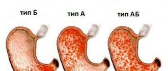
In order to save money, the bacterium Helicobacter pylori is not always detected during fibrogastroduodenoscopy. Often, clinicians do not attach any importance to the presence of gastritis, of which there are many varieties: some of them are corrected quite simply, others require long-term treatment and regular monitoring, as they can seriously increase the risk of stomach cancer.
Symptoms that make doctors wary of cancer: progressive loss of body weight, the presence of enlarged peripheral lymph nodes and blood in the feces indicate an advanced disease, and its treatment may be delayed.
COMPLAINTS for gastrointestinal diseases
We recommend that you consult a doctor if you have the following complaints, which can be divided into three main groups:
Main local complaints:
- dysphagia (impaired swallowing).
Dysphagia can be associated with certain neurological diseases or emotional issues with a completely healthy esophagus and stomach. As well as mechanical, chemical damage to the esophagus, the presence of a foreign body or tumor.
- gastric dyspepsia—by which we mean heaviness and pain in the epigastric region, heartburn, as well as belching, nausea, and vomiting.
- Heartburn is a burning sensation behind the sternum, in the epigastric region, resulting from the reflux of gastric contents into the esophagus and irritation of the esophageal mucosa with acidic gastric contents. The reason for this may be insufficiency of the cardiac sphincter, increased gastric motility, and increased acidity of gastric juice.
- vomiting is a complex reflex act in which the contents of the stomach are expelled.
The causes of vomiting can be, firstly, increased irritation (excitation) of various reflexogenic zones of internal organs, these can be pathological processes in the brain (for example, vomiting can occur during a concussion) and toxic effects on the vomiting center. Vomiting can be bloody, and then we are talking about bleeding from the gastrointestinal tract.
- intestinal dyspepsia - splashing, rumbling, abdominal pain, bloating, diarrhea or constipation.
- constipation is a long-term retention of feces in the intestines, associated with slowing of its peristalsis, mechanical obstacles in the intestines (tumor, diverticula) and nutritional factors. Constipation can be spastic, atonic and organic.
Spastic - associated with spasm, which can have a wide variety of causes.
Atonic - associated with intestinal atony. This is often observed in fairly elderly people.
Organic - associated with the presence of a mechanical obstacle (tumor).
- Diarrhea is loose, unformed stool that can be accompanied by increased bowel movements. Diarrhea is divided into enteral, colitic, gastric and pancreatic. There may be defecation disorders - false urges (tenesmus), pain and increased frequency of bowel movements.
- bloody stool.
Main common complaints:
- decreased appetite.
- weight loss (up to cachexia).
Additional general pathological complaints: increased fatigue, decreased performance, muscle weakness, neurotic disorders.
Comprehensive laboratory diagnostics to assess the functioning of the digestive system
Medical center » Diagnostics » Analyzes » Comprehensive laboratory diagnostics » Comprehensive laboratory diagnostics assessing the functioning of the digestive system
Prices Doctors Info
Sign up Comprehensive laboratory diagnostics involves conducting a number of tests for the purpose of preventing, diagnosing and monitoring the development of diseases of the gastrointestinal tract. This approach optimizes the provision of medical care and allows for objective monitoring of the dynamics of changes in the patient’s condition.
Diagnostic programs for diseases of the digestive system are aimed at identifying common pathologies. The complexes are based on general clinical and biochemical blood tests for substances that are markers of the development of diseases of the stomach, intestines, pancreas and liver.
When undergoing a comprehensive laboratory examination at SM-Clinic, the patient is guaranteed to receive discounts. Tests within the program are cheaper than individual tests. A person submits biomaterial only once, and at the same time receives the results of all studies provided for within the program.
Comprehensive laboratory diagnostics of the digestive tract at SM-Clinic allows you to quickly and comfortably undergo an examination at the best price, and obtain accurate results in the shortest possible time.
Advantages of comprehensive laboratory diagnostics:
- Availability of programs to assess the condition of almost all body systems
- Significant time savings, you complete the necessary research in 1 time
- Taking tests as a package is up to 15% cheaper
All prices are indicated without taking biomaterial into account.
“Intestinal dysbiosis” - promotional price 1,850 rubles.
Assessment of the composition of intestinal microflora, digestive processes, identification of possible causes of disruption of the large intestine.
Composition of the survey:
- Intestinal dysbiosis and sensitivity to an extended spectrum of antibiotics and bacteriophages
- Protein in stool
- General analysis of stool - coprogram
Completion time: 4-5 days
Laboratory screening of the digestive system - promotional price 6,200 rubles.
It is a laboratory examination that allows you to exclude or identify at an early stage the development of most diseases of the digestive system, timely prevent the development of pathology and, if necessary, treat.
Composition of the survey:
- Study of biochemical blood parameters (total bilirubin, direct bilirubin, Alanine aminotransferase - ALT, Aspartate aminotransferase - AST, Gamma-glutamyltransferase - GGT, Iron - tests allow you to get an idea of the condition of the liver, pancreas, gall bladder, changes in fat metabolism).
- Gastropanel (allows you to establish a diagnosis of atrophic gastritis, determine the risk of developing stomach cancer and peptic ulcers; localization of the pathological process and assess the nature of the changes, as well as the presence of Helicobacter pylori infection).
- Clinical blood test.
“Taking care of the liver. Standard" - promotional price RUB 1,258.
A set of laboratory diagnostics for identifying liver diseases at an early stage.
Recommended for suspected hepatitis, cholelithiasis, cirrhosis of the liver.
With decreased appetite, drowsiness, pain in the right side, yellowing of the eyes. Contents of the survey (service code A311406):
- Alanine aminotransferase - ALT
- Aspartate aminotransferase - AST
- Total bilirubin
- Direct bilirubin
- Bilirubin indirect
- Gamma glutamyl transferase - GGT
- Alkaline phosphatase
Completion time: 1-2 days
“Parasitic infections” - promotional price RUB 4,225.
A set of studies to detect infection of the body by parasites (helminths) and damage to the main functions of the liver and biliary tract.
Composition of the survey:
- IgG antibodies to helminths: (opisthorchis, trichinella, toxocara, echinococcus)
- Giardiasis, IgG antibodies to Giardia lamblia, blood, quality/quantity
- Alanine aminotransferase - ALT
- Total bilirubin
- Total protein
- Clinical blood test (CBC, LF, ESR) with smear microscopy in the presence of pathological changes, blood, quantity.
- Ascariasis, IgG antibodies to Ascaris llumbricoides, blood, quality/count.
- Helicobacter, IgG antibodies to Helicobacter pylori, blood, quality/count.
- Aspartate aminotransferase - AST
- Direct bilirubin
- Alkaline phosphatase
Completion time: 4-6 days
“Parasitic infestations” (screening of 11 indicators) - promotional price RUB 5,117.
This set of tests is one of the most effective methods for determining the presence of helminths at the early stage of parasitism.
Recommended for digestive problems, dermatological diseases, and weight loss. Pet owners need to be screened regularly. For ascariasis, opisthorchiasis, echinococcosis, toxocariasis, giardiasis, trichinosis and toxoplasmosis. Composition of the survey:
- Leukocyte formula with mandatory smear microscopy
- IgG antibodies to Helicobacter pylori
- IgG antibodies to Opisthorchis felineus
- IgG antibodies to Toxocara canis
- IgG antibodies to Trichinella spiralis
- Immunoglobulin E
- Complete blood count without ESR and leukocyte formula
- Total antibodies to Giardia lamblia
- IgG antibodies to Echinococcus granulosus
- IgG antibodies to Ascaris llumbricoides
- IgG antibodies to Toxoplasma gondii
Completion time: 6 days
Statistics
In Japan, among all operated patients with gastric cancer, patients with early cancer account for 50-55%. This is explained by the fact that categories of the population predisposed to stomach cancer undergo regular examination there. In Russia, from 60 to 90% of patients with newly diagnosed gastric cancer have stages III-IV, which determines the corresponding prognosis.
Our Center specializes in the diagnosis of early forms of malignant lesions of the gastrointestinal tract (esophagus, stomach, small intestine, colon) and their minimally invasive endoscopic treatment.
The special skills of our endoscopists allow us to identify tumors with a diameter of 1 mm and suspect their malignant nature, and timely surgical treatment using the endoscopic method allows us to cure the patient completely and in a timely manner.
During the endoscopic procedure, special techniques are used: chromoscopy, magnifying endoscopy, examination in a narrow spectrum of NBI light. This tactic undoubtedly improves the quality of histological verification, allows for targeted identification of foci of change and correct biopsy, which leads to an increase in the detection of various gastrointestinal pathologies, incl. oncological
Modern technologies in the diagnosis of gastroenterological diseases
Leites Yu.G., Marchenko E.V.
Research in recent years has revealed a clear trend towards an increase in the incidence of digestive organs. In this regard, there is an urgent need to constantly improve existing diagnostic methods, as well as to create and develop new, previously unused methods that allow identifying diseases in the early stages, assessing the degree of organ damage, and monitoring the results of therapy.
Methods for diagnosing gastroenterological diseases.
All diagnostic methods can be divided into instrumental and non-instrumental.
Non-instrumental methods.
The clinician can obtain a lot of useful and valuable information during a conversation with the patient, as well as during his examination. A careful study of complaints, life history and illness, external examination, palpation, percussion, and auscultation sometimes provide more information than instrumental methods and allow you to choose the right direction of the diagnostic search. Thus, the diagnosis of all diseases, including the digestive organs, should always begin with non-instrumental methods and end with instrumental ones, with the help of which the diagnosis is confirmed by evidence.
Symptoms of the disease (complaints).
The patient may present a large number of complaints, but each of them will have its own characteristics for various pathologies. When studying a pain syndrome, it is necessary to clarify the nature of the pain, its localization, irradiation, conditions of occurrence, and the means taken by the patient to reduce it.
Appetite - its loss or decrease (and sometimes an aversion to a certain type of food, for example, meat) can be observed with the development of a malignant tumor.
Patients with diseases of the liver and biliary tract complain of bitterness in the mouth. A sour taste in the mouth and heartburn often occur in patients with diseases of the stomach and esophagus.
Belching air more often occurs in patients suffering from neuroses and neuropathies. Rotten belching on an empty stomach occurs with pyloric stenosis and gastric atony. Belching with a fecal odor occurs in patients with intestinal obstruction.
Nausea and vomiting are quite common symptoms of diseases of the digestive system that occur with lesions of the stomach (gastritis, peptic ulcer, tumors), chronic cholecystitis and pancreatitis, and intestinal obstruction.
Dysphagia is a symptom characteristic of impaired passage of food masses through the esophagus of a functional or organic nature.
Bowel movement disorders can manifest as constipation, diarrhea, or an alternation of both. Both of them can be symptoms of acute or chronic, functional or organic damage to the digestive organs.
Bloating, rumbling, and transfusion in the abdomen indicate a violation of intestinal tone, secretion and absorption processes, and the state of the intestinal microflora.
History of the patient's life.
The conditions in which the patient lives, dietary habits (regularity, consumption of excessively hot and spicy foods), alcohol abuse and smoking actively affect the functioning and condition of the digestive organs. The emergence of functional disorders and exacerbation of chronic diseases is often facilitated by irregular working hours in combination with mental stress, conflict situations at work and at home, and insufficient rest. Occupational hazards also play a role in the occurrence of pathology of the digestive system. During the collection of a life history, the clinician identifies the prerequisites for the development of the disease, the reasons why painful changes occurred in the patient’s body. A correctly collected life history makes a great contribution to the formulation of the diagnosis, and most importantly, it allows us to identify the causative factors in the development of the disease.
History of the disease.
When collecting a patient’s medical history, the doctor tries to find out in detail how the disease developed from the first manifestations to a clear clinical picture:
- what, in the patient’s opinion, could provoke his painful condition (errors in diet, physical activity, alcohol abuse, use of poor-quality food products, taking medications, etc.);
- what complaints arose and what the patient did to reduce them;
- how events further developed (addition of other complaints, changes or intensification of existing ones) until the moment of seeking medical help.
If the patient has been suffering from this disease for a long time and has already received treatment, it is necessary to find out what therapy was prescribed previously (names of drugs, their doses, effectiveness of use), whether surgical interventions were performed (name of operations, course of the postoperative period). Having all the data, it is much easier for the doctor to make an assumption about the cause of the patient’s poor health and decide on tactics for further diagnostic search.
Objective clinical examination of the patient.
Inspection, percussion, palpation, auscultation and digital examination per rectum are the main diagnostic methods in gastroenterology after studying complaints and anamnesis.
During examination, pay attention to the skin, an increase in the volume of the abdomen, bulging, tension; during percussion - for pain, the nature of the sound and the presence of resistance from the abdominal wall. The mucous membranes are examined: first of all, the eyes, mouth, attention is paid to the color of the tongue, its moisture, the presence/absence of plaque, etc.
Auscultation is a valuable research and diagnostic method. During auscultation of the abdomen in various pathologies, one can hear noises of a different nature or a characteristic absence of noises throughout the abdomen.
Palpation is one of the main methods of physical examination of the abdominal organs. Palpation can be superficial (approximate) and deep. Palpation allows you to determine: the tone of the abdominal wall, the position of the organs, their size, shape, degree of density, the nature of the surface, accessibility of palpation, mobility, sensitivity, soreness of the organs, the presence of a tumor, accumulation of fluid and gases.
2. Laboratory diagnostics.
Laboratory methods allow us to assess changes in the body's homeostasis. When studying the hemogram of patients with gastroenterological pathology, the following can often be detected: leukocytosis with a shift in the leukocyte formula to the left, eosinophilia, an increase in ESR, which indicates an inflammatory process. In severe forms of malabsorption or bleeding, anemia may develop. The biochemical blood test also contains groups of indicators characteristic of certain pathologies. When diagnosing diseases of the liver and biliary tract, attention is paid to: indicators of blood bilirubin (direct and indirect fractions), hepatic aminotransferases in the blood serum, alkaline phosphatase, blood protein levels, iron metabolism indicators, etc. Of particular interest is the long-known method of diagnosing pancreatitis using establishing alpha-amylase activity in the blood and urine. Determination of the activity of alpha-amylase isoenzymes - isoamylase, pancreatic proteases (trypsin, chymotrypsin, elastase, carboxypeptidase) is also of diagnostic importance.
To detect H. Pylori (HP) in diseases of the stomach and duodenum, the following are used: serological method (since colonization of HP causes a systemic immune response). IgG antibodies directed against various bacterial antigens appear in the serum of infected people.
A breath test is used (its principle is based on the fact that after oral administration of a solution of urea labeled with radioactive carbon, urease HP metabolizes the labeled urea and releases labeled carbon dioxide, which is determined in the exhaled air within 10-30 minutes).
Tests that detect markers of autoimmune damage can detect the presence of antibodies to cellular components of the host body that are associated with certain diseases.
In recent years, the method of determining the types of microorganisms using polymerase chain reaction (PCR diagnostics) has become widespread. The method is based on the complementary completion of a section of genomic DNA or RNA of the pathogen, carried out in vitro using the enzyme thermostable DNA polymerase. Using PCR diagnostics, some representatives of microflora with intracellular or membrane localization are determined. This method is used mainly for verification of infectious pathology.
Biochemical analysis of feces reflects the content of metabolites of the intestinal microflora, which allows a more accurate assessment of disorders, diagnosis and differentiation of gastrointestinal pathology. Nowadays, the most commonly used method is the determination of short-chain fatty acids in the intestinal contents, which are metabolites of sugar and proteolytic intestinal microflora and are used as an integral indicator of its condition.
The method for determining short-chain fatty acids in various media is successfully used to assess the state of microflora as a diagnostic test for various gastrointestinal pathologies. These include: a clarifying test for the presence of pathology, a test for monitoring the prevalence and activity of the process in nonspecific ulcerative colitis, assessment of the detoxification function of the liver in chronic hepatitis and cirrhosis, determination of markers of the intensity of portosystemic shunting and portal hypertension, a diagnostic test for the presence and stage of hepatic encephalopathy. This allows you to evaluate the effectiveness of the therapy and individual choice of drugs.
As a result, it should be noted that laboratory diagnostics is the most dynamically developing branch of medicine, which has enormous diagnostic significance. Most known diseases of infectious and non-infectious nature have special markers that can be identified during laboratory tests, which makes an invaluable contribution to diagnosis.
Instrumental methods
Ultrasound diagnostics.
The method of ultrasound examination (ultrasound) of the abdominal organs has become widespread. This is a non-invasive, highly sensitive diagnostic method that is safe for the patient.
The operation of ultrasonic scanning devices is based on the effect of converting electrical energy into acoustic energy. As a result of recording reflected signals, it is possible to determine the location, size of the organs being studied, the nature and extent of the pathological process. Currently, equipment is used that operates in one-dimensional and two-dimensional scanning modes (depth scanning). During one-dimensional scanning, partial reflection of sound signals occurs at the density interface, which are recorded on the screen and can be recorded on photographic film. Much more information can be obtained using two-dimensional ultrasound (B-scan), when information is presented in depth and width. This produces an image in the form of a slice of the examined area. Real-time gray scale ultrasound is also used, in which the image is immediately visible and moving objects can be observed.
Dopplerography is an ultrasound method that uses a change in the length of the ultrasonic wave when reflected from moving blood (the Doppler effect) to determine the direction and speed of blood flow. Using color Doppler ultrasound, you can determine the patency and direction of blood flow in both arteries and veins. This test is used to diagnose thrombosis, portal hypertension and other blood flow disorders.
X-ray diagnostics.
At the end of the twentieth century, after the advent of ultrasound diagnostics and endoscopy, X-ray diagnostics in gastroenterology faded into the background. Until about 30 years ago, radiology was the only and main imaging modality. But even today, X-ray diagnostics remains the main method in diagnosing certain diseases of the gastrointestinal tract (primarily diseases of the small, large intestine and esophagus). With the advent of the possibility of computer processing of X-ray data (including computed tomography and multislice), the diagnostic value of the method has increased markedly, and X-ray diagnostics is again regaining its leading position.
The following diagnostic methods are used in the diagnosis of diseases of the digestive system:
1. Plain radiography of the abdominal cavity without contrast - used to diagnose intestinal obstruction. Signs of an intestinal obstruction include levels of fluid in the intestine and "arches" - loops of intestine filled with gas.
2. Contrast studies. These studies use radiopaque contrast agents containing a suspension of barium sulfate particles. When taking a barium suspension orally, the pharyngeal and esophageal phases of the swallowing process, contrasting the esophagus and stomach are assessed, and stenosis of the gastric outlet is diagnosed. If mechanical intestinal obstruction is suspected, oral barium intake is used with dynamic monitoring. After 6 and 8 hours from the moment of taking the barium drug, a series of images are taken.
Iodine-containing (water-soluble) radiocontrast preparations are usually used to diagnose esophageal perforations. When entering the mediastinum, iodine preparations are much safer than barium preparations. The contrast of iodine preparations is lower, so they are less able to detect small and isolated perforations. If no contrast agent is detected outside the esophagus, a repeat barium study is indicated.
The X-ray method has particular diagnostic value in cases of suspected perforation of the walls of hollow organs and, first of all, the duodenum and stomach. In this case, the role of a contrast agent is played by a gas that enters the abdominal cavity from a hollow organ and accumulates in the suprahepatic space in the form of a “sickle”, which is clearly visualized by radiography.
3. X-ray of the upper gastrointestinal tract.
The sensitivity of radiography of the upper gastrointestinal tract is much lower than that of the endoscopic method, so it is used only when endoscopy is not possible. Using the X-ray method, it is possible to verify ulcerative lesions of the stomach and duodenum. In this case, direct (morphological) and indirect (functional) signs are identified.
4. Examination of the small intestine.
Examination of the small intestine using an x-ray method is only possible with oral administration of a barium suspension. In this case, a series of photographs are taken (no earlier than after 4-6 hours). During the study, a mechanical obstacle to the movement of barium suspension can be detected.
5. Examination of the large intestine.
5.1. Irrigoscopy with double contrast - is carried out to assess the anatomical structure of the colon (the presence of additional loops and bends), identify intraluminal formations (tumors and polyps), inflammatory changes (narrowing of the lumen, disappearance of folds), diverticula, foreign bodies, etc. In the first half of the study the colon is filled with a barium suspension, and its anatomy is studied. Then, after the intestine has emptied the barium suspension, the organ is filled with air. In this case, a double contrast of the organ under study occurs with barium residues on the mucosa and with air. With double contrast, it is possible to study the relief of the mucous membrane.
5.2. Multislice computed tomography of the intestine with the construction of a virtual image of the intestine.
This test is performed after the colon has been filled with air. A multislice CT scan is performed, and on the computer screen the researcher receives a three-dimensional image of the intestine with the ability to create a virtual intraluminal picture. This method began to be used in our country quite recently, and is still in the development stage.
Computer diagnostics.
Computed tomography allows you to evaluate in detail the structure of the organs being examined and to safely insert a needle into hard-to-reach areas of the abdominal cavity. CT scanning makes it possible to more accurately assess the structure of the organ under study, identify and diagnose characteristic signs of damage. Ingestion of a contrast agent allows additional assessment of the condition of hollow organs, and its intravenous administration allows visualization of the vascular network, which increases the reliability of assessing the condition of various organs (for example, identifying an area of necrosis in the pancreas). CT with intravenous contrast is also a reliable method for identifying and evaluating tumors and tumor-like formations. In many clinics, computed arterial portography (CAPG) is used to enhance liver contrast - computed tomography with the introduction of a radiopaque agent through a catheter into the superior mesenteric artery. Clearer contrasting of the liver vascular system provides more accurate localization of metastasis within the segment.
Magnetic resonance imaging (MRI).
MRI uses the magnetic properties of protons distributed throughout the body. The T1 image measures the rate at which protons collide after a radio wave pulse, while the T2 image measures the rate at which the energy of the radio wave decreases. MRI allows precise differentiation of tissues containing different amounts of fat and water. This method allows a very accurate assessment of blood flow, so MRI is the diagnostic method of choice for confirming vascular changes, in particular hemangiomas.
Using moving detectors and an X-ray tube, a picture of cross-sectional targeted thin tomographic sections of the entire body at selected levels is obtained.
Radioisotope scanning.
The method is based on the ability of various cells (hepatocytes, malignant tumor cells, areas of inflammation) to capture specific radiopharmaceuticals (RP) labeled with radionuclides. Isotope scintigraphy is used in the diagnosis of almost all gastrointestinal diseases. With its help, you can assess the patency of the afferent loop of the gastroenteroanastomosis, anastomoses of the biliary tract with the lumen of the gastrointestinal tract, determine the time of passage of food through the esophagus, identify and evaluate gastroesophageal reflux, aspiration, space-occupying formations of the abdominal cavity, determine the stage or identify relapse of the tumor, detect the source of bleeding, localization of the inflammatory process.
New diagnostic technologies include single-photon emission computed tomography, which allows visualization of tissue sections by the distribution of radioisotopes, as well as positron emission tomography, which provides information about blood flow and tissue metabolism.
Functional diagnostic methods.
When examining a patient with diseases of the digestive organs, in addition to collecting data on anatomical (organic) pathological changes, it is very important to obtain information about the functional state of the organ. Using modern diagnostic methods (ultrasound, endoscopy, X-ray diagnostics, CT and MRI), it is possible to obtain a visual image of any organ, but none of these methods reflects their function.
The main functions of the digestive organs are the movement of food masses through the intestinal tube, their digestion under the influence of digestive juices containing enzymes, and the absorption of digested nutrients. Assessing the motor function of the digestive organs, the degree of digestion and absorption of food is a difficult task. Researchers have only recently begun to tackle such problems.
Electrogastroenterography is carried out using the Gastroscan-GEM and Gastroscan-M5 devices. This study makes it possible to study the motor-evacuation function of the gastrointestinal tract by recording electrical activity with measuring electrodes attached to the patient's skin. Peripheral computerized EGEG pursues certain goals - determining the type of disorder (functional or mechanical), identifying the location of the lesion (gastrointestinal tract), choosing a treatment method and selecting corrective therapy. Indications for research by this method are the presence in patients of various signs of impaired motor activity of the digestive tract.
EGDS.
The introduction of fiber endoscopes into clinical practice has opened up great opportunities for studying the pathology of the upper digestive tract. When carrying out this examination method, it becomes possible to examine in detail the surface of the mucous membrane of the esophagus, stomach and duodenal bulb. Endoscopic examination is the most reliable and reliable method for confirming or rejecting the diagnosis of peptic ulcer, establishing the location of the ulcer, its shape, size and monitoring the healing or scarring of the ulcer, and assessing the effect of treatment. The method for identifying the phenomena of intestinal metaplasia has become widespread. In this case, methylene is used, as a result of which the foci of intestinal metaplasia are painted an intense blue color. When performing an endoscopy, biopsy material is also taken.
Colonoscopy.
During this study, the large intestine is examined and examined along its entire length, to the level of its transition to the small intestine. It allows you to assess the condition of its mucous membrane (color, the nature of the vascular pattern, features of folding), identify the presence of cellular metaplasia and tumor formations, and determine the level and degree of obstruction. Fistula tracts and diverticula are also detected. The colonoscopy method is indispensable when it is necessary to visually examine the intestinal lumen and obtain biopsy material from pathologically altered areas of the mucous membrane.
Sigmoidoscopy.
Rigid sigmoidoscopy is a common and publicly available method of endoscopic examination, which allows visual assessment of the inner surface of the rectum and distal third of the sigmoid colon to a level of 20-25 cm from the anus. There are practically no contraindications to examining the intestine through a sigmoidoscope. When performing this study, attention is paid to the color, shine, moisture, elasticity and relief of the mucous membrane, the nature of its folding, features of the vascular pattern, the presence of pathological changes, and the tone and motor function of the examined sections are assessed.
Assessment of the acid-producing function of the stomach.
Each of the available methods has its own characteristics that can be used in a certain situation and provide additional volume of necessary information.
1. Aspiration-titration method.
The aspiration-titration method is based on the extraction of gastric contents using a gastric tube and its subsequent titration in vitro. This makes it possible to evaluate not only the acid production of the stomach, but also, if necessary, to carry out a detailed analysis of the chemical composition of the secretion and determine the activity of enzymes.
2. Intracavitary pH-metry.
Determination of intragastric pH is a fundamentally new research method, aimed not at analyzing the aspirated gastric juice, but at determining the pH of the intragastric environment by contacting the measuring electrode of a pH-metric probe with the mucous membrane. To carry out pH measurements, acidogastrometers (AGM-03, Gastroscan-5M, Gastroscan-24) are mainly used. The main types of intragastric pH-metry are:
2.1. Express pH-metry.
Express pH-metry is performed to study the acid production of the stomach over a short period of time. To conduct the study, oral pH probes with a cutaneous silver chloride reference electrode are used.
2.2. Monitoring acid formation.
Daily pH monitoring makes it possible to study the acid-producing function of the stomach under conditions as close as possible to physiological ones, to study the influence of various endogenous and exogenous factors and, in particular, medications on acid production, as well as to accurately record duodenogastric and gastroesophageal refluxes.
2.3. Endoscopic pH-metry.
This method allows you to determine the acidity on the surface of the mucous membrane of various parts of the gastrointestinal tract under visual control. Parietal pH-metry significantly increases the information content of endoscopic examination and allows you to fully characterize not only visual changes in the mucous membrane of the upper digestive tract, but also fasting acidity and alkalizing function of the antrum of the stomach.
Biopsy.
Intravital pathomorphological study of altered tissues makes it possible to solve many interrelated clinical issues. First of all, this method is important for recognizing the nature of neoplasms. Microscopic confirmation of the diagnosis of cancer is necessary to avoid unnecessary operations for inflammatory diseases and benign tumors. Histological examination of tumor tissue determines its structure and degree of differentiation of cellular elements, which allows you to correctly select the scope of surgical intervention. This method can help the doctor in recognizing inflammatory and functional bowel diseases. In inflammatory diseases, a biopsy makes it possible to objectively assess the severity of the pathological process and monitor the effectiveness of the therapy.
Biopsy of space-occupying formations is carried out using the method of fine-needle aspiration, which allows one to obtain groups of cells (and sometimes small fragments of tissue) for cytomorphological examination, or the method of puncture biopsy, which allows one to obtain cylindrical fragments of tissue with preserved architecture 1-2 cm long for histomorphological examination.
Direct histological visualization of H. pylori (HP) after serial biopsy sections is the gold standard for diagnosis. A rapid urease test (CLO test) is also used, in which the biopsy is placed in agar containing urea and a pH indicator. Hydrolysis of urea under the influence of Helicobacter urease leads to an increase in the pH of the agar and causes a color change.
Bacteriological method - by growing HP on selective media. This method is necessary when determining the sensitivity of HP to antibiotics and other antibacterial drugs, especially in cases of resistance to therapy.
Endoscopic ultrasound.
Endoscopic ultrasound allows you to clearly visualize all layers of the wall of the organ under study and adjacent structures when there is insufficient data obtained by endoscopy. Basically, EUS is used to identify and determine the degree of invasion of tumor lesions of various organs of the gastrointestinal tract. Direct contact of the ultrasound probe with the intestinal wall and the use of high-frequency ultrasound provide detailed visualization of the intestinal wall and adjacent structures. This allows for a differential diagnosis of submucosal tumors and compression of the intestine by adjacent structures or pathological formations with an accuracy of up to 95%.
Endocapsule.
The capsule is a small device (size: 11 x 30 mm), which contains a camera, a light source and an image transmitter. To begin the study, you must swallow the capsule. The image from a weak transmitter of a capsule moving through the digestive tract is transmitted to a receiver attached to the patient’s belt. After a few hours, the data is collected and the researcher can begin to analyze it.
The article was published on the website https://www.gastroscan.ru
