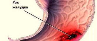
Esophageal carcinoma
– a malignant tumor that forms from the overgrown and degenerated epithelium of the esophageal wall. Clinically, esophageal cancer is manifested by progressive swallowing disorders and, as a consequence, loss of body weight as a result of malnutrition. Initially, as a rule, a tumor formation is detected by radiography, endoscopic examination, CT or ultrasound. The diagnosis of esophageal cancer is established after a histological examination of a biopsy of the neoplasm to detect malignant cells.
Like any malignant neoplasm, esophageal cancer has an even more unfavorable prognosis the later the disease is detected. Early detection of cancer contributes to a more pronounced effect; tumors at stages 3-4 usually cannot be completely cured.
When a cancerous tumor grows in the walls of the esophagus, the surrounding tissues of the mediastinum, trachea, bronchi, large vessels, and lymph nodes are affected. The tumor is prone to metastasis to the lungs, liver, and can spread through the digestive tract to the stomach and intestines.
Risk factors for developing esophageal cancer
Currently, the mechanisms of development of esophageal cancer are not fully understood. Factors contributing to the occurrence of a malignant tumor are:
- smoking;
- alcohol abuse;
- eating too hot and too cold food;
- occupational hazards (inhalation of toxic gases);
- content of heavy metals in drinking water;
- chemical burns of the esophagus when swallowing caustic substances.
Regular inhalation of air containing a dust suspension of harmful substances (when living in a smoky area, working in unventilated areas with a high concentration of industrial dust) can also contribute to the development of cancer.
Diseases that contribute to the development of esophageal cancer are gastroesophageal disease, obesity, and keratoderma. Esophageal hernia, achlosia (relaxation of the lower esophageal sphincter) contribute to regular reflux - the reflux of stomach contents into the esophagus, which in turn leads to the development of a specific condition: Barrett's disease .
Barrett's disease (Barrett's esophagus) is characterized by degeneration of the epithelial lining of the esophagus similar to the gastric epithelium. This condition is considered precancerous, like most epithelial dysplasias (disorders of tissue development).
It has been noted that esophageal cancer occurs more often in people over 45 years of age; men suffer from it three times more often than women.
The development of cancer is promoted by a diet containing insufficient amounts of vegetables, herbs, protein, minerals and vitamins. Irregular eating and a tendency to overeat also have a negative effect on the walls of the esophagus, which can contribute to a decrease in protective properties.
One of the factors in the malignancy of precancerous formations is decreased immunity.
Sources
- Zhao H., Misariu AM., Ramirez-GarciaLuna JL., Nobel T., Mueller C., Cools-Lartigue J., Spicer J., Molena D., Bains M., Swisher S., Hofstetter W., Ferri L. Synchronous Esophageal and Lung Cancers – Is Combined Anatomic Resection Appropriate? // Ann Thorac Surg - 2021 - Vol - NNULL - p.; PMID:33905733
- Liao X., Gao Y., Liu J., Tao L., Wang D., Xie D., Mo S. Corrigendum: Combination of Tanshinone IIA and Cisplatin Inhibits Esophageal Cancer by Downregulating NF-κB/COX-2/VEGF Pathway . // Front Oncol - 2021 - Vol11 - NNULL - p.670798; PMID:33905466
- Ali O., Challa SR., Siddiqui OM., Ali S., Kim RE. A rare cause of esophagopleural fistula due to intensity-modulated proton therapy: a case report and review of literature. // Clin J Gastroenterol - 2021 - Vol - NNULL - p.; PMID:33905092
- Xu ZJ., Lin YD. Reply to Zhang et al. // Eur J Cardiothorac Surg - 2021 - Vol - NNULL - p.; PMID:33904932
- Jessen NH., Jensen H., Helsper CW., Falborg AZ., Glerup H., Gronbaek H., Vedsted P. Cancer suspicion, referral to cancer patient pathway and primary care interval: a survey and register study exploring 10 different types of abdominal cancer. // Fam Pract - 2021 - Vol - NNULL - p.; PMID:33904928
- Zhang X., Jiang Y., Zhang L. Nodal skip metastasis in oesophageal cancer: different definition and different prognostic role. // Eur J Cardiothorac Surg - 2021 - Vol - NNULL - p.; PMID:33904927
- Odze R., Spechler SJ., Podgaetz E., Nguyen A., Konda V., Souza RF. Histologic Study of the Esophagogastric Junction of Organ Donors Reveals Novel Glandular Structures in Normal Esophageal and Gastric Mucosae. // Clin Transl Gastroenterol - 2021 - Vol12 - N5 - p.e00346; PMID:33904522
- Conio M., Filiberti RA., Siersema PD., Manta R., Blanchi S., De Ceglie A. A new designed self-expandable metal stent for the management of benign radiotherapy-induced hypopharyngeal or cervical esophageal strictures. // Surg Endosc - 2021 - Vol - NNULL - p.; PMID:33903933
- Davern M., Donlon NE., Power R., Hayes C., King R., Dunne MR., Reynolds JV. The tumor immune microenvironment in oesophageal cancer. // Br J Cancer - 2021 - Vol - NNULL - p.; PMID:33903730
- Lan T., Xue X., Dunmall LC., Miao J., Wang Y. Patient-derived xenograft: a developing tool for screening biomarkers and potential therapeutic targets for human esophageal cancers. // Aging (Albany NY) - 2021 - Vol13 - NNULL - p.; PMID:33903283
Classification of esophageal cancer
Esophageal cancer is classified according to the international TNM nomenclature for malignant neoplasms:
- by stage ( T0 – precancer, carcinoma, non-invasive epithelial tumor, T1 – cancer affects the mucosa, T2 – tumor grows into the submucosal layer, T3 – layers up to the muscular layer are affected, T4 – tumor penetration through all layers of the esophageal wall into the surrounding tissues);
- by the distribution of metastases in regional lymph nodes ( N0 – no metastases, N1 – there are metastases);
- by the spread of metastases in distant organs ( M1 – yes, M0 – no metastases).
Cancer can also be classified into stages from first to fourth, depending on the extent of the tumor in the wall and its metastasis.
Currently, esophageal cancer, despite relatively low incidence rates (2.4% in men and 0.6% in women), ranks eighth in the structure of mortality from cancer in the Russian Federation [1]. Squamous cell carcinoma of the esophagus is the most common histological type with an extremely malignant course and early metastasis.
Unfortunately, more than 75% of cases of esophageal cancer are diagnosed only at stages II-III of the disease, and the five-year survival rate of patients is extremely low and varies from 4 to 25% [2]. The aggressiveness index, calculated as the ratio of deaths to new cases, in esophageal cancer is extremely high and amounts to about 95% [3]. Therefore, diagnosing esophageal cancer in the early stages and adequate treatment can lead to increased survival and decreased mortality.
Endoscopic diagnosis of early esophageal cancer and high-grade dysplasia is a challenging task in modern endoscopy. Routine white light endoscopic examination has low diagnostic yield due to the difficulty of detecting minimal signs of early neoplastic changes. Chromoendoscopy with Lugol's solution is the “gold standard” for diagnosing early esophageal cancer. For this purpose, 20-50 ml of 2-3% Lugol's solution is used, which is an absorbent dye containing potassium iodine and pure iodine, which reacts with non-keratinized epithelial cells containing glycogen [4]. When applying Lugol's solution, the normal mucous membrane of the esophagus takes on a dark brown or greenish-brown color, and areas that do not contain glycogen (neoplastic and dysplastic areas, columnar epithelium, in some cases - inflammatory changes) are not stained. In contrast to inflammatory changes, in the case of esophageal cancer, the color of the area not stained during chromoscopy may change: immediately after staining it is yellow, and after 2-3 minutes it becomes pink. The pink color sign phenomenon occurs due to the complete absence of glycogen in squamous cell carcinoma cells, which demonstrates high sensitivity and specificity (88 and 95%, respectively) in the diagnosis of esophageal cancer. However, the use of Lugol's solution is associated with some side effects such as chest pain, cough and allergic reactions.
The use of optical technologies, such as narrow-spectrum endoscopy, has made it possible to simplify the task of searching for areas suspicious for dysplasia and cancer, and to make the study safer and faster. The finding of “brownish areas” in the esophageal mucosa is specific for early esophageal cancer and is due to the presence of dilated irregular vessels on a brown background. This allows not only to detect the early form of cancer, but also to accurately determine its boundaries. Moreover, sometimes the combined use of chromoscopy with Lugol's solution and narrow-spectrum endoscopy reveals the phenomenon of metallic silver sign in areas of neoplasia not stained by chromoscopy.
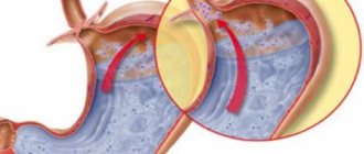
However, one of the most effective diagnostic techniques is magnifying endoscopy, which allows a detailed assessment of the morphology of intrapapillary capillary loops (ICPLs). According to the most common classification by Inoue et al. [5], according to the characteristics of capillary loops, it is possible not only to predict the histological structure of the area and distinguish between benign and malignant areas, but also to diagnose the level of neoplasia invasion and, as a result, choose treatment tactics (see figure). In accordance with this classification, there are five main types of intrapapillary capillary loops, which make it possible to characterize in detail changes in the squamous epithelium of the esophagus from normal (type I) mucosa to squamous cell carcinoma (type V). Neoplastic changes are characterized by the presence of four main changes in capillary loops: dilation, tortuosity, changes in caliber and differences in shape. The presence of one or two signs (mainly consisting of lengthening of the loops) in the area stained during chromoscopy corresponds to type II and occurs with inflammatory changes, in particular with gastroesophageal reflux disease. The absence of changes in capillary loops in the unstained area corresponds to type III and defines a borderline condition that can include both inflammation and low-grade dysplasia. Type IV has three of the four characteristics of neoplasia and is a marker of high-grade dysplasia. Intrapapillary capillary loops of type V can also differ in the severity of irregularity and destruction, which correlates with the degree of invasion (V1 - m1 invasion, V2 - m2 invasion, V3 - m3 and sm1 invasion, Vn - sm2 invasion and deeper). M. Arima et al. [6] proposed an additional characteristic of esophageal cancer - the presence of a so-called avascular zone, the extent of which strictly correlates with the depth of invasion. However, in addition to changes in capillary loops, another important sign is a change in the background color of the mucosa (the area between the microvessels). In the zone of neoplasia, the background color of the mucous membrane is different (becomes darker) compared to the surrounding unchanged tissue. This is due to the presence of extravascular hemoglobin directly in the cytosol of tumor cells [7], as well as the thinning of the keratin layer of the mucous membrane [8].
Types of patterns of intrapapillary capillary loops according to H. Inoue (adapted from [5]). a — type I, normal mucous membrane; b — type II, inflammatory changes; c — type III, borderline condition, observation required; d — type IV, high-grade dysplasia or carcinoma in situ, endoscopic resection is required; e — type V1 (expansion, tortuosity, changes in caliber and difference in shape), squamous cell carcinoma, degree of invasion m1, endoscopic resection/dissection is absolutely indicated; e — type V2 (elongation of loops of type V1), squamous cell carcinoma, degree of invasion m2, endoscopic resection/dissection is absolutely indicated; g — type V3 (significant destruction of the capillary pattern), squamous cell carcinoma, degree of invasion m3 or sm1, endoscopic resection/dissection is relatively indicated; h — type Vn (formation of new tumor vessels), squamous cell carcinoma, degree of invasion sm2 and deeper, surgical treatment is indicated.
Detection of early esophageal cancer or high-grade dysplasia allows for minimally invasive endoscopic treatment, which shows high effectiveness with long-term follow-up. According to the Japanese Society of Esophageal Diseases, the five-year survival rate of patients with early esophageal cancer with m1 and m2 invasion without metastases to regional lymph nodes after endoscopic resection reaches 95% [9]. Similar figures are demonstrated by a study conducted in Germany, according to which the seven-year survival rate of patients with high-grade dysplasia and early squamous cell carcinoma after endoscopic resection was 77% [10]. Currently, the technique of choice for early neoplastic processes is dissection in the submucosal layer, since only with this technique is it possible to remove the tumor en bloc [11]. The indication for endoscopic dissection in the submucosal layer is early squamous cell carcinoma that grows to the level of the muscular plate of the mucous membrane (mm) and even invades the superficial layers of the submucosal layer (less than 200 μm - sm1) [12]. However, dissection in the submucosal layer in early esophageal cancer has a number of difficulties and features. Firstly, the technique of dissection in the submucosal layer in the esophagus is labor-intensive and requires the endoscopist to have extensive experience in performing endoscopic resections. The complexity of the operations is due to the relatively narrow lumen of the esophagus, as well as transmission movements from nearby organs (respiratory movements and heart pulsation), which limits the possibilities of manipulation. Secondly, complications such as perforation are slightly more common than, for example, with dissection in the stomach and can cause pneumomediatinum and subcutaneous emphysema. This is due to the fact that the wall of the esophagus is much thinner than the wall of the stomach. In addition, quite often, in more than half of the cases, pneumomediastinum is diagnosed when the muscular layer of the esophagus is damaged, even in the absence of perforation, which, however, does not lead to life-threatening complications. Thirdly, when a pathological area that extends to more than 2/3 of the circumference of the esophagus is removed, strictures are often formed in the surgical area, for the prevention of which in these cases the use of per os
in the postoperative period [13].
On the other hand, there are several features of the technique of endoscopic dissection in the esophagus, associated with the characteristics of the submucosal layer, which simplifies the operation at some stages. The submucosal layer of the esophagus practically does not contain large vessels, which significantly facilitates hemostasis at the dissection stage, and the injection of saline solution is carried out without significant effort and complications. Moreover, the pathological area is localized, as a rule, in a tangential direction, so the submucosal layer of the esophagus is easy to recognize and subsequently separate it from the muscular layer.
Thus, the use of modern endoscopic technologies allows not only to diagnose the earliest forms of esophageal cancer, but also to effectively treat identified formations, and the widespread use of these techniques in clinical practice can lead to increased survival and reduced mortality from esophageal cancer.
Symptoms of esophageal cancer
Early-stage esophageal cancer most often does not manifest itself clinically; symptoms begin to appear when there is already a fairly large tumor that interferes with the passage of food.
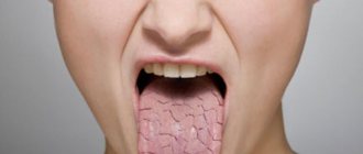
The most common symptom of esophageal cancer is a swallowing disorder - dysphagia . Patients tend to take liquid food; harder food gets stuck in the esophagus, creating a feeling of a “lump” behind the sternum.
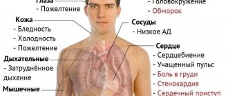
As the tumor progresses, pain may occur behind the sternum and in the pharynx. The pain may radiate to the upper back.
Reduced esophageal patency contributes to vomiting.
As a rule, prolonged lack of nutrition (associated with difficulty eating) leads to general dystrophy: loss of body weight, dysfunction of organs and systems.
Often, esophageal cancer is accompanied by a constant dry cough (occurs reflexively as a result of irritation of the trachea), hoarseness (chronic laryngitis). In the terminal stages of tumor development, blood can be detected in vomiting and coughing.
All clinical manifestations of esophageal cancer are nonspecific, but require immediate medical attention.
Patients suffering from Barrett's disease require regular follow-up with a gastroenterologist, as they are at high risk of developing esophageal cancer.
Symptoms
The initial stages of esophageal cancer occur without symptoms. The disease manifests itself only when there is a disruption in the process of swallowing and moving food through the esophagus. This disorder occurs when the lumen of the esophagus is partially blocked by a tumor growing inside. The presence of a small tumor can cause spasm of the wall and, as a result, choking on food. With further growth of the tumor and blocking most of the lumen of the esophagus, the patient may lose the ability to eat normally, which without special measures leads to exhaustion of the body. Retention of food above the site of narrowing leads to esophageal vomiting, regurgitation of saliva and mucus.
Low-intensity pain behind the sternum with irradiation into the interscapular area when food passes and/or drooling are late symptoms and are often associated with concomitant esophagitis or tumor invasion into neighboring organs. When cancer is localized in the area of the cardia (the transition of the esophagus to the stomach), the first sign may not be a violation of swallowing and moving food, but constant regurgitation of air.
When a tumor grows beyond the esophagus, it can compress the airways and impair breathing. It can also compress or grow into the nerve trunks located next to the wall of the esophagus, leading to hoarseness of voice, cough, development of Horner's syndrome (drooping of the upper eyelid, narrowing of the pupil, weakening of the pupil's reaction to light, dilatation of the vessels of the conjunctiva of the eye, retraction of the eyeball, impaired sweating on the face and redness of the facial skin).
Diseases with similar symptoms
When swallowing (dysphagia), but these disorders tend to progress, albeit very slowly. This condition is also accompanied by a night cough, regurgitation (movement in the opposite direction) or aspiration (inhalation) of food masses, and sometimes increased mucus secretion during regurgitation, which is associated with stagnation in the esophagus. The periodic occurrence of swallowing disorders during a painful attack behind the sternum indicates a spasm of the esophagus. Dysphagia also accompanies the early stage of damage to the esophagus with scleroderma, but reflux (reflux) of stomach contents into the esophagus develops. In addition, additional signs of scleroderma may be thinning of the skin with translucency of subcutaneous vessels, and a decrease in the amplitude of respiratory movements.
Diagnosis of esophageal cancer
Like any neoplasm, a tumor of the esophagus can be called malignant only after a biopsy and detection of cancer cells. To visualize the tumor, the following are used:
- X-ray of the lungs (sometimes you can see the formation of cancer foci in the lungs and mediastinum);
- Barium contrast radiography can detect tumor formation on the walls of the esophagus.
Endoscopic examination ( esophagoscopy ) allows you to examine in detail the inner wall, mucous membrane, detect a neoplasm, examine its size, shape, surface, the presence or absence of ulcerations, necrotic areas, and bleeding. Endoscopic ultrasound can determine the depth of tumor growth into the wall of the esophagus and surrounding tissues and organs. Ultrasound of the abdominal cavity provides information about the presence of metastases of esophageal cancer. MSCT and magnetic resonance imaging make it possible to obtain detailed images of internal organs, identify changes in lymph nodes, mediastinal organs, and the condition of blood vessels and adjacent tissues.
Positron emission tomography ( PET ) complements these studies by identifying malignant tissue.
A laboratory blood test determines the presence of tumor markers.
Diagnostics
If symptoms are present, the doctor will begin a special X-ray test, called a barium swallow. In this exam, the patient drinks a liquid containing barium. This allows the esophagus to show up well on x-ray images.
The doctor may also use an esophagoscope (a thin, flexible tube with a front light) to view the inside of the esophagus. This test is called esophagoscopy. In this exam, an esophagoscope is inserted through the mouth and throat into the esophagus. Before the examination, a local anesthetic (a substance that causes temporary local anesthesia) is sprayed into the throat area so that the patient does not feel pain.
If your doctor finds tissue that doesn't look normal, he or she may take a small piece to examine it under a microscope to look for changes. This process is called a biopsy. The biopsy is usually performed during an esophagoscopy, with local anesthesia still in effect so there is no pain.
Treatment of esophageal cancer
Treatment tactics for esophageal cancer depend on its location, size, degree of infiltration of the esophageal wall and surrounding tissues by the tumor, the presence or absence of metastases in the lymph nodes and other organs, and the general condition of the body.
As a rule, several specialists take part in the choice of therapy: gastroenterologist, oncologist, surgeon, radiation therapy specialist.
In most cases, all three main methods of treating malignant tumors are combined: surgical removal of the tumor and affected tissue, radiotherapy and chemotherapy.
Surgical treatment of esophageal cancer involves resection of part of the esophagus with the tumor and adjacent tissues, and removal of nearby lymph nodes. After which the remaining section of the esophagus is connected to the stomach. In this case, for plastic surgery of the esophagus, both the tissue of the stomach itself and the intestinal tube can be used. If the tumor cannot be completely removed, then it is partially excised to free the lumen of the esophagus.
In the postoperative period, patients are fed parenterally until the ability to eat food in the usual way is restored. To prevent the development of infection in the postoperative period, patients are prescribed antibiotic therapy. To destroy the remaining malignant cells, a course of radiation therapy is additionally possible.
Anticancer radiation therapy involves irradiating the affected area of the body with high-intensity X-rays. There is external radiation therapy (irradiation is carried out from an external source into the projection area of the irradiated organ) and internal radiotherapy (irradiation with radioactive implants introduced into the body).
Radiation therapy often becomes the treatment of choice when surgical removal of the tumor is not possible.
Chemotherapy for esophageal cancer is used as an adjuvant method to suppress the activity of cancer cells. Chemotherapy is carried out using strong cytotoxic drugs.
For patients who cannot undergo surgical correction of the lumen of the esophagus, photodynamic therapy is indicated to facilitate swallowing. This technique involves injecting a light-sensitive substance into the tumor tissue, after which a laser is applied to the cancer, destroying it. However, it is impossible to achieve complete destruction of a malignant tumor using this technique - this is palliative therapy.
At the end of the course of antitumor treatment, all patients must be registered with oncologists and regularly undergo comprehensive examinations.
Reasons ↑
A common cause of squamous cell cancer is precancerous diseases that have not received proper attention and subsequent treatment. Such pathologies include:
- leukoplakia - the process of disease formation occurs through keratinization of pharyngeal epithelial cells, taking on the appearance of white plaques with a gray coating. Visually, the patient may not notice any changes, but feel discomfort in the form of a foreign body in the throat. The spots do not rise above the surface of the pharyngeal mucosa, and their sizes vary within six millimeters;
- pachydermia is characterized by the appearance of growths on the pharyngeal mucosa as a result of a long-term inflammatory process in it, without proper treatment. The growths can be either multiple or single. The main signs of the disease include hoarseness;
- Laryngeal papillomatosis has the appearance of papillary growths in the pharynx, with varying degrees of keratinization, and after their removal there are frequent relapses. The symptoms of papillomas depend on their location. With dislocation in the area of the vocal cords, hoarseness of the voice, sometimes aphonia, is observed. If the papillomas are localized in the supraglottic space, patients note a feeling of a foreign body in the pharynx, under the vocal cords - a feeling of tickling, coughing and sometimes, with a widespread process, difficulty breathing. Papillomas must be removed without fail.
Leukoplakia, papillomatosis and pachydermia are diseases that have a high degree of malignancy; they are also called obligate pathologies. If they are identified, the issue of therapy should be immediately addressed.
There are also optional diseases that have a lower degree of possible malignancy, but theoretically, without measures taken, this fact remains possible. Such diseases include:
- contact granulomas;
- scar formations after the chronic course of specific infections;
- epithelial mucosal dysplasia;
- contact fibromas.
In addition to precancerous diseases that can degenerate into squamous cell carcinoma of the larynx, there are also factors that provoke the development of oncology in the pharynx. The most dangerous are smoking and drinking alcohol. Their carcinogenic substances damage the integrity of the mucous membrane of the pharynx, depleting it and leading to the development of serious mutation processes. Also, along with drinking alcohol and smoking, working in chemical plants, the paint and varnish industry and the production of heavy metals, in addition to living in dusty cities, poses a danger.
Complications of esophageal cancer and its treatment
The main complication of esophageal cancer is weight loss resulting from progressive dysphagia. Various nutritional disorders (nutritional dystrophy, hypovitaminosis) may also occur as a consequence of malnutrition. A malignant tumor can be complicated by the addition of a bacterial infection. In addition, the tumor may ulcerate and bleed.
Chemotherapy is often accompanied by severe side effects: baldness (alopecia), nausea and vomiting, diarrhea, general weakness, headaches.
Patients who have undergone photodynamic therapy should avoid direct sunlight in the coming months as their skin becomes particularly sensitive to light.
Prognosis for esophageal cancer
Cure of esophageal cancer is possible in the early stages, when the malignant process is limited to the wall of the esophagus. In such cases, surgical excision of the tumor in combination with radio-radiation therapy gives a very positive effect, there is every chance of a complete recovery.
When cancer is detected already at the metastasis stage and the tumor grows into the deeper layers, the prognosis is unfavorable, and a complete cure is usually impossible.
In severe cases, old age and when complete tumor removal is not possible, palliative treatment is used to restore normal nutrition. The survival rate of patients with unoperated esophageal cancer does not exceed 5%.
For foreign citizens we offer a preliminary free consultation
Leading specialists of the Center will answer your questions.
To get a consultation
Esophageal cancer is a malignant disease in which tumor cells appear in the tissues of the esophagus.
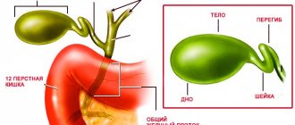
The esophagus is a hollow muscular tube that carries solid and liquid food from the throat to the stomach.
The wall of the esophagus consists of several layers of tissue, namely:
- mucous;
- muscular;
- connective tissue.
Esophageal cancer, as a rule, begins its development from the inner mucous membrane and, as it grows, spreads to other layers. The stomach and esophagus belong to the upper parts of the digestive tract.