1.General information
A bezoar is a concretory formation in the stomach, that is, in fact, an analogue of kidney, gallstones or fecal stones. Some sources indicate that the term comes from the Arabic “bâzahr”, while others consider it to be a translation from the French “bézoard”.
The absolute frequency of occurrence in terms of the general population is not given in the literature and is usually not even approximately estimated. As a rule, only the specific shares of various types (see below) in the total flow of bezoars included in the register, or the share of bezoars (approx. 1%) among the causes of intestinal obstruction, are statistically analyzed. In any case, the formation of bezoar stones is interpreted as a rare or, rather, rarely diagnosed phenomenon. Often without showing any clinical symptoms, bezoars can exist in the stomach for years and are discovered only when there is a tendency to grow, the appearance of discomfort, during an autopsy, or simply by chance (for example, during an examination performed for other indications). Wikipedia reports that by the end of the twentieth century, only about 400 cases of detection of bezoars in humans were described in the world medical literature. However, this information is hardly accurate. According to Perm gastroenterologists, who recently carried out a deep statistical and bibliographic search, domestic doctors alone reported at least 300 cases of bezoar stones in the stomach (since 1912, when such an observation was published for the first time). However, this does not fundamentally change the overall picture: even if the real frequency is hundreds of times higher than the estimated frequency, in relation to the population size it will still remain very small. Another question is that the difficulties of objective diagnosis, as well as the lack of sufficient personal experience among therapists and gastroenterologists in this area (when analyzing a specific case, doctors do not immediately remember that the cause may be a bezoar, and not a tumor, inflammation, etc.) – cause a very large proportion of incorrect diagnoses: according to the results of the above-mentioned statistical analysis, at the first stages of the examination, a bezoar is identified correctly only in every tenth case.
In different historical eras and in different cultures (including modern fantasy art), miraculous and protective properties were attributed to bezoar stones: most often they were considered an antidote or even a panacea. In ruminants and some marine animals, bezoars are much more common than in humans.
A must read! Help with treatment and hospitalization!
Introduction
Bezoars of the gastrointestinal tract (GIT) are a pathology that is extremely rare in the practice of a surgeon. According to their origin, bezoars are divided into phyto-, stibo (sebo)-, trichobezoars, as well as foreign bodies of organic (resin, tar, mineralization of a blood clot in the lumen of the stomach) or embryonic origin [1, 2]. In this case, the most common finding should be considered phytobezoars (up to 70-75% of observations), which, as a rule, are noted in the general population, while trichobezoars, which are a conglomerate of hair and food masses, are a consequence of trichotillomania and are most often recorded in a contingent with emotional lability, as well as sluggish schizophrenia [1, 3-6]. In the case of trichotillomania, trichophagia is detected in 1/3 of patients, and the risk of the formation of conglomerates and the development of complications in the form of obstruction, organ perforation occurs with a frequency not exceeding 0.064% [3]. As a rule, this pathology is more common in females who have an irresistible desire to bite (bite) and swallow their own hair. Gradually, conglomerates of intertwined hair and food masses of varying sizes and density form in the stomach, reaching the consistency of felt [1].
According to summary data, by 1991, about 400 descriptions of trichobezoars of gastric localization were registered in the world press [7], and in Pub Med as of February 2021, a little more than 1300 literary references were identified, with the combination of the terms “gastrictrichobezoar” and “Rapunzelsyndrome” (extreme variant of trichobezoars of the gastrointestinal tract) is registered only in 106 literary sources.
To highlight the problem of foreign bodies in the gastrointestinal tract, to the descriptions of this rarity, if not the casuistic nature of the pathology, let us add an expressive example of a giant trichobezoar of the stomach in a young woman of 24 years old.
Clinical example
Patient S.
, 24 years old, applied on June 14, 2016 as planned to the surgical clinic of the State Budgetary Healthcare Institution of the City Clinical Hospital named after. V.V. Veresaeva with complaints of heaviness in the epigastric region, dyspepsia, and a feeling of rapid satiety. The above-described complaints are noted by the patient for 3-4 months. Fibrogastroduodenoscopy was performed on an outpatient basis on 05/31/16, and a trichobezoar up to 20 cm in length was detected in the lumen of the stomach, occupying almost the entire lumen of the organ and consisting of hair and food masses (Fig. 1),
Rice.
1. a, b. Endoscopic photo of a trichobezoar in the lumen of the stomach. upon instrumental palpation it showed a loose consistency. A decision was made to inpatient treatment and endoscopic extraction of the foreign body. On objective examination: the patient has satisfactory nutrition and proper physique. Emotionally labile. No hemodynamic or respiratory disorders were noted. The abdomen is of regular shape, soft, painless, pathological mass formations are not palpable. The patient was consulted by a psychiatrist on an outpatient basis at the prehospital stage: no organic pathology was detected, a picture of a hysteroid personality type with pronounced emotional lability. A clinical blood test showed signs of mild hypochromic anemia (blood Hb 107 g/l, RBC 4.12×10¹²/l, color index 0.78).
Taking into account the findings, the patient was prepared for endoscopic extraction of the foreign body. Esophagogastroduodenoscopy from 06/15/16: almost the entire lumen of the stomach is occupied by a trichobezoar measuring 18.0×5.0×5.5 cm, consisting on the outside of many long hairs with inclusions of food masses. The gastric mucosa is patchily hyperemic. An overtube is installed into the esophagus using a flexible endoscope to protect the oropharynx and esophagus. A two-legged gripper was passed through the working channel of the endoscope to remove foreign bodies. Attempts have been made to fragment the trichobezoar using various instruments and various methods - without achieving results. With a deeper instrumental grasp and palpation of the bezoar with an instrument, its extremely dense consistency is noted at a depth of about 1.0 cm from the surface along its entire length, and therefore, given the size of the formation and the impossibility of its intraluminal endoscopic removal, the study was stopped.
Taking into account the impossibility of endoscopic extraction, the patient was offered surgical removal of the foreign body using video laparoscopic technologies. Laparoscopy of the abdominal cavity from June 16, 2016: the stomach is enlarged throughout its entire length, palpation reveals a dense, displaceable formation, occupying up to 75-80% of the volume of the organ cavity. A gastrotomy up to 5-6 cm long was performed along the greater curvature; a foreign body in the form of an organ cast, consisting of hair and food masses, rocky in depth and loose at the periphery, was identified in the stomach cavity. The foreign body is placed in an endocontainer, the latter is removed from the abdominal cavity through a minilaparotomy (4 cm) incision in the area of the optical trocar. The gastrostomy opening was sutured with a double-row atraumatic intracorporeal suture (Vicryl 2/0), the abdominal cavity was sanitized, and drained in the subhepatic space. Desufflation. Sutures of trocar wounds. The macropreparation is represented by a conglomerate of hair with food masses, rocky density down to a cloth roll in the depths of the conglomerate, with areas of softening on the periphery, measuring 18.0×6.0×6.5 cm (Fig. 2)
Rice. 2. The removed macropreparation is a trichobezoar of the stomach. with the smell of fermentation. The mass of the removed conglomerate is slightly more than 1.5 kg.
The course of the postoperative period is smooth, the patient’s early activation. The drainage was removed on the 2nd day after surgery. She was discharged on the 5th day for outpatient follow-up treatment. The sutures were removed on the 10th day, the trocar wounds and the perprima minilaparotomy approach were healing.
Examination six months later: the patient has satisfactory nutrition, has no complaints, recovery. During endoscopic examination, foreign bodies are not detected in the gastric cavity.
2. Reasons
Despite a certain similarity with stones formed in the urinary tract, biliary ducts, intestines, etc., the etiopathogenesis of gastric bezoars is still different; in addition, to date it has not been sufficiently studied.
If in the first case the main etiopathogenetic role is played by the deposition of salts of various organic acids, then the trigger factor for the formation of bezoars is the ingestion of indigestible substances into the stomach: hair (especially if the patient has an obsessive habit of gnawing or biting hair), casein, synthetic fibers, etc.
There are many such variants known (pyxo-, tricho-, sebobezoars, and many others). Thus, bezoars that had the property of burning were repeatedly extracted from the stomachs of alcoholics who regularly consumed alcohol-containing chemical compounds.
However, three quarters of bezoar stones are of plant origin (phytobezoars). In this regard, it is quite well known that the most dangerous products are those that are dense, fibrous, leathery, with a high content of astringent, sticky, tannin, resinous substances (nuts, dates, figs, wild plums, sunflower seeds, etc.), and the leader Persimmons always appear on this list - most often unripe, or completely ripe, but consumed on an empty stomach and in large quantities.
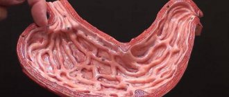
Such lumps of plant fiber, especially if it is poorly chewed, become the center of compacted formations (usually round in shape and with a smooth surface), which are then often compressed to stone hardness. In general, stone-hard bezoars are more common - sometimes such a stone cannot be broken with a hammer - however, much less dense formations are also known, with a consistency reminiscent, for example, of thick dough.
As a rule, bezoar stones have a foul odor. The dimensions are usually several centimeters, but giant bezoars have also been encountered, representing almost a complete internal cast of the gastric space. In approximately 15% of cases, the stone is formed not in the stomach, but in the small intestine.
Risk factors are disturbances in the evacuation of gastric contents, the vital activity of yeast fungi (for which the bezoar substance serves as a very favorable nutrient medium), hyposecretion of gastric juice, viscous mucus, insufficient primary processing of food in the oral cavity (due to dental problems, constant haste, or simply a bad swallowing habit). large pieces, practically without chewing them), as well as some previous surgical interventions, for example, vagotomy with pyloroplasty.
Visit our Gastroenterology page
Preventing the formation of stones in the stomach
Since the most common type of bezoar is a trichobezoar, prevention in this case is aimed at sedative therapy in psychiatric patients and the formation of correct behavior in children (the child should be explained why this cannot be done in calm conditions), just as the method can be applied to short haircuts in children. these risk groups.
Special protective equipment (masks, respirators) are necessary for people of various professions who deal with hair, wool and fibers of other nature.
Photo: https://pixabay.com/photos/smoothies-fruit-fruits-vegetables-3809508/
In cases of the formation of phytobezoar and steobezoar stones, we are talking about diet. That is, limiting certain types of food and eating patterns.
People after surgical interventions should also remember about the diet (gentle cooking and fractional meals) in order to prevent complications in an already weak stomach or intestines.
3. Symptoms and diagnosis
The clinical picture depends on a number of factors - the number of bezoars (they can be multiple), their size, location, composition, “age”, mobility - and therefore varies widely. In the first stages, symptoms may be completely absent, and for a very long period. Then, as gastric functions become impaired, nonspecific symptoms appear: nausea, vomiting, loss of appetite, belching, abdominal pain, general weakness, weight loss, sensations of a full stomach or, if large, a “ball” inside, etc.
If the formation of multiple bezoars is recurrent in nature and if they are mobile enough to be evacuated into the duodenum before reaching a significant size (more often the stone remains in the stomach), the patient’s condition can change in waves, but there are also cases when a bezoar becomes the cause of obstruction and obstruction Gastrointestinal tract.
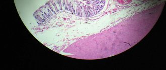
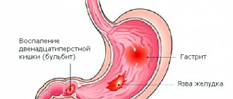
It is necessary to clearly differentiate a bezoar stone from a malignant tumor, gastroenteritis and other types of symptomatically similar pathology. The most informative diagnostic method is FEGDS; sometimes radiography and ultrasound can also help establish the correct diagnosis. Careful examination of all available anamnestic information is essential.
About our clinic Chistye Prudy metro station Medintercom page!
What is a bezoar?
A bezoar, forming in the stomach, is a lump that subsequently interferes with the digestion of food and contributes to the retention of any elements (food, hair, etc.), thereby increasing in size more and more.
In some sources, the term gastric bezoar does not imply the presence of a stone in the intestine, which, on the one hand, is correct. But there are cases when a bezoar can be in the stomach and continue in the intestines, so the article uses the wording bezoars of the stomach and small intestine. Stone formation always begins in the stomach!
4.Treatment
As a rule, an attempt is first made to eliminate the problem using conservative methods, for example, a soda solution in combination with a special massage, which in many cases is sufficient for the destruction and subsequent evacuation of the bezoar in parts. If the desired effect is absent, endoscopic destruction or removal is tried. If this does not produce results, they resort to surgical removal; in the presence of severe complications (ulceration, obstruction with a large stone, etc.), the operation is performed urgently, urgently or as an emergency.
The prognosis is usually favorable, especially if the diagnosis is made timely and accurately (for this, first of all, it is necessary to promptly seek help with the complaints described above).
Classification
Bezoars can be of different origins, that is, have different compositions.
So, what could such a stone consist of:
- trichobezoar - a stone made of hair, wool or fiber (thread);
- phytobezoar - a stone made of cellulose (dietary fiber);
- steobezoar – a stone made from crystallized fats (tard);
- organic bezoar - a stone made from blood clots treated with gastric juice;
- embryonic bezoar - a stone in a newborn child (meconium stones and stones from a cyst formed in the prenatal period);
- mixed stones.
Reviews from our patients
- Feedback on the treatment of urolithiasis and cholelithiasis
Review of the treatment of urolithiasis and cholelithiasis June 25, 2021The patient suffered from pain in the lower abdomen. She visited various medical institutions. There the pain syndrome was relieved, after which they were sent home. At the Medicine 24/7 clinic, she was examined, which showed the presence of stones in the kidneys and gall bladder. Two operations were performed. Problem solved.
read more
- Feedback on the treatment of cholelithiasis
Feedback on the treatment of gallstone disease May 14, 2021
Review of treatment at the Medicine 24/7 clinic. Patient Nina Iosifovna was taken to the clinic in serious condition. After stabilizing her condition in the intensive care unit, the patient underwent surgery to remove stones in the bile duct. Thanks to the efforts of resuscitators and surgeons, Nina Iosifovna feels well, moves independently and takes care of herself, vital signs...
read more
Possible complications
Complications develop, as a rule, in patients with large bezoars, with undiagnosed bezoars (a sudden manifestation is possible, that is, everything begins with a complication) and when patients refuse surgical interventions (not immediately, after some time).
A bezoar, localized on one of the walls of the stomach, can provoke a so-called bedsore of the wall. With the pressure created by the stone (bezoar), the stomach wall is poorly supplied with blood and does not receive enough oxygen. Areas of ulceration (ulcers and erosions) may form in this area.
In the future, when the process is started, there may be perforation of the wall, that is, a hole (hole) is formed at the site of the ulcer and the contents of the stomach are poured into the abdominal cavity, which entails the development of peritonitis (purulent melting) and sepsis (contamination of various organs by bacteria through the blood vessels). All these complications are acute surgical pathologies and require emergency medical care (specialized surgical care).
And another complication is intestinal obstruction. It develops in cases where a bezoar penetrates the cavity of the small intestine and “grows” there. Intestinal obstruction can also trigger the development of peritonitis and sepsis and also requires immediate medical attention.